Review
. 2014 Dec 17;6(12):5047-76.
doi: 10.3390/v6125047.

Architectural insight into inovirus-associated vectors (IAVs) and development of IAV-based vaccines inducing humoral and cellular responses: implications in HIV-1 vaccines

Affiliations
Review

Architectural insight into inovirus-associated vectors (IAVs) and development of IAV-based vaccines inducing humoral and cellular responses: implications in HIV-1 vaccines

Kyriakos A Hassapis et al. Viruses. .

Abstract

Inovirus-associated vectors (IAVs) are engineered, non-lytic, filamentous bacteriophages that are assembled primarily from thousands of copies of the major coat protein gp8 and just five copies of each of the four minor coat proteins gp3, gp6, gp7 and gp9. Inovirus display studies have shown that the architecture of inoviruses makes all coat proteins of the inoviral particle accessible to the outside. This particular feature of IAVs allows foreign antigenic peptides to be displayed on the outer surface of the virion fused to its coat proteins and for more than two decades has been exploited in many applications including antibody or peptide display libraries, drug design, and vaccine development against infectious and non-infectious diseases. As vaccine carriers, IAVs have been shown to elicit both a cellular and humoral response against various pathogens through the display of antibody epitopes on their coat proteins. Despite their high immunogenicity, the goal of developing an effective vaccine against HIV-1 has not yet materialized. One possible limitation of previous efforts was the use of broadly neutralizing antibodies, which exhibited autoreactivity properties. In the past five years, however, new, more potent broadly neutralizing antibodies that do not exhibit autoreactivity properties have been isolated from HIV-1 infected individuals, suggesting that vaccination strategies aimed at producing such broadly neutralizing antibodies may confer protection against infection. The utilization of these new, broadly neutralizing antibodies in combination with the architectural traits of IAVs have driven the current developments in the design of an inovirus-based vaccine against HIV-1. This article reviews the applications of IAVs in vaccine development, with particular emphasis on the design of inoviral-based vaccines against HIV-1.


Figures

Figure 1
Figure 1
A 3D scale schematic model of an end-to-end Ff (fd) inoviral virion. The model is based on published physical data including the determined helical parameters of the major coat protein gp8 and the X-ray structure of the N1-N2 domains of the minor coat protein gp3. The model shows the relative location of the circular single-stranded DNA (cssDNA) genome (6408 nucleotides long, illustrated as blue ribbons), some structural details of the outer virion capsid (major coat protein pg8) and the four minor coat proteins (gp6, gp3, gp7 and gp9) present at the ends of the virion. On top, a digital scanning transmission electron micrograph (STEM) of unstained fd virus, prepared by the wet-film technique according to previously established procedures of the Brookhaven STEM facility [32,33,34]. The ends of one complete virion are designated by arrows. The data were collected in collaboration with L.A. Day and J. S. Wall at the Brookhaven National Laboratory, Upton New York. Under these STEM conditions fd virions are about 8800 Å long and about 65 Å in diameter [3]. In the middle, a proposed end-to-end scale 3D diagram of fd virion is presented. The entire fd virion is composed of about 2700 subunits of gp8 with the exception of its two ends. Architectural details of an axial slab 176 Å long (about 1/50 of the virion length) consisting entirely of subunits of major coat protein gp8. The structure of the 50-amino-acid-long and extended α-helical gp8, shown below in both surface and ribbon images, is presented in the virion model as a cylindrical stack of 25 gray disks about 70 Å long and 10 Å in diameter. The images of gp8 were derived from coordinates of RCSB PDB database accession number 2cOW [35] using PyMOL [36]. The gp8 subunits are arranged with a helical symmetry that includes a two-fold screw axis and a five-fold rotation axis, consisting of two pentamers of pg8 [28,29,30,35]. The two pentamers are architecturally related to each other by a translation of about 16 Å along the virion axis and a rotation of 36° about the axis [28]. The proximal end of the virion, shown on the left, is composed of five copies of each of the minor coat proteins gp6 and gp3 (for a recent review see [4]). The proximal end is modeled based on partial information known about the structures of gp6 and gp3. Specifically, the N-terminal portion of gp6 was modeled following the helical parameters of gp8, based on protein sequence homology between the two [23,24,25]. Five copies of gp3 subunits were modeled based on structural information of the N1-N2 domains. The images of the N1-N2 domains of gp3 are shown below and were derived from coordinates of RCSB PDB database accession number 1g3p [36] using PyMOL. The domain organization of gp3 is also shown. The distal end of the virion (right) consists of five copies of each minor coat proteins gp7 and gp9, modeled following the helical parameters of gp8 according to a previously published model [25].
Figure 2
Figure 2
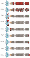
Schematic representations of antigen display on the surface of Ff inovirus-associated vectors (IAVs). Foreign antigens are shown as red spheres. The designation on the left denotes the inoviral gene, which can be genetically modified to express an antigen on the outer architecture of the virion. IAVs that contain both the wild type and antigen display capsid proteins are designated by “m” which indicates that the virion is a mosaic. Each Ff virion contains about 2700 copies of major capsid protein gp8, and five copies of each of the minor capsid proteins, gp3, gp6, gp7 and gp9.
Figure 3
Figure 3
Surface lattice diagrams of wild-type Ff the Ff.g8 inovirus-associated vector. Left, virion surface models of axial slabs about 130 Å long of wild-type Ff (top) and Ff.g8 inovirus-associated vector (bottom) showing the major coat protein gp8 subunits arranged with a combined five-fold rotation axis and an approximate two-fold screw axis [28]. Right, the corresponding surface lattices, identical to those previously published [30]. The lattice diagrams show the relative position of each gp8 subunit on the outer virion surface. The five gp8 subunits of each of the two interlocking pentamers constituting the helical symmetry of the virion are indicated by blue and green dots respectively. The relative virion surface area (about 1400 Å2) associated with each gp8 subunit is marked in yellow. The virion perimetrical (azimuthial) distance is calculated based on a virion diameter of about 65 Å. The displayed antigens, represented by red spheres, are arranged on the surface of the Ff.g8 inovirus-associated vector according to helical symmetry of the virion outer architecture (bottom).

References

    1. Day L.A. Inoviruses. In: Mahy B.W.J., Regenmortel M.H.V.V., editors. Encyclopedia of Virology. 3rd ed. Academic Press; Oxford, UK: 2008. pp. 117–124.
    1. Marvin D.A., Symmons M.F., Straus S.K. Structure and assembly of filamentous bacteriophages. Prog. Biophys. Mol. Biol. 2014;114:80–122. - PubMed
    1. Day L.A., Marzec C.J., Reisberg S.A., Casadevall A. DNA packing in filamentous bacteriophages. Annu. Rev. Biophys. Biophys. Chem. 1988;17:509–539. - PubMed
    1. Rakonjac J., Bennett N.J., Spagnuolo J., Gagic D., Russel M. Filamentous bacteriophage: Biology, phage display and nanotechnology applications. Curr. Issues Mol. Biol. 2011;13:51–76. - PubMed
    1. Brown L.R., Dowell C.E. Replication of coliphage m-13. I. Effects on host cells after synchronized infection. J. Virol. 1968;2:1290–1295. - PMC - PubMed

Publication types