Review
. 2015 Mar;22(3):398-407.
doi: 10.1038/cdd.2014.204. Epub 2014 Dec 19.

Autophagy in the physiology and pathology of the central nervous system

Affiliations
Review

Autophagy in the physiology and pathology of the central nervous system

V Nikoletopoulou et al. Cell Death Differ. 2015 Mar.

Abstract

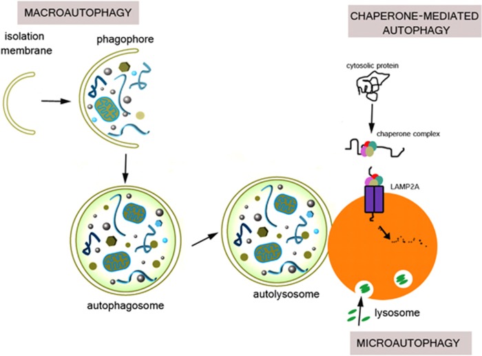
Neurons are highly specialized postmitotic cells that depend on dynamic cellular processes for their proper function.These include among others, neuronal growth and maturation, axonal migration, synapse formation and elimination, all requiring continuous protein synthesis and degradation. Therefore quality-control processes in neurons are directly linked to their physiology. Autophagy is a tightly regulated cellular degradation pathway by which defective or superfluouscytosolic proteins, organelles and other cellular constituents are sequestered in autophagosomes and delivered to lysosomes for degradation. Here we present emerging evidence indicating that constitutive autophagic fluxin neurons has essential roles in key neuronal processes under physiological conditions.Moreover, we discuss how perturbations of the autophagic pathway may underlie diverse pathological phenotypes in neurons associated with neurodevelopmental and neurodegenerative diseases.


Figures

Figure 1
Figure 1
Schematic representation of the three types of autophagy: Microautophagy, CMA, and macroautophagy. In microautophagy, invaginations of the lysosomal membrane directly engulf portions of the cytoplasm. By contrast, CMA involves the chaperone Hsc70 and its co-chaperones that recognize and unfold substrate proteins and then bind to the lysosomal protein LAMP2A and are translocated across the lysosomal membrane for degradation. In macroautophagy, substrates are sequestered by an isolation membrane (known as the phagophore), which elongates and eventually seals to surround the substrate, forming a double membranous structure, the autophagosome. Autophagosomes then fuse with the lysosome to form autolysosomes
Figure 2
Figure 2
Impairment of autophagy leads to neurodegeneration. Schematic representation of (a) a healthy and (b) an autophagy-deficient neuron. Note that autophagy deficiency leads to aberrant aggregation of ubiquitinated proteins within inclusion bodies, as well as the accumulation of defective organelles such as mitochondria. Autophagy-deficient neurons display increased axonal diameter (swelling) and axonal degeneration, ultimately leading to age-dependent loss of neurons

References

    1. Boya P, Reggiori F, Codogno P. Emerging regulation and functions of autophagy. Nat Cell Biol. 2013;15:713–720. - PMC - PubMed
    1. Mizushima N, Komatsu M. Autophagy: renovation of cells and tissues. Cell. 2011;147:728–741. - PubMed
    1. Orenstein SJ, Cuervo AM. Chaperone-mediated autophagy: molecular mechanisms and physiological relevance. Semin Cell Dev Biol. 2010;21:719–726. - PMC - PubMed
    1. Lee JA. Neuronal autophagy: a housekeeper or a fighter in neuronal cell survival. Exp Neurobiol. 2012;21:1–8. - PMC - PubMed
    1. Singh R, Cuervo AM. Autophagy in the cellular energetic balance. Cell Metab. 2011;13:495–504. - PMC - PubMed

Publication types