. 2015 Aug;22(8):1275-86.
doi: 10.1038/cdd.2014.209. Epub 2014 Dec 19.

Anoikis evasion in inflammatory breast cancer cells is mediated by Bim-EL sequestration

Affiliations

Anoikis evasion in inflammatory breast cancer cells is mediated by Bim-EL sequestration

C L Buchheit et al. Cell Death Differ. 2015 Aug.

Abstract

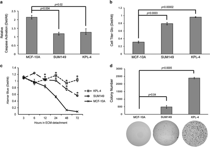
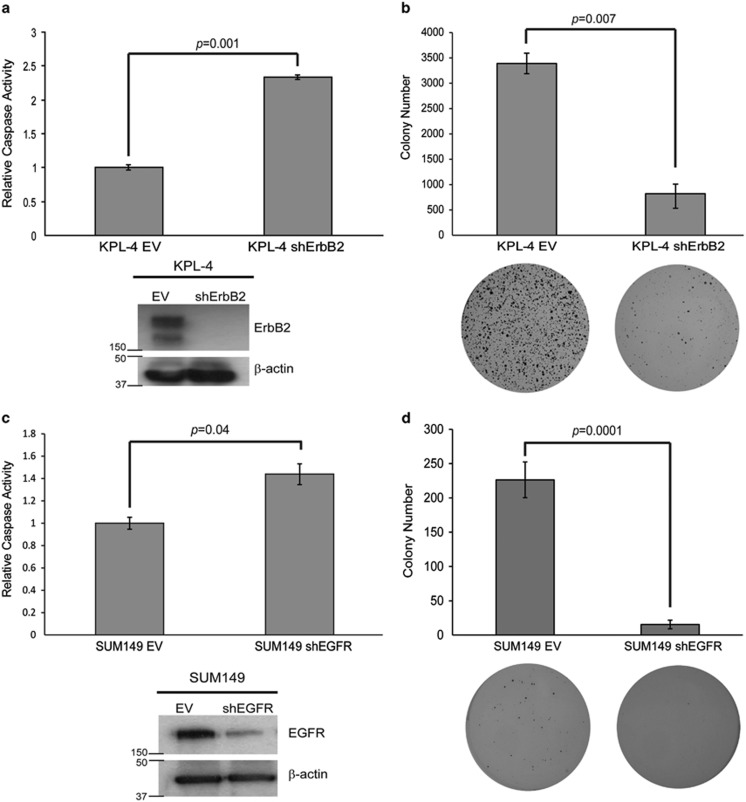
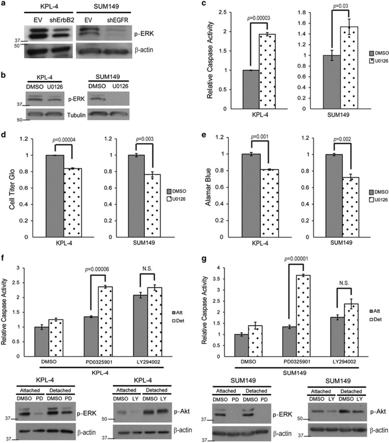
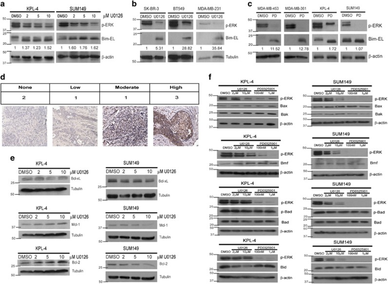
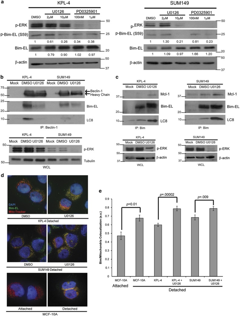
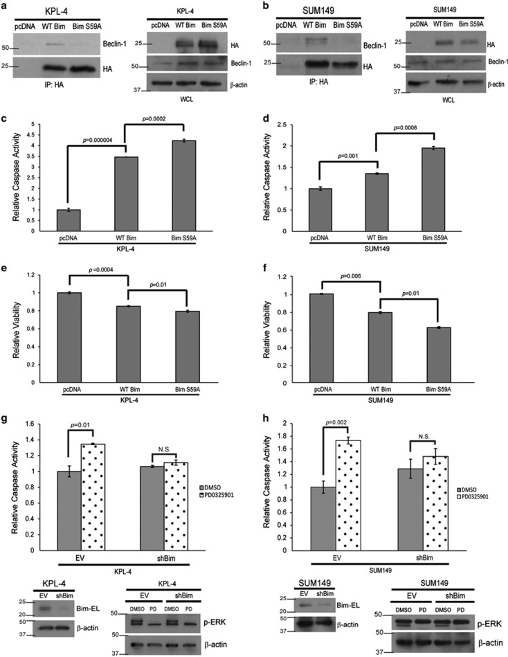
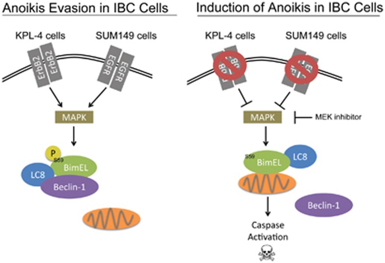
Inflammatory breast cancer (IBC) is a rare and highly invasive type of breast cancer, and patients diagnosed with IBC often face a very poor prognosis. IBC is characterized by the lack of primary tumor formation and the rapid accumulation of cancerous epithelial cells in the dermal lymphatic vessels. Given that normal epithelial cells require attachment to the extracellular matrix (ECM) for survival, a comprehensive examination of the molecular mechanisms underlying IBC cell survival in the lymphatic vessels is of paramount importance to our understanding of IBC pathogenesis. Here we demonstrate that, in contrast to normal mammary epithelial cells, IBC cells evade ECM-detachment-induced apoptosis (anoikis). ErbB2 and EGFR knockdown in KPL-4 and SUM149 cells, respectively, causes decreased colony growth in soft agar and increased caspase activation following ECM detachment. ERK/MAPK signaling was found to operate downstream of ErbB2 and EGFR to protect cells from anoikis by facilitating the formation of a protein complex containing Bim-EL, LC8, and Beclin-1. This complex forms as a result of Bim-EL phosphorylation on serine 59, and thus Bim-EL cannot localize to the mitochondria and cause anoikis. These results reveal a novel mechanism that could be targeted with innovative therapeutics to induce anoikis in IBC cells.


Figures

Figure 1
Figure 1
IBC cells survive in ECM-detached conditions. (a) Cells were plated on either normal or poly-HEMA coated plates, and caspase activation was measured at 48 h using the Caspase-Glo assay. (b) Cells were plated in attached or detached conditions, and cell viability was measured at 48 h post-plating via Cell Titer Glo Assay. (c) Cells were plated on either normal or poly-HEMA coated plates, and cell viability was measured at the indicated times via alamarBlue assay. (d) MCF-10A and SUM149 cells were plated at 100 000 cells/well, and KPL-4 cells were plated at 60 000 cells/well in soft agar. Colonies were stained at either 14 days (KPL-4) or 30 days (SUM149, MCF-10A) using 0.01% INT-violet and quantified with ImageJ. Error bars represent S.E.M. *P-value<0.05 compared with MCF-10A
Figure 2
Figure 2
ErbB2 and EGFR are necessary for anoikis protection in KPL-4 and SUM149 cells, respectively. (a) KPL-4 cells were transduced with a lentivirus containing either an empty vector (EV) or an ErbB2 shRNA-containing vector (shErbB2), and western blotting analysis confirmed the knockdown. KPL-4 EV and shErbB2 cells were plated on poly-HEMA-coated plates, and caspase activation was quantified at 48 h as previously described. (b) KPL-4 EV and shErbB2 cells were plated at 60 000 cells/well in soft agar as described above. Colonies were stained at 14 days with 0.01% INT-violet and quantified with ImageJ. (c) SUM149 cells were transduced with a lentivirus containing either an empty vector (EV) or an EGFR shRNA-containing vector (shEGFR), and western blotting analysis confirmed the knockdown. SUM149 EV and shEGFR cells were plated on poly-HEMA-coated plates, and caspase activation was quantified at 48 h as previously described. (d) SUM149 EV and shEGFR cells were plated at 100 000 cells/well in soft agar as previously described. Colonies were stained after 30 days using 0.01% INT-violet and quantified with ImageJ. Error bars represent S.E.M.
Figure 3
Figure 3
ERK/MAPK signaling is necessary for anoikis protection. (a) KPL-4 EV and shErbB2 and SUM149 EV and shEGFR cells were plated on poly-HEMA-coated plates for 48 h, and p-ERK levels were measured via immunoblotting. (b and c) KPL-4 and SUM149 cells were plated on poly-HEMA-coated plates in the presence of DMSO or U0126 (10 μM). Caspase activation was measured at 48 h as previously described. Inhibition of the ERK/MAPK pathway was confirmed via western blotting analysis. (d and e) KPL-4 and SUM149 cells were plated in ECM detachment with DMSO or U0126 (10 μM). Cellular viability was measured with the Cell Titer Glo Assay (d) or alamarBlue assay (e). (f and g) KPL-4 (f) and SUM149 (g) cells were plated in attached or detached conditions with DMSO, PD0325901 (1 μM), or LY294002 (25 μM). Caspase activation was measured at 24 h. Cell lysates were prepared and normalized, and protein levels were analyzed via western blotting analysis to confirm inhibitor efficacy. Error bars represent S.E.M. NS, not signifcant
Figure 4
Figure 4
MEK inhibition does not affect Bcl-2 family member protein levels in IBC cells. (a) Cells were plated on poly-HEMA-coated plates with DMSO or increasing amounts of U0126 for 48 h, and protein content was analyzed via immunoblotting. (b) Cells were plated in ECM-detached conditions with DMSO or U0126 (10 μM) for 48 h. Cell lysates were prepared for immunoblotting. (c) Cells were plated on poly-HEMA-coated plates with DMSO or PD0325901 (1 μM) for 48 h, and lysates were prepared for immunoblotting. (d) Seven IBC patient tissue samples were obtained, and IHC was utilized to visualize Bim protein levels in the tissue. Images were blind scored for staining intensity. (e) Cells were plated in detachment and treated with DMSO or increasing amounts of U0126 for 48 h. Lysates were prepared and normalized, and protein levels were analyzed via immunoblotting. (f) KPL-4 and SUM149 cells were plated in detachment with the indicated doses of U0126 or PD0325901 for 48 h. Protein levels were analyzed via western blotting
Figure 5
Figure 5
ERK/MAPK signaling leads to the sequestration of Bim-EL from the mitochondria to prevent anoikis. (a) Cells were plated on poly-HEMA-coated plates with DMSO or the indicated dose of U0126 or PD0325901 for 6 h. Lysates were prepared and normalized, and protein levels were analyzed via western blotting. (b) Cells were plated in detachment and treated with DMSO or U0126 (10 μM) for 3 h. Cell lysates were prepared, and immunoprecipitation was performed with a Beclin-1 antibody. Western blotting analysis was utilized to identify interacting proteins and confirm equivalent protein content across samples. (c) Cells were plated in detachment and treated with DMSO or U0126 (10 μM) for 3 h. Cell lysates were prepared and normalized, and immunoprecipitation was performed with a Bim antibody. Interacting proteins were identified via immunoblotting, and equivalent protein content across samples was confirmed. (d and e) Cells were plated on poly-HEMA-coated plates and treated with either DMSO or U0126 (10 μM) and 20 μM z-VAD-fmk for 24 h. Cells were fixed and stained with Mito-Tracker Red (200 nM), DAPI (5 μg/ml), and Bim-EL and imaged using an Applied Precision DeltaVision OMX fluorescent microscope. Co-localization was measured using the Applied Precision softWoRx software. Error bars represent S.E.M.
Figure 6
Figure 6
Bim is necessary and sufficient for anoikis in IBC cells (a and b) KPL-4 (a) and SUM149 (b) cells were transfected with pcDNA EV, HA-WT Bim, or HA-Bim S59A constructs using Lipofectamine LTX. Lysates were prepared and normalized. Coimmunoprecipitation experiments were conducted with an HA antibody. Immunoblotting was used to identify interacting proteins. (c and d) KPL-4 (c) and SUM149 (d) cells were transfected with pcDNA EV, HA-WT Bim, or HA-Bim S59A constructs using Lipofectamine LTX. Cells were plated in suspension and caspase activity was measured as described above. (e and f) KPL-4 (e) and SUM149 (f) cells were transfected as previously described. Cells were plated in suspension, and cellular viability was measured using Cell Titer Glo. (g and h) KPL-4 (g) and SUM149 (h) cells were transduced with shRNA-containing lentivirus to create stable Bim knockdowns (shBim). EV and shBim cells were plated in ECM detachment and treated with DMSO or PD0325901 (1 μM). Caspase activation was measured at 48 h, and western blotting analysis confirmed the Bim knockdown and inhibitor efficacy. NS, not signifcant
Figure 7
Figure 7
Model for anoikis evasion in IBC cells

References

    1. Gonzalez-Angulo AM, Hennessy BT, Broglio K, Meric-Bernstam F, Cristofanilli M, Giordano SH, et al. Trends for inflammatory breast cancer: is survival improving. Oncologist. 2007;12:904–912. - PubMed
    1. Woodward WA, Cristofanilli M. Inflammatory breast cancer. Semin Radiat Oncol. 2009;19:256–265. - PubMed
    1. Harris EE, Schultz D, Bertsch H, Fox K, Glick J, Solin LJ. Ten-year outcome after combined modality therapy for inflammatory breast cancer. Int J Radiat Oncol Biol Phys. 2003;55:1200–1208. - PubMed
    1. Lerebours F, Bieche I, Lidereau R. Update on inflammatory breast cancer. Breast Cancer Res. 2005;7:52–58. - PMC - PubMed
    1. Yamauchi H, Woodward WA, Valero V, Alvarez RH, Lucci A, Buchholz TA, et al. Inflammatory breast cancer: what we know and what we need to learn. Oncologist. 2012;17:891–899. - PMC - PubMed

Publication types

MeSH terms