. 2014 Dec 19;9(12):e114263.
doi: 10.1371/journal.pone.0114263. eCollection 2014.

Integrated analysis of whole genome and transcriptome sequencing reveals diverse transcriptomic aberrations driven by somatic genomic changes in liver cancers

Affiliations

Integrated analysis of whole genome and transcriptome sequencing reveals diverse transcriptomic aberrations driven by somatic genomic changes in liver cancers

Yuichi Shiraishi et al. PLoS One. .

Abstract

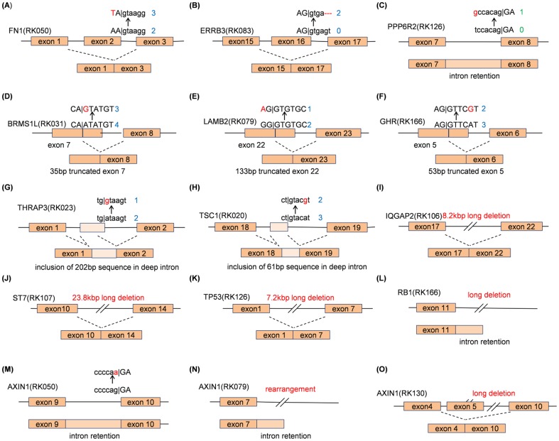
Recent studies applying high-throughput sequencing technologies have identified several recurrently mutated genes and pathways in multiple cancer genomes. However, transcriptional consequences from these genomic alterations in cancer genome remain unclear. In this study, we performed integrated and comparative analyses of whole genomes and transcriptomes of 22 hepatitis B virus (HBV)-related hepatocellular carcinomas (HCCs) and their matched controls. Comparison of whole genome sequence (WGS) and RNA-Seq revealed much evidence that various types of genomic mutations triggered diverse transcriptional changes. Not only splice-site mutations, but also silent mutations in coding regions, deep intronic mutations and structural changes caused splicing aberrations. HBV integrations generated diverse patterns of virus-human fusion transcripts depending on affected gene, such as TERT, CDK15, FN1 and MLL4. Structural variations could drive over-expression of genes such as WNT ligands, with/without creating gene fusions. Furthermore, by taking account of genomic mutations causing transcriptional aberrations, we could improve the sensitivity of deleterious mutation detection in known cancer driver genes (TP53, AXIN1, ARID2, RPS6KA3), and identified recurrent disruptions in putative cancer driver genes such as HNF4A, CPS1, TSC1 and THRAP3 in HCCs. These findings indicate genomic alterations in cancer genome have diverse transcriptomic effects, and integrated analysis of WGS and RNA-Seq can facilitate the interpretation of a large number of genomic alterations detected in cancer genome.


Conflict of interest statement

Competing Interests: The authors have declared that no competing interests exist.

Figures

Figure 1
Figure 1. The outline of theRNA-Seq study integrated with whole genome sequencing.
(A) First, we detected various types of genomic and transcriptomic changes from RNA-Seq data of 22 HCCs. The characterized changes detected by each analysis were compared to reveal the effects of somatic genomic changes on transcriptomic aberrations. (B) The four types of splicing aberrations defined in this study. Green lines and arrows indicate normal transcription whereas red lines and arrows indicate aberrant transcriptions.
Figure 2
Figure 2. Several examples of genomic changes other than essential splice-site mutations causing splicing aberrations obtained from our comparative whole genome and transcriptome sequencing analyses.
Exonic and intronic sequences are designated by capital and small letters, respectively. Red sequences are somatic mutations in HCCs. Blue and green numbers on the side of sequences are edit distances from splicing donor motif (AG|GTRAGT, [38]) and splicing acceptor motif (YYYYNCAG|G), respectively. Most somatic mutations changed the edit distance to splicing donor motifs so that the corresponding alteration can be enhanced.
Figure 3
Figure 3. The expression profiles of 22 HCCs and non-cancerous liver samples for eight over-expressed genes.
Blue and red bars show the FKPMs for HCCs and the corresponding non-cancerous liver, respectively, which is calculated by RNA-Seq data. Red circles indicate samples with HBV integrations on the loci of the overexpressed genes. Green circles indicate those with gene fusions and/or SVs that can drive gene over-expression.
Figure 4
Figure 4. HBV integrations and fusion events in 22 HCCs.
(A) Seven HBV-TERT fusion transcripts were detected in RK010. One transcript was an un-spliced transcript having the same breakpoint as the genomic integration breakpoint. The others existed in spliced forms and GT-AG splicing motifs were observed at the breakpoints of all but one. In addition to HBV fusion splicing hotspot (458 bp), 3 fusion transcripts were spliced at the coordinate of 1634 bp coordinates in HBV sequences. One fusion transcript included a newly generated 87 bp pseudo-exon sequence as well as subsequence exonic sequences. (B) HBV integrations in the MLL4 loci and their resultant fusion transcripts in five samples. Green triangles on the genome sequence show the HBV integration sites. Most fusion transcripts shared breakpoints with those of genomic HBV integration coordinates for both sides, and thus, they appear to exist in un-spliced forms. The fusion transcripts for RK141 and RK159 were validated to be concatenated (Figure S11). (C) HBV-FN1 fusion transcripts for 7 adjacent non-cancerous liver samples. Almost all the fusion transcripts had the breakpoint at the HBV fusion splicing hotspot. The other fusion transcripts which had breakpoints at intronic regions appear to be un-spliced transcripts around the integration sites.
Figure 5
Figure 5. RNA editing candidates in 22 HCCs.
(A) The number of cancer-specific RNA mutation events (RNA editing candidates) and their substitution patterns for each sample. (B) Scatter plot between the number of A:T>G:C RNA-editing events and ADAR expression value (FKPM) calculated by whole transcriptome sequence data. There is a significant correlation (P-value  = 2.38×10⁻⁷ by Wilcoxon rank sum test) between the number of A:T>G:C events and ADAR expression levels.
Figure 6
Figure 6. The status of genomic and transcriptomic alterations of representative genes, detected by WGS and RNA-Seq of 22 HBV-related HCCs.
The list of genes were extracted by (1) significantly mutated genes in WGS analysis, (2) having no less than 3 mutations (point mutations or indels in coding regions, or GMTAs), (3) having no less than 2 GMTAs and registered in cancer gene census . (4) involved in WNT signaling pathway, (5) TERT or MLL4.

References

    1. El-Serag HB (2011) Hepatocellular carcinoma. N Engl J Med 365:1118–1127. - PubMed
    1. Farazi PA, DePinho RA (2006) Hepatocellular carcinoma pathogenesis: from genes to environment. Nat Rev Cancer 6:674–687. - PubMed
    1. Meyerson M, Gabriel S, Getz G (2010) Advances in understanding cancer genomes through second-generation sequencing. Nat Rev Genet 11:685–696. - PubMed
    1. Fujimoto A, Totoki Y, Abe T, Boroevich KA, Hosoda F, et al. (2012) Whole-genome sequencing of liver cancers identifies etiological influences on mutation patterns and recurrent mutations in chromatin regulators. Nat Genet 44:760–764. - PubMed
    1. Huang J, Deng Q, Wang Q, Li KY, Dai JH, et al. (2012) Exome sequencing of hepatitis B virus-associated hepatocellular carcinoma. Nat Genet 44:1117–1121. - PubMed

Publication types