Review
. 2014 Dec 17;15(12):23448-500.
doi: 10.3390/ijms151223448.

Protecting the melatonin rhythm through circadian healthy light exposure

Affiliations
Review

Protecting the melatonin rhythm through circadian healthy light exposure

Maria Angeles Bonmati-Carrion et al. Int J Mol Sci. .

Abstract

Currently, in developed countries, nights are excessively illuminated (light at night), whereas daytime is mainly spent indoors, and thus people are exposed to much lower light intensities than under natural conditions. In spite of the positive impact of artificial light, we pay a price for the easy access to light during the night: disorganization of our circadian system or chronodisruption (CD), including perturbations in melatonin rhythm. Epidemiological studies show that CD is associated with an increased incidence of diabetes, obesity, heart disease, cognitive and affective impairment, premature aging and some types of cancer. Knowledge of retinal photoreceptors and the discovery of melanopsin in some ganglion cells demonstrate that light intensity, timing and spectrum must be considered to keep the biological clock properly entrained. Importantly, not all wavelengths of light are equally chronodisrupting. Blue light, which is particularly beneficial during the daytime, seems to be more disruptive at night, and induces the strongest melatonin inhibition. Nocturnal blue light exposure is currently increasing, due to the proliferation of energy-efficient lighting (LEDs) and electronic devices. Thus, the development of lighting systems that preserve the melatonin rhythm could reduce the health risks induced by chronodisruption. This review addresses the state of the art regarding the crosstalk between light and the circadian system.


Figures

Figure 1
Figure 1
General overview of the functional organization of the circadian system in mammals. Inputs: environmental periodical cues can reset the phase of the central pacemaker so that the period and phase of circadian rhythms coincide with the timing of the external cues; Central pacemakers: the suprachiasmatic nuclei (SCN) is considered to be the major pacemaker of the circadian system, driving circadian rhythmicity in other brain areas and peripheral tissues by sending them neural and humoral signals (such as melatonin, secreted by the pineal gland (P)). The SCN receives light-dark cycle information through the retinohypothalamic tract (RHT). Peripheral oscillators: most peripheral tissues and organs contain circadian oscillators. Usually, they are under the control of the SCN; however, under some circumstances (e.g., restricted feeding, jet lag and shift work), they can desynchronize from the SCN; Outputs: central pacemakers and peripheral oscillators are responsible for the daily rhythmicity observed in most physiological and behavioral functions. Some of these overt rhythms (physical exercise, core temperature, sleep-wake cycle and feeding time), in turn, provide feedback, which can modify the function of the SCN and peripheral oscillators, (redrawn from [11]).
Figure 2
Figure 2
Molecular clock of mammals. Circadian locomotor output cycles kaput (CLOCK)/brain and muscle aryl hydrocarbon receptor nuclear translocator-like (BMAL1) heterodimers (red and green ovals) bind the DNA of clock target genes at E-boxes or E’-boxes and permit their transcription. The resulting period (PER) and cryptochrome (CRY) proteins (blue and yellow) dimerize in the cytoplasm and translocate to the nucleus where they inhibit CLOCK/BMAL1 proteins from initiating further transcription (redrawn from [16]).
Figure 3
Figure 3
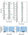
Absence and presence of circadian photoreception in two totally blind subjects. A and B correspond to the sleep-wake pattern and the results of melatonin suppression test in a 70-year old blind patient with congenital glaucoma who reported no conscious light perception and whose electroretinogram (ERG) and visually evoked potential (VEP) responses were not detectable. In (A), the sleep-wake pattern is double-plotted according to time of day (abscissa) and study day (ordinate). It is evident that the subject’s circadian system was not entrained to the light-dark cycle, and the core body temperature rhythm (circle) exhibited a non-24-h period; (B) shows the null effect of light (white bar) on melatonin secretion; C and D correspond to a 21-year-old woman with Leber’s congenital amaurosis, a type of retinal dystrophy. The ERG was undetectable, but an abnormal VEP was recorded. As represented in C, her circadian system was normally entrained (24-h period) and melatonin secretion was suppressed when she was exposed to light. Both results indicated that this patient, despite her lack of conscious light perception, preserved the retina-SCN-pineal pathway (reproduced from [122]).
Figure 4
Figure 4
Schematic view of brain regions and circuits inervated by intrinsically photosensitive retinal ganglion cells (ipRGCs). The location of their somas, axons and main targets are represented in blue. Projections of ipRGCs to the SCN (orange) allow photic entrainment of the circadian clock. The red pathway with green nuclei represents a polysynaptic circuit originating in the SCN, which photically regulates melatonin release by the pineal gland (P) through sympathetic innervation. Synaptic links in this pathway include the paraventricular nucleus (PVN) of the hypothalamus, the intermediolateral nucleus (IML) of the spinal cord and the superior cervical ganglion (SCG). The olivary pretectal nucleus (OPN) is another direct target of ipRGCs, and is a crucial link in the circuitry underlying the pupillary light reflex, shown in brown (fibers) and purple (nuclei). Synapses in this parasympathetic circuit are found at the Edinger-Westphal nucleus (EW), the ciliary ganglion (CG) and the iris muscles (I). Other targets of the ipRGCs include two components of the lateral geniculate nucleus of the thalamus, the ventral division (LGNv) and the intergeniculate leaflet (IGL) (reproduced from [138]).
Figure 5
Figure 5
Example of a pupillographic recording in response to a 5-s bright white light stimulus in a normal human subject. The response waveform during the constriction phase has two components. When the light is turned ON, there is transient phase characterized by a short-latency, high-velocity maximal change in pupil size. Thereafter, the pupil partly redilates, or escapes, to a state of partial pupil constriction that represents the sustained phase of the pupil light reflex. When the light stimulus ends, the pupil starts to recover its original size after a period (which does not always occur) in which some degree of contraction persists after the light stimulus (modified from [155]).
Figure 6
Figure 6
Spectral responses of the pupillary light reflex (PLR). Comparison of the PLR to 480 and 620 nm monochromatic long-duration (5 min) stimulations at 6 different irradiances in a single subject. After the initial and rapid pupil constriction, the steady state equilibrium and the persistent responses are present in all except the lowest irradiances, thus depending on wavelength and light intensity. The amplitude of the steady state equilibrium response is rapidly attained and particularly robust at 480 nm for the highest irradiances used. The persistent responses are also greater at 480 nm, as compared to 620 nm at equivalent irradiances. Note that for the higher irradiances, the pupil has not yet returned to the baseline within 5 min after extinction of the stimulus (reproduced from [134]).
Figure 7
Figure 7
Age-related losses in retinal illumination due to decreasing crystalline lens light transmission and pupil area. The percentage of loss per decade is reasonably uniform and most prominent at shorter violet (400–440 nm) and blue (440–500 nm) wavelengths (reproduced from [294]).
Figure 8
Figure 8
Short wavelength light sensitivity for melatonin suppression. Comparison of the effects of 460 nm (blue circles) and 555 nm (green circles) light exposure on melatonin suppression in a blind man. The graph represents the direct effects for melatonin suppression of exposure to green (555 nm) and blue (460 nm) monochromatic light on the male subject. Exposure to 555 nm light caused no suppression of melatonin as compared to the corresponding clock time the previous day, whereas exposure to 460 nm light suppressed melatonin and maintained the suppression effect throughout the entire 6.5 h exposure (reproduced from [140]).

References

    1. Haim A., Portnov B.A. Light Pollution as a New Risk Factor for Human Breast and Prostate Cancers. Springer Netherlands; Dordrecht, The Netherlands: 2013.
    1. De Beaune S.A., White R. Ice Age Lamps. Sci. Am. 1993;206:108–113. doi: 10.1038/scientificamerican0393-108. - DOI
    1. Nordhaus W.D., Nordhaus W.D. Do real-output and real-wage measures capture reality? The history of lighting suggests not. In: Brenahan T.F., Gordon J., editors. The Economics of New Goods. The University of Chicago Press; Chicago, IL, USA: 1994. pp. 27–70.
    1. Cinzano P., Falchi F., Elvidge C.D. The first world atlas of the artificial night sky brightness. Mon. Not. R. Astron. Soc. 2001;328:689–707. doi: 10.1046/j.1365-8711.2001.04882.x. - DOI
    1. Navara K.J., Nelson R.J. The dark side of light at night: Physiological, epidemiological, and ecological consequences. J. Pineal Res. 2007;43:215–224. doi: 10.1111/j.1600-079X.2007.00473.x. - DOI - PubMed

Publication types

LinkOut - more resources