

Proximal tibiofibular dislocation: a case report and review of literature
- PMID: 25526858
- PMCID: PMC4278967
- DOI: 10.1007/s11751-014-0209-8
Proximal tibiofibular dislocation: a case report and review of literature
Abstract
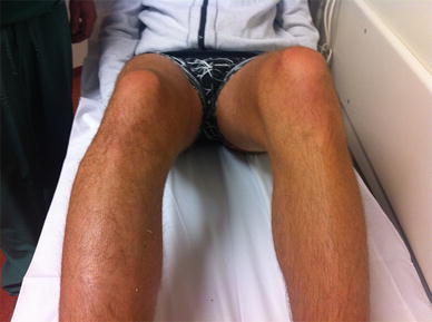
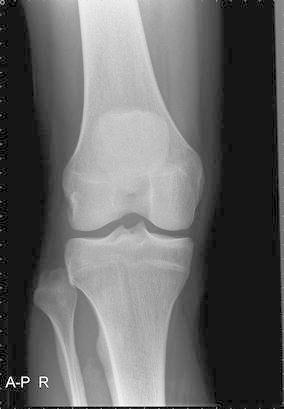
An isolated dislocation of the proximal tibiofibular joint is uncommon. The mechanism of this injury is usually sports related. We present a case where initial X-rays did not show the tibiofibular joint dislocation conclusively. It was diagnosed after comparative bilateral AP X-rays of the knees were obtained. A closed reduction was performed and followed by unrestricted mobilization after 1 week of rest. A review of the literature was conducted on PubMed MEDLINE. Thirty cases of isolated acute proximal tibiofibular joint dislocations were identified in a search from 1974. The most common direction of the dislocation was anterolateral, and common causes were sports injury or high velocity accident related. More than 75 % of the cases were successfully treated by closed reduction. Complaints, if any, at the last follow-up (averaging 10 months, range 0-108) were, in the worst cases, pain during sporting activities. We advise comparative knee X-rays if there is a presentation of lateral knee pain after injury and diagnosis is uncertain. Closed reduction is usually successful if a dislocation of the proximal tibiofibular joint is diagnosed. There is no standard for after-care, but early mobilization appears safe if there are no other knee injuries.
Figures


References
-
- Ogden JA (1974) The anatomy and function of the proximal tibiofibular joint. Clin Orthop Relat Res (101):186–191 - PubMed
-
- Nelaton A. Eléments de pathologie chirurgicale. 2. Paris: Librairie Germer Ballière; 1874. p. 282.
-
- Sekiya JK, Kuhn JE. Instability of the proximal tibiofibular joint. J Am Acad Orthop Surg. 2003;11:120–128. - PubMed
-
- Ogden JA. Subluxation and dislocation of the proximal tibiofibular joint. J Bone Joint Surg Am. 1974;56(1):145–154. - PubMed
LinkOut - more resources
Full Text Sources
Other Literature Sources
