

Endogenous assessment of chronic myocardial infarction with T(1ρ)-mapping in patients
- PMID: 25526973
- PMCID: PMC4272542
- DOI: 10.1186/s12968-014-0104-y
Endogenous assessment of chronic myocardial infarction with T(1ρ)-mapping in patients
Abstract
Background: Detection of cardiac fibrosis based on endogenous magnetic resonance (MR) characteristics of the myocardium would yield a measurement that can provide quantitative information, is independent of contrast agent concentration, renal function and timing. In ex vivo myocardial infarction (MI) tissue, it has been shown that a significantly higher T(1ρ) is found in the MI region, and studies in animal models of chronic MI showed the first in vivo evidence for the ability to detect myocardial fibrosis with native T(1ρ)-mapping. In this study we aimed to translate and validate T(1ρ)-mapping for endogenous detection of chronic MI in patients.
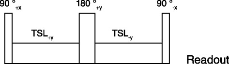
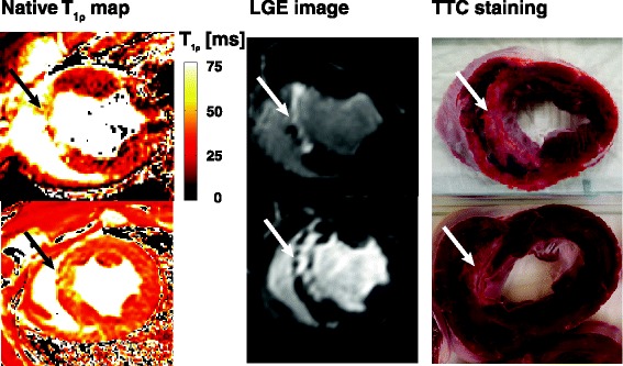
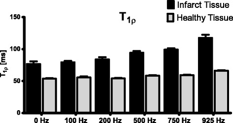
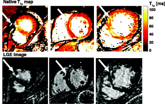
Methods: We first performed a study in a porcine animal model of chronic MI to validate the implementation of T(1ρ)-mapping on a clinical cardiovascular MR scanner and studied the correlation with histology. Subsequently a clinical protocol was developed, to assess the feasibility of scar tissue detection with native T(1ρ)-mapping in patients (n = 21) with chronic MI, and correlated with gold standard late gadolinium enhancement (LGE) CMR. Four T1ρ-weighted images were acquired using a spin-lock preparation pulse with varying duration (0, 13, 27, 45 ms) and an amplitude of 750 Hz, and a T(1ρ)-map was calculated. The resulting T(1ρ)-maps and LGE images were scored qualitatively for the presence and extent of myocardial scarring using the 17-segment AHA model.
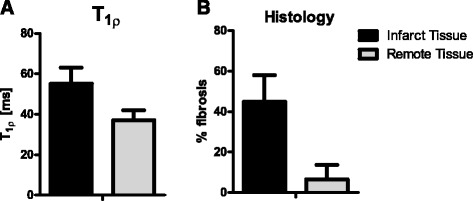
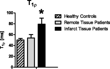
Results: In the animal model (n = 9) a significantly higher T(1ρ) relaxation time was found in the infarct region (61 ± 11 ms), compared to healthy remote myocardium (36 ± 4 ms) . In patients a higher T(1ρ) relaxation time (79 ± 11 ms) was found in the infarct region than in remote myocardium (54 ± 6 ms). Overlap in the scoring of scar tissue on LGE images and T(1ρ)-maps was 74%.
Conclusion: We have shown the feasibility of native T(1ρ)-mapping for detection of infarct area in patients with a chronic myocardial infarction. In the near future, improvements on the T(1ρ)-mapping sequence could provide a higher sensitivity and specificity. This endogenous method could be an alternative for LGE imaging, and provide additional quantitative information on myocardial tissue characteristics.
Figures
References
-
- Kwong RY, Chan AK, Brown K a, Chan CW, Reynolds HG, Tsang S, Davis RB. Impact of unrecognized myocardial scar detected by cardiac magnetic resonance imaging on event-free survival in patients presenting with signs or symptoms of coronary artery disease. Circulation. 2006;113:2733–43. doi: 10.1161/CIRCULATIONAHA.105.570648. - DOI - PubMed
-
- Müller K a L, Müller I, Kramer U, Kandolf R, Gawaz M, Bauer A, Zuern CS. Prognostic Value of Contrast-enhanced Cardiac Magnetic Resonance Imaging in Patients with Newly Diagnosed Non-Ischemic Cardiomyopathy: Cohort Study. PLoS One. 2013;8:e57077. doi: 10.1371/journal.pone.0057077. - DOI - PMC - PubMed
-
- El Aidi H, Adams A, Moons KGM, Den Ruijter HM, Mali WPTM, Doevendans PA, Nagel E, Schalla S, Bots ML, Leiner T. Cardiac magnetic resonance imaging findings and the risk of cardiovascular events in patients with recent myocardial infarction or suspected or known coronary artery disease: a systematic review of prognostic studies. J Am Coll Cardiol. 2014;63:1031–45. doi: 10.1016/j.jacc.2013.11.048. - DOI - PubMed
Publication types
MeSH terms
Substances
LinkOut - more resources
Full Text Sources
Other Literature Sources
Medical
