. 2015 Jan 30;290(5):2798-811.
doi: 10.1074/jbc.M114.620906. Epub 2014 Dec 19.

PINK1 kinase catalytic activity is regulated by phosphorylation on serines 228 and 402

Affiliations

PINK1 kinase catalytic activity is regulated by phosphorylation on serines 228 and 402

Liesbeth Aerts et al. J Biol Chem. .

Abstract

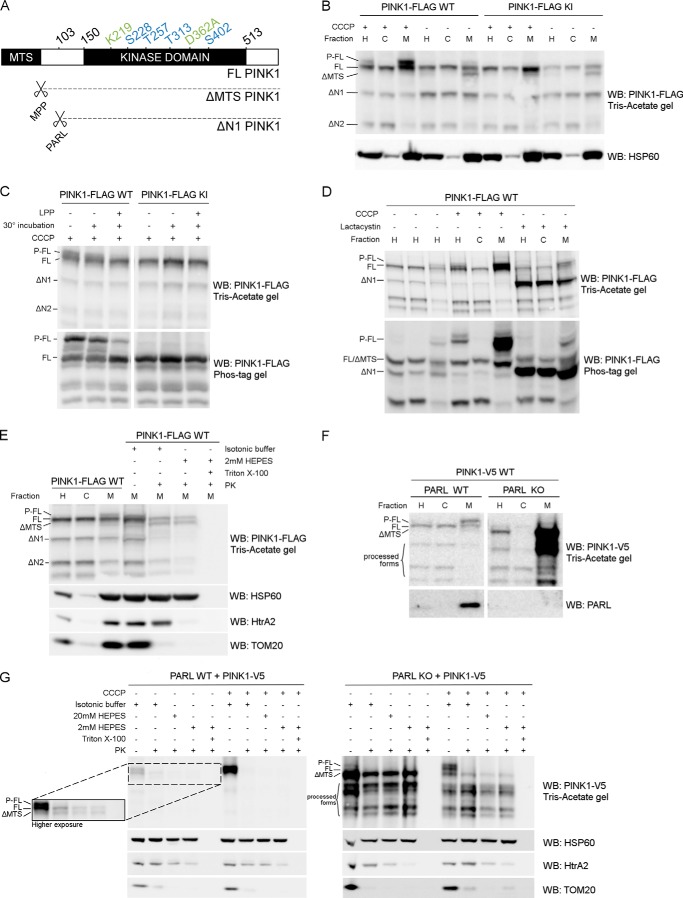
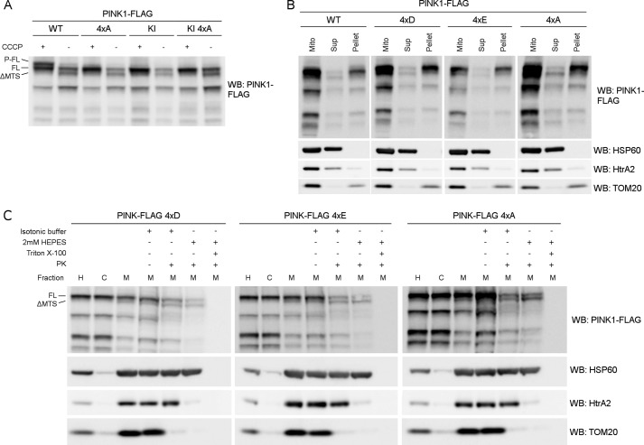
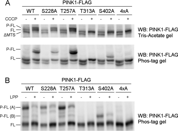
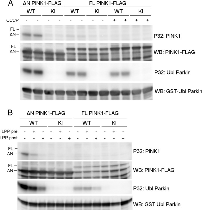
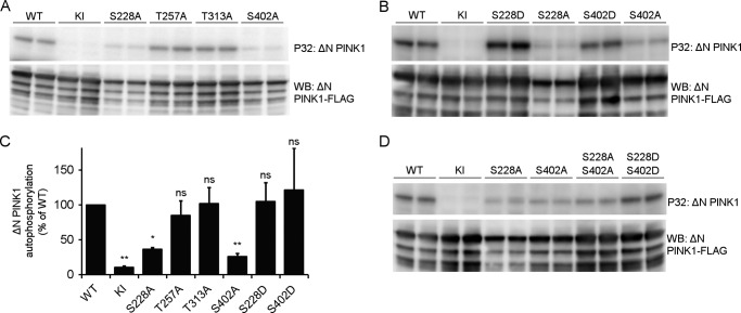
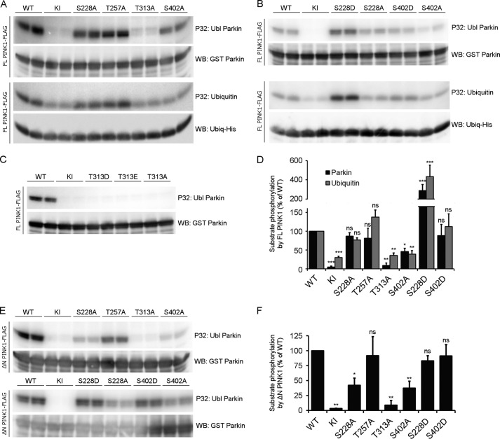
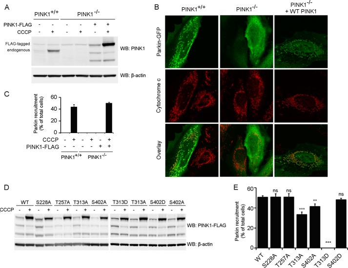
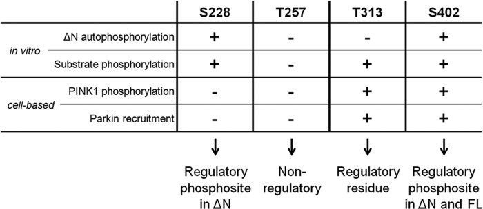
Mutations in the PINK1 gene cause early-onset recessive Parkinson disease. PINK1 is a mitochondrially targeted kinase that regulates multiple aspects of mitochondrial biology, from oxidative phosphorylation to mitochondrial clearance. PINK1 itself is also phosphorylated, and this might be linked to the regulation of its multiple activities. Here we systematically analyze four previously identified phosphorylation sites in PINK1 for their role in autophosphorylation, substrate phosphorylation, and mitophagy. Our data indicate that two of these sites, Ser-228 and Ser-402, are autophosphorylated on truncated PINK1 but not on full-length PINK1, suggesting that the N terminus has an inhibitory effect on phosphorylation. We furthermore establish that phosphorylation of these PINK1 residues regulates the phosphorylation of the substrates Parkin and Ubiquitin. Especially Ser-402 phosphorylation appears to be important for PINK1 function because it is involved in Parkin recruitment and the induction of mitophagy. Finally, we identify Thr-313 as a residue that is critical for PINK1 catalytic activity, but, in contrast to previous reports, we find no evidence that this activity is regulated by phosphorylation. These data clarify the regulation of PINK1 through multisite phosphorylation.

Keywords: Mitochondria; Neurodegenerative Disease; PTEN-induced Putative Kinase 1 (PINK1); Parkinson Disease; Posttranslational Modification (PTM); Protein Phosphorylation; Serine/Threonine Protein Kinase.


Figures

FIGURE 1.
FIGURE 1.
Phosphorylated PINK1 is present at the outer mitochondrial membrane. A, schematic representing FL PINK1, including the kinase domain and MTS. MPP cleaves off the MTS, resulting in ΔMTS PINK1, and further processing by PARL at residue 103 results in the ΔN1 PINK1 product. The four phosphorylation sites analyzed in this study (blue) and the sites mutated in KI (green) PINK1 are indicated. B, HEK cells transiently transfected with WT or KI FLAG-tagged human PINK1 and treated with 10 μm CCCP for 3 h were fractionated in postnuclear or homogenate fraction (H), and subsequent H fraction was further centrifuged at 7000 × g to obtain the mitochondrially depleted or cytosol-enriched (C) and mitochondrially enriched (M) fractions, respectively. Expression of PINK1 was evaluated after SDS-PAGE on 7.5% Tris acetate gel and immunoblotted using anti-FLAGM2. Immunoblotting for the mitochondrial marker HSP60 served as a fractionation quality control. For WT, but not KI, a doublet was observed that corresponded to the phosphorylated and non-phosphorylated FL form of PINK1 (P-FL and FL, respectively). Further processed PINK1 forms are indicated as ΔMTS, ΔN1, and ΔN2. WB, Western blot. C, mitochondrially enriched fractions, obtained from HEK cells transfected with WT and KI PINK1-FLAG and incubated with 10 μm CCCP for 3 h, were treated with LPP at 30 °C. Samples were analyzed by SDS-PAGE on a 7.5% Tris acetate gel and 7.5% Phos-Tag gel and immunoblotted with anti-FLAGM2 antibody for PINK1 detection. A control for loss of phosphorylation by 30 °C incubation alone was included. Phosphatase treatment confirmed that the upper band (P-FL) was a phosphorylated form of PINK1. D, fractionation of HEK cells treated for 3 h with either 10 μm CCCP or 10 μm lactacystin was analyzed by SDS-PAGE on a 7.5% Tris acetate or 7.5% Phos-Tag gel and further immunoblotted for PINK1 detection using anti-FLAGM2 antibody. Mitochondrial PINK1 was phosphorylated under control (DMSO) conditions but accumulated upon CCCP-induced depolarization. Proteasomal blockage with lactacystin also led to a modest increase of P-FL PINK1 in addition to accumulation of the ΔN1 processed form. E, mitochondrially enriched fractions of MEF cells stably expressing FL PINK1-FLAG were subjected to a PK sensitivity assay to assess submitochondrial localization of P-FL PINK1. P-FL is PK-sensitive, whereas non-phosphorylated FL PINK1 and PINK1 lacking the mitochondrial targeting sequence (ΔMTS) are at least partially protected from PK even under hypotonic conditions (2 mm HEPES). As a control for protein digestion under each condition, fractions were also immunoblotted using antibodies against the outer membrane protein TOM20, intermembrane space protein HtrA2, and matrix protein HSP60. F, MEFs derived from PARL WT and PARL KO mice stably expressing WT PINK1-V5 were fractionated and expression of PINK1 was evaluated after SDS-PAGE and anti-V5 immunoblotting. ΔMTS PINK1 accumulates in PARL KO cells and further N-terminal processing is altered leading to an accumulation of PINK1 C-terminal fragments in the mitochondria (M) of PARL KO cells. G, mitochondrially enriched fractions from PARL WT and KO MEFs stably expressing WT PINK1-V5 were subjected to PK treatment under isotonic and hypotonic (20 mm and 2 mm HEPES) conditions in the presence or absence of detergent. Equal amounts were loaded for PARL WT and KO. A higher exposure inset for PARL WT allows better analysis of PINK1 expression under these conditions. ΔMTS PINK1 was cleaved by PARL and accumulated in PARL KO mitochondria but was only digested by PK in the presence of detergent. Depolarization induced by CCCP led to the accumulation of P-FL PINK1, sensitive to proteinase K under isotonic conditions. Therefore, the accumulation caused by the absence of PARL or by CCCP-induced depolarization concerns different PINK1 forms present in different submitochondrial compartments. As a control, the PK sensitivity of the outer membrane protein TOM20, intermembrane space protein HtrA2, and matrix protein HSP60 was evaluated by immunoblotting.
FIGURE 2.
FIGURE 2.
Mutation of Ser-228, Thr-257, Thr-313, and Ser-402 affects phosphorylation without interfering with localization or processing. A, mitochondrially enriched fractions of DMSO (control) or 10 μm CCCP-treated HEK cells transiently expressing WT and KI PINK1-FLAG WT with and without the quadruple mutation of the putative phosphorylation sites Ser-228, Thr-257, Thr-313, and Ser-402 (4×A) were analyzed by SDS-PAGE and immunoblotting against anti-FLAGM2 for PINK1 detection. Results show that P-FL PINK1 no longer accumulates for 4×A quadruple mutant PINK1. WB, Western blot. B, mitochondrially enriched fractions from MEF cells stably expressing WT, phosphomimetic (4×D and 4×E) or phosphodead (4×A) quadruple PINK1-FLAG mutants were treated with Na2CO3 (pH 11.5). The majority of PINK1 was not extracted by Na2CO3 indicating that the WT and the quadruple mutants are membrane-associated proteins. Expression of soluble HSP60 and HtrA2 and membrane-associated TOM20 was evaluated as a control for Na2CO3 extraction. Mito, mitochondria; Sup, supernatant. C, Proteinase K sensitivity was tested on mitochondrially enriched fractions from MEF cells stably expressing either 4×D, 4×E, and 4×A quadruple mutant PINK1-FLAG. The distribution of FL and processed PINK1 forms was not altered because the pattern for mitochondrial fractions and PK sensitivity under isotonic and hypotonic (2 mm HEPES) conditions was unchanged. H, post-nuclear or homogenate fraction; C, mitochondria-depleted or cytosolic fraction; M, mitochondria-enriched fraction. As a control for protein digestion under each condition, the PK sensitivity of the outer membrane protein TOM20, intermembrane space protein HtrA2, and matrix protein HSP60 was evaluated. H, post-nuclear or homogenate fraction; C, mitochondria-depleted or cytosolic fraction; M, mitochondria-enriched fraction.
FIGURE 3.
FIGURE 3.
Thr-313 and Ser-402 are required for PINK1 phosphorylation at the mitochondrial outer membrane. A, mitochondrially enriched fractions from HEK cells transiently transfected with different phosphomutant forms of PINK1-FLAG and treated with DMSO or 10 μm CCCP were analyzed by SDS-PAGE on 7.5% Tris acetate and 7.5% Phos-Tag gels. The presence of P-FL, FL, and ΔMTS PINK1 was assessed by immunoblotting using anti-FLAGM2 antibody. Although FL PINK1 accumulated upon CCCP treatment for every evaluated mutant, P-FL PINK1 was not detected or altered for T313A, S402A, and the 4×A quadruple mutant PINK1. WB, Western blot. B, mitochondrial fractions from CCCP-treated HEK cells transfected with different phosphomutant PINK1-FLAG forms were treated with LPP and further probed with anti-FLAGM2 antibody for PINK1 detection. The bands P-FL(A) and P-FL(B) show sensitivity to LPP, indicating that they are both phosphorylated forms of PINK1 (P-FL A and B).
FIGURE 4.
FIGURE 4.
Full-length PINK1 shows lack of autophosphorylation activity in vitro. A, an in vitro phosphorylation assay using [γ-32P]ATP was performed with purified WT and KI PINK1-FLAG and purified GST-Ubl Parkin as substrate. Although WT ΔN PINK1 shows autophosphorylation and is able to phosphorylate the substrate Parkin, the autoradiogram shows no detectable autophosphorylation for FL PINK1, although FL PINK1 phosphorylates the Ubl-domain of Parkin. WB, Western blot. B, PINK1 was dephosphorylated using LPP prior (pre) to incubation with [γ-32P]ATP and Ubl Parkin in an in vitro phosphorylation. This dephosphorylation did not reveal an increase in detectable FL PINK1 autophosphorylation. The same LPP treatment of PINK1 and Parkin after (post) completion of the kinase assay did lead to a considerable amount of Parkin and ΔN PINK1 dephosphorylation, indicating that protein dephosphorylation was successful under the applied conditions.
FIGURE 5.
FIGURE 5.
Autophosphorylation of ΔN PINK1 occurs at residues Ser-228 and Ser-402. A, an in vitro phosphorylation assay using [γ-32P]ATP was performed with purified human ΔN PINK1 harboring phosphodead mutations on four putative phosphosites. The phosphomutants S228A and S402A showed reduced phosphorylation, whereas mutation of Thr-257 and Thr-313 did not affect ΔN PINK1 autophosphorylation when compared with WT. B, in vitro phosphorylation assay using [γ-32P]ATP and purified ΔN PINK1 with phosphomimetic and phosphodead mutant PINK1 for residues Ser-228 and Ser-402 shows that a phosphomimetic mutant PINK1 can restore PINK1 phosphorylation levels observed for the corresponding phosphodead mutation. C, quantification of ΔN PINK1 autophosphorylation relative to WT (mean ± S.E., n = 3 independent experiments). Statistical significance was calculated between each mutant and WT ΔN PINK1 using Dunnett's test. *, p < 0.05; **, p < 0.01; ns, nonspecific. D, in vitro phosphorylation assay using [γ-32P]ATP and purified ΔN PINK1 with a combined S228A and S402A mutation shows decreased ΔN PINK1 autophosphorylation levels comparable with the levels for the single mutations, indicating the existence of residual phosphorylated residue(s) on PINK1. The combined phosphomimetic mutation S228D and S402D shows increased in vitro autophosphorylation levels, showing that phosphorylation of these two residues increases PINK1 kinase activity and phosphorylation of the residual phosphoresidue(s). Immunoblot analysis using anti-FLAGM2 shows that equal amounts of PINK1 were applied.
FIGURE 6.
FIGURE 6.
The residues Thr-313 and Ser-402 are important for substrate phosphorylation. A, an in vitro phosphorylation assay using [γ-32P]ATP was performed on purified FL PINK1 with purified Ubl Parkin or His-tagged Ubiquitin. Autoradiographic exposure shows that, for FL PINK1, mutation of Ser-228 and Thr-257 does not affect Parkin or Ubiquitin phosphorylation. Mutation of Thr-313 completely abrogates substrate phosphorylation, whereas the S402A mutation leads to a substantial decrease. There is still phosphorylation detectable for KI PINK1, indicating the presence of a contaminating kinase capable of phosphorylating Ubiquitin at low levels. Immunoblot analysis using anti-GST or anti-His confirms that equal amounts of Parkin and Ubiquitin were applied. WB, Western blot. B, an in vitro phosphorylation assay using [γ-32P]ATP, FL PINK1, and Ubl Parkin or Ubiquitin shows that the S228D and S402D mutations increase substrate phosphorylation. The S228D mutation increases substrate phosphorylation beyond WT levels, which are comparable with that obtained for S228A PINK1. The decreased Parkin phosphorylation for FL S402A PINK1 can be rescued by a phosphomimetic mutation, S402D. C, an in vitro phosphorylation assay using [γ-32P]ATP was performed on purified FL PINK1 mutated at the Thr-313 residue with purified Ubl Parkin. Phosphomimetic (T313D or T313E) mutation of Thr-313 does not rescue the decrease in Parkin phosphorylation observed for T313A. D, quantification of Ubl Parkin and Ubiquitin phosphorylation relative to WT. Statistical significance was calculated between each mutant and WT FL PINK1 using Dunnett's test. *, p < 0.05; **, p < 0.01; ***, p < 0.001; ns, nonspecific. Data are mean ± S.E. (n = 3 independent experiments). E, an in vitro phosphorylation assay using [γ-32P]ATP was performed using purified ΔN PINK1 and purified Ubl Parkin. Similar to FL PINK1, Parkin phosphorylation is abolished upon T313A mutation in ΔN PINK1, whereas the S228A and S402A mutations reduce it significantly. The S228D and S402D phosphomimetic mutations restore Parkin phosphorylation back to WT levels. F, quantification of Ubl Parkin phosphorylation by ΔN PINK1. Statistical significance was calculated between each mutant and WT ΔN PINK1 using Dunnett's test. *, p < 0.05; **, p <0.01; ns, nonspecific. Data are mean ± S.E. (n = 3 independent experiments).
FIGURE 7.
FIGURE 7.
Thr-313 and Ser-402 are important for Parkin recruitment by PINK1. A, WT (PINK1+/+) and PINK1 KO (PINK1−/−) HeLa cells were treated with 10 μm CCCP for 3 h and analyzed for PINK1 expression via Western blot (WB) analysis. No endogenous PINK1 was detected in PINK1−/− cells. B, HeLa cells were transfected with Parkin-GFP and treated with CCCP for 3 h. Immunohistochemistry for Parkin-GFP (green) and the mitochondrial marker cytochrome c (red) shows that WT (PINK1+/+) but not PINK1 KO (PINK1−/−) HeLa cells display mitochondrial Parkin recruitment. Expression of WT PINK1 in PINK1 KO cells rescues Parkin recruitment. Immunoblot analysis using anti-β-actin shows equal loading. C, quantification of Parkin recruitment in WT (PINK1+/+), KO (PINK1−/−), and KO HeLa cells rescued with human WT PINK1 after 3 h of DMSO or 10 μm CCCP treatment. Statistical significance was calculated between HeLa WT and PINK1 KO rescued cells using Student's t test. Data are mean ± S.E. (n = 6 independent experiments). D, PINK1 KO HeLa cells were transiently transfected with WT and mutant PINK1 and subsequently treated with DMSO or 10 μm CCCP for 3 h. PINK1 expression levels were analyzed by Western blot analysis. Immunoblot analysis using anti-β-actin shows equal loading. E, quantification of Parkin recruitment in PINK1 KO HeLa cells rescued with human WT or mutant PINK1 after 3 h of 10 μm CCCP treatment. Statistical significance was calculated between each mutant and WT PINK1 using Dunnett's test. **, p <0.01; ***, p < 0.001; ns, nonspecific. Data are mean ± S.E. (n = 4 independent experiments).
FIGURE 8.
FIGURE 8.
Overview of the role of Ser-228, Thr-257, Thr-313, and Ser-402 residues in PINK1. Ser-228 is an autophosphorylation site in ΔN PINK1, and it can regulate substrate phosphorylation in vitro. However, both in vitro and in cells, Ser-228 phosphorylation plays no role in the regulation of WT FL PINK1 activity. Nevertheless, we propose it as a regulatory phosphosite for processed PINK1. We found no implication for Thr-257 as a (regulatory) phosphosite in any of our experimental setups and, therefore, propose that this putative phosphosite has no functional role for PINK1 activity. Although Thr-313 is an essential residue for PINK1, its function is not regulated through phosphorylation because autophosphorylation is not affected upon Thr-313 mutation, and phosphomimetics rescue none of the observed functional defects. Like Ser-228, Ser-402 is an autophosphorylation site in ΔN PINK1, but it also regulates FL PINK1 in vitro and in cells. We propose this residue as a regulatory phosphosite for both FL and processed PINK1.

References

    1. Nuytemans K., Theuns J., Cruts M., Van Broeckhoven C. (2010) Genetic etiology of Parkinson disease associated with mutations in the SNCA, PARK2, PINK1, PARK7, and LRRK2 genes: a mutation update. Hum. Mutat. 31, 763–780 - PMC - PubMed
    1. Valente E. M., Abou-Sleiman P. M., Caputo V., Muqit M. M., Harvey K., Gispert S., Ali Z., Del Turco D., Bentivoglio A. R., Healy D. G., Albanese A., Nussbaum R., González-Maldonado R., Deller T., Salvi S., Cortelli P., Gilks W. P., Latchman D. S., Harvey R. J., Dallapiccola B., Auburger G., Wood N. W. (2004) Hereditary early-onset Parkinson's disease caused by mutations in PINK1. Science 304, 1158–1160 - PubMed
    1. Yang Y., Gehrke S., Imai Y., Huang Z., Ouyang Y., Wang J.-W., Yang L., Beal M. F., Vogel H., Lu B. (2006) Mitochondrial pathology and muscle and dopaminergic neuron degeneration caused by inactivation of Drosophila Pink1 is rescued by Parkin. Proc. Natl. Acad. Sci. U.S.A. 103, 10793–10798 - PMC - PubMed
    1. Exner N., Treske B., Paquet D., Holmström K., Schiesling C., Gispert S., Carballo-Carbajal I., Berg D., Hoepken H.-H., Gasser T., Krüger R., Winklhofer K. F., Vogel F., Reichert A. S., Auburger G., Kahle P. J., Schmid B., Haass C. (2007) Loss-of-function of human PINK1 results in mitochondrial pathology and can be rescued by parkin. J. Neurosci. 27, 12413–12418 - PMC - PubMed
    1. Clark I. E., Dodson M. W., Jiang C., Cao J. H., Huh J. R., Seol J. H., Yoo S. J., Hay B. A., Guo M. (2006) Drosophila pink1 is required for mitochondrial function and interacts genetically with parkin. Nature 441, 1162–1166 - PubMed

Publication types