. 2014 Dec;26(12):4680-701.
doi: 10.1105/tpc.114.133496. Epub 2014 Dec 19.

The root hair "infectome" of Medicago truncatula uncovers changes in cell cycle genes and reveals a requirement for Auxin signaling in rhizobial infection

Affiliations

The root hair "infectome" of Medicago truncatula uncovers changes in cell cycle genes and reveals a requirement for Auxin signaling in rhizobial infection

Andrew Breakspear et al. Plant Cell. 2014 Dec.

Abstract

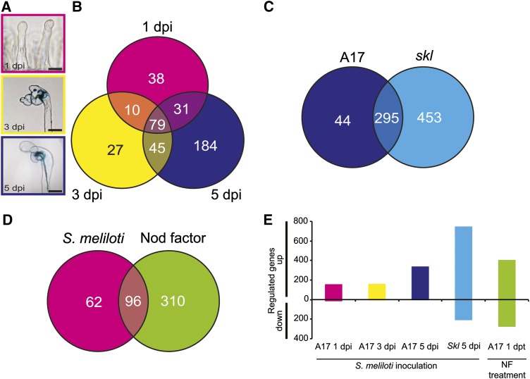
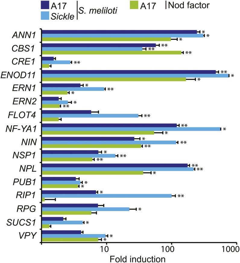
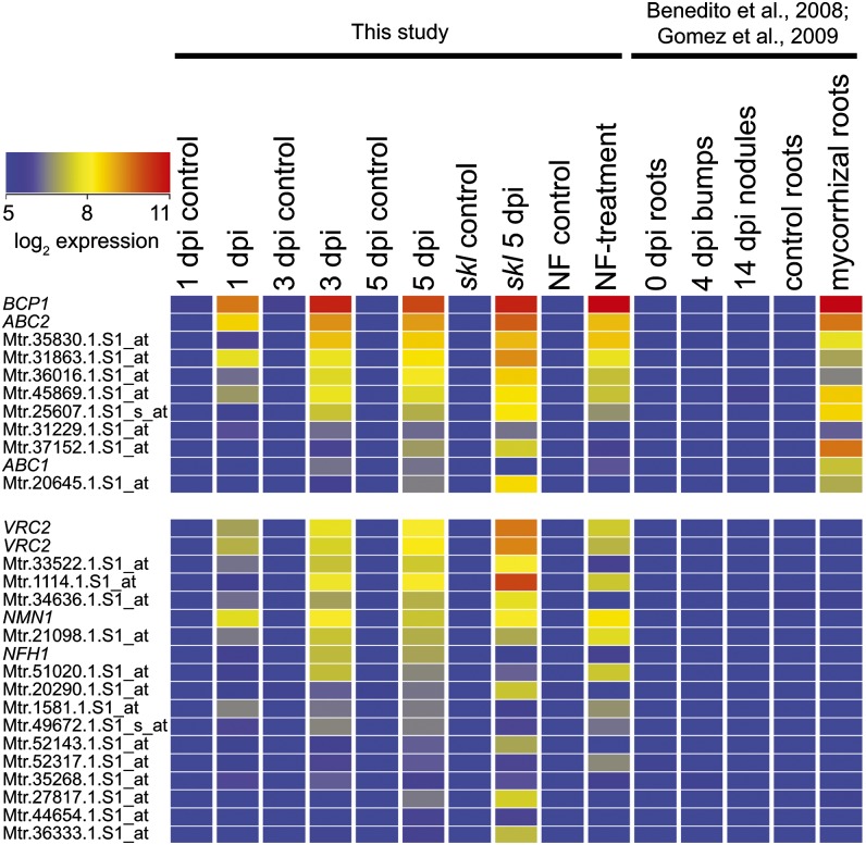
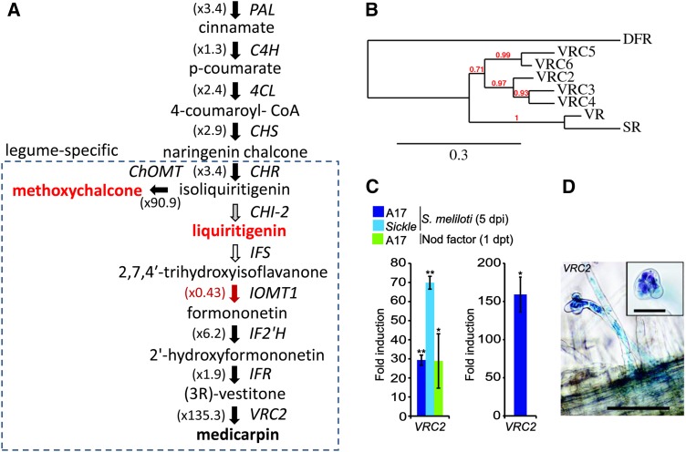
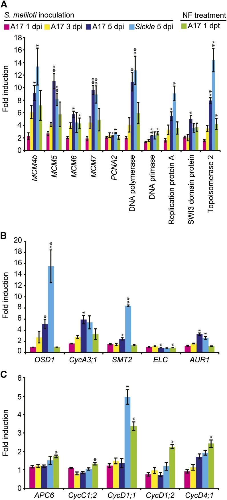
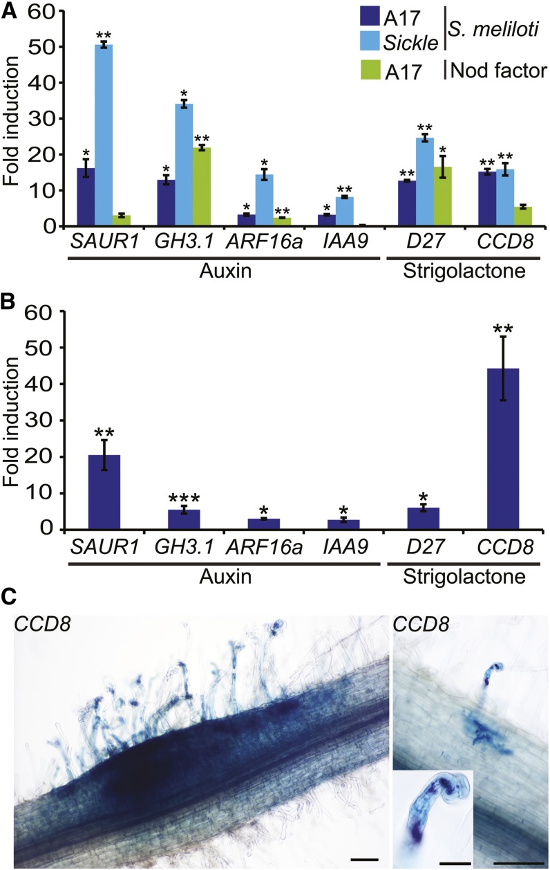
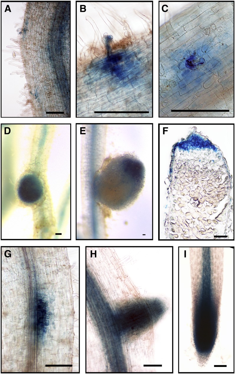
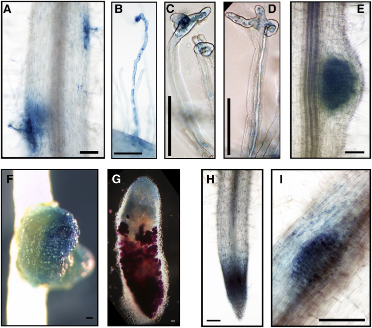
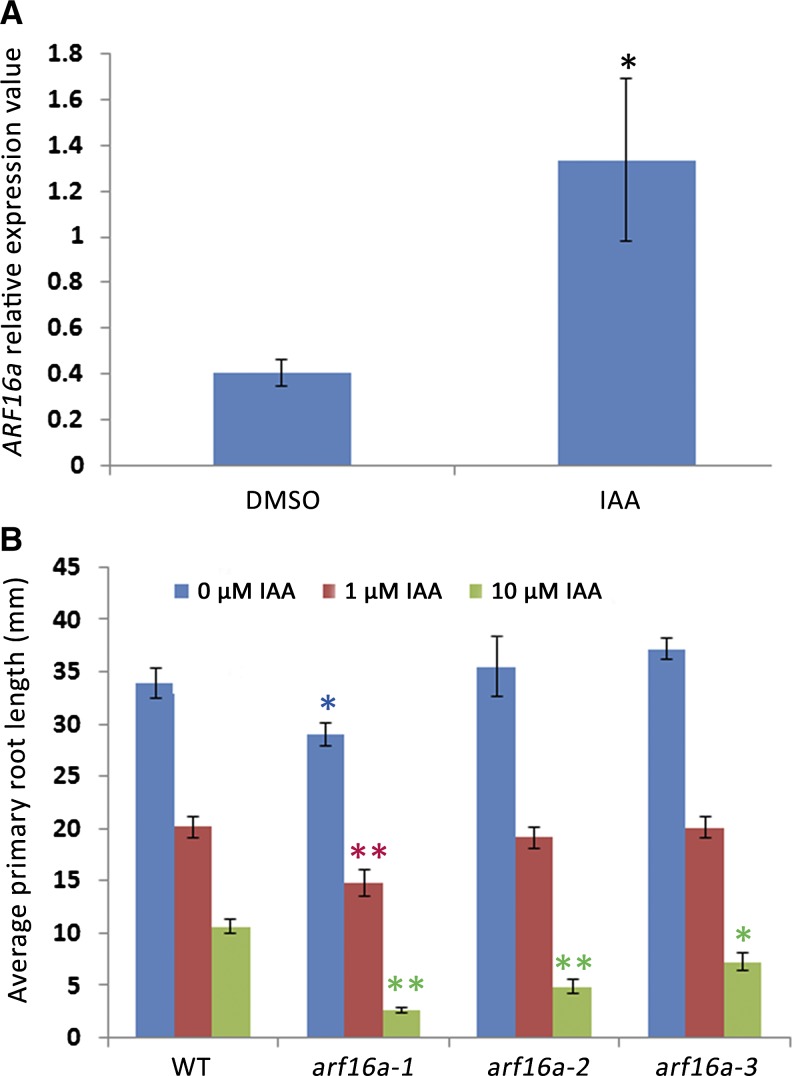
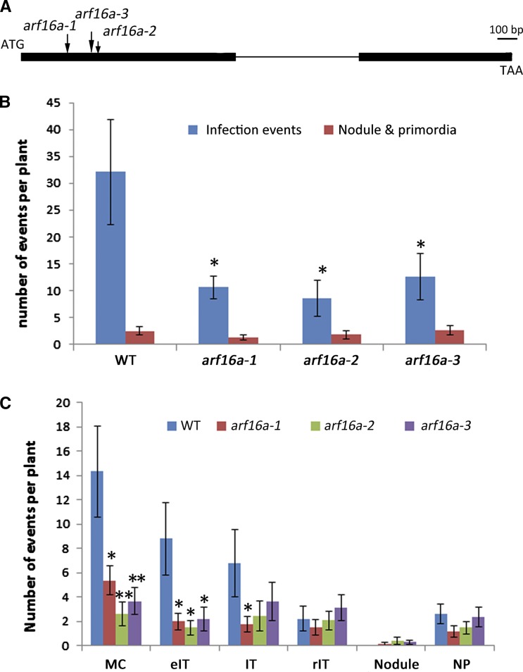
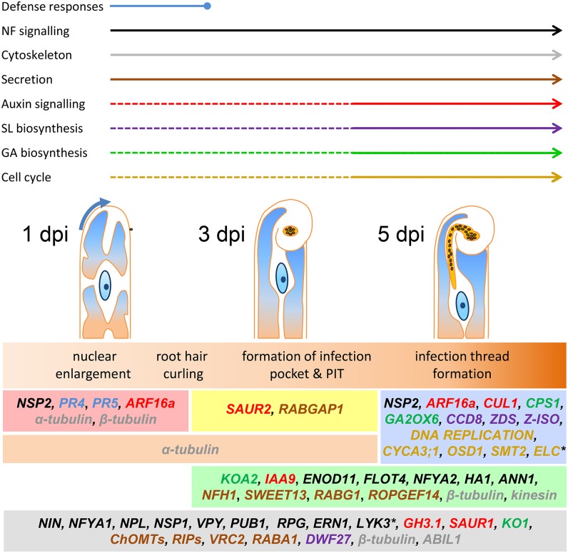
Nitrogen-fixing rhizobia colonize legume roots via plant-made intracellular infection threads. Genetics has identified some genes involved but has not provided sufficient detail to understand requirements for infection thread development. Therefore, we transcriptionally profiled Medicago truncatula root hairs prior to and during the initial stages of infection. This revealed changes in the responses to plant hormones, most notably auxin, strigolactone, gibberellic acid, and brassinosteroids. Several auxin responsive genes, including the ortholog of Arabidopsis thaliana Auxin Response Factor 16, were induced at infection sites and in nodule primordia, and mutation of ARF16a reduced rhizobial infection. Associated with the induction of auxin signaling genes, there was increased expression of cell cycle genes including an A-type cyclin and a subunit of the anaphase promoting complex. There was also induction of several chalcone O-methyltransferases involved in the synthesis of an inducer of Sinorhizobium meliloti nod genes, as well as a gene associated with Nod factor degradation, suggesting both positive and negative feedback loops that control Nod factor levels during rhizobial infection. We conclude that the onset of infection is associated with reactivation of the cell cycle as well as increased expression of genes required for hormone and flavonoid biosynthesis and that the regulation of auxin signaling is necessary for initiation of rhizobial infection threads.


Figures

Figure 1.
Figure 1.
Experimental Overview of Transcriptome Analysis Performed and Numbers of Significantly Regulated Genes Identified. (A) Infection time course of wild-type (A17) M. truncatula root hairs from seedlings inoculated with S. meliloti expressing lacZ. Major developmental milestones of infection: 1 dpi represents preinfection before visible root hair curling, at 3 dpi microcolonies had formed within curled root hairs, and at 5 dpi elongating infection threads were seen. Bar = 20 μm. (B) Genes upregulated in the wild type 1, 3, and 5 dpi with Sm1021. (C) Genes upregulated 5 dpi with Sm1021 in the wild type and in the hyperinfected skl mutant. (D) Genes upregulated in wild-type 1 dpi with Sm1021 and 1 d post-treatment with Nod factor. (E) Summary of up- and downregulated genes for each treatment. dpt, days post-treatment. Genes with significant regulation relative to control experiments are shown (>2-fold change; P < 0.05).
Figure 2.
Figure 2.
Induction of Known Symbiotic Genes. Fold induction of known symbiotic genes 5 dpi with Sm1021 or 1 d post-treatment with Nod factor. References for genes not cited elsewhere in the text: ANN1 (De Carvalho-Niebel et al., 2002), ERN2 (Andriankaja et al., 2007; Cerri et al., 2012), and ENOD11 (Journet et al., 2001). Significant inductions relative to control experiments are shown (*P < 0.05, **P < 0.01). Error bars = se (n = 3).
Figure 3.
Figure 3.
Transcriptome Analysis of Purified Root Hairs Reveals Genes with Common Symbiotic and Nodulation Specific Expression. Heat map displays probe sets that are significantly (P < 0.05) induced at least 2-fold in root hairs 5 dpi with S. meliloti but remain at low levels in a nodule time course (Benedito et al., 2008; Gomez et al., 2009). Names are provided for genes mentioned in the text; otherwise, probe sets are given. Further details including probe sets and corresponding gene models are provided in Supplemental Data Set 4 (Infection genes).
Figure 4.
Figure 4.
Expression of Genes Involved in Production of Phenylpropanoid Compounds with Activity as S. meliloti nod Gene Inducers and Phytoalexins Are Increased Following Rhizobial Inoculation. (A) Genes encoding enzymes involved in biosynthesis of the S. meliloti nod gene-inducing compounds liquiritigenin and methoxychalcone and the phytoalexin medicarpin are shown. Genes with expression altered by S. meliloti rhizobia and/or Nod factor are indicated using filled arrows (upregulated in solid black and downregulated are in solid red). Maximum fold increase expression (all treatments) compared with controls is indicated in brackets. (B) A phylogenetic tree showing vestitone reductase (VR) and five close homologs (Vestitone Reductase Cluster2-6) located within a 71-kb region on M. truncatula chromosome 7. Pea sophorol reductase (SR), which produces the closely related pterocarpin pisatin, is included as a reference. PAL, phenylalanine ammonia lyase; C4H, cinnamic acid 4-hydroxylase; C3H, coumarate 3-hydroxylase; 4CL, 4-coumarate:coA ligase; CHS, chalcone synthase; ChOMT, chalcone O-methyltransferase; CHI, chalcone isomerase; IFS, isoflavone synthase; IF2'H, isoflavone 2'-hydroxylase; IFR, isoflavone reductase. (C) Analysis of VRC2 expression in root hairs following inoculation with S. meliloti or addition of purified Nod factors as determined by microarray analysis (left). Induction of VRC2 by S. meliloti in wild-type root hairs was confirmed by qPCR (right). (D) Analysis of pVRC2:GUS expression in hairy roots transformed by A. rhizogenes. Inset shows curled root hairs; rhizobia are stained using magenta-gal to indicate rhizobia within a curled root hair tip. Bars in main image = 100 μm and in inset = 20 μm.
Figure 5.
Figure 5.
Cell Cycle-Related Genes Are Regulated during Infection. (A) Fold induction of replication fork components including mini chromosome maintenance (MCM) genes. (B) Fold induction of cell cycle regulation genes. (C) Nod factor treatments induce the expression of the anaphase promoting complex (APC6), a C-type cyclin, and genes encoding three D-type cyclins. Significant inductions relative to control experiments are shown (*P < 0.05, **P < 0.01). Error bars = se (n = 3).
Figure 6.
Figure 6.
Auxin Signaling and SL Biosynthesis Genes Are Induced by S. meliloti and by Nod Factors. (A) Microarray-based quantification of expression of auxin signaling genes SAUR1, GH3.1, ARF16a, and IAA9 and SL biosynthetic genes D27 and CCD8 at 5 dpi with S. meliloti or 1 d post-treatment with Nod factors in the wild type and skl. (B) qPCR confirmation of the induction of auxin signaling and SL biosynthetic genes at 5 dpi with S. meliloti. (C) Analysis of expression of the SL biosynthesis gene CCD8 using a promoter-GUS fusion in A. rhizogenes-induced transgenic roots stained for GUS activity (image on left). The image on the right shows staining of both CCD8:GUS (blue) and S. meliloti (lacZ) (magenta/purple). Bars in main images = 100 μm; insets = 20 μm. Significant induction relative to control experiments are shown (*P < 0.05, **P < 0.01, and ***P < 0.001). Error bars = se (n = 3).
Figure 7.
Figure 7.
DR5 Is Expressed throughout the Infection Zone after Rhizobial Inoculation. DR5:GUS expression in stable transgenic M. truncatula seedlings inoculated with S. meliloti. The blue color in (A) and (B) indicates staining of GUS activity. Arrows (A) indicate infection pockets containing microcolonies, and the arrowhead (B) indicates a root hair containing an infection thread. Bars = 50 μm. (A) 3 dpi. The inset is a magnification of the boxed region. (B) 5 dpi.
Figure 8.
Figure 8.
Expression of GH3.1-GUS Promoter Fusion during Rhizobial Infection and Development of Nodules and Lateral Roots. (A) to (D) ProGH3.1:GUS expression in A. rhizogenes-induced transgenic hairy roots showing expression at infection sites ([A] and [B]) and in nodules ([C] and [D]), 3 weeks after inoculation with S. meliloti carrying pXLGD4 (lacZ). (E) to (G) ProGH3.1:GUS expression in a lateral root tip (E), a lateral root primordium (F), and an emerging lateral root (G). (H) ProGH3.1:GUS expression in zone I and zone II in a mature nodule (3 weeks after inoculation with S. meliloti). The blue color ([A] to [H]) indicates GUS staining, and the magenta color indicates LacZ staining of S. meliloti. Bars = 100 μm.
Figure 9.
Figure 9.
SAUR1-GUS Promoter Fusion Expression during Rhizobial Infection, and Development of Nodules and Lateral Roots. A. rhizogenes-induced hairy roots were used to analyze ProSAUR1:GUS expression: (A) to (C) during rhizobial infection; (D) to (F) during nodule development; (G) during the formation of a lateral root primordium; (H) in an emerging lateral root; and (I) in a root tip. The blue color ([A] to [I]) indicates GUS staining of ProSAUR1:GUS activity, and the magenta color (C) indicates LacZ staining of S. meliloti. Bars = 100 μm.
Figure 10.
Figure 10.
ARF16a-GUS Promoter Fusion Expression during Rhizobial Infection and in Nodules and Lateral Roots. A. rhizogenes-induced hairy roots were used to analyze ProARF16a:GUS expression: (A) to (D) during rhizobial infection ([A] and [B], 3 weeks after inoculation; [C] and [D], 3 and 5 dpi); (E) in a young nodule; (F) in an elongated nodule; (G) in a mature nodule (median longitudinal section); (H) in a lateral root tip; and (I) and in a lateral root primordium. The blue color ([A] to [I]) indicates staining of GUS activity, and the magenta color (G) indicates LacZ activity of S. meliloti carrying pXLGD4. Bars = 100 μm.
Figure 11.
Figure 11.
ARF16a Responds to Auxin Treatment. (A) qPCR of relative expression of ARF16a upon treatment with DMSO or 1 μM IAA. (B) The inhibition of primary root growth by 1 or 10 μM IAA in the wild type (R108), arf16a-1 (NF12634), arf16a-2 (NF13450), and arf16a-3 (NF16027). The histograms show averages (±se) of primary root length 7 d after germination, based on 10 plants for each treatment of each genotype. Bar = se. Significant (Student’s t test) differences between the wild type and mutants are marked with asterisks (*P ≤ 0.05 and **P ≤ 0.01).
Figure 12.
Figure 12.
Nodulation Phenotype of M. truncatula arf16a Mutants. (A) Gene structure of ARF16a gene. The bold line indicates the exons and thin line indicates an intron. The locations of Tnt1 insertions for the mutant alleles are indicated by arrows. Bar = 100 bp. (B) Rhizobial infections and formation of nodule primordia in the wild type (R108), arf16a-1(NF12634), arf16a-2 (NF13450), and arf16a-3 (NF16027). (C) Quantification of different stages of infection and development of nodule primordia in the wild type and arf16a mutants 7 dpi with S. meliloti. Infection events and nodule primordia were scored 7 dpi with S. meliloti 1021 carrying pXLGD4 (lacZ) after LacZ staining. The histograms show averages (±se) based on 14 plants for each line. Significant (Student’s t test) differences between the wild type and mutants are marked with asterisks (*P ≤ 0.05 and **P ≤ 0.01). IT, fully elongated infection thread in root hair; eIT, elongating infection thread in root hair; MC, microcolony; rIT, ramified infection thread in cortex; NP, nodule primordium.
Figure 13.
Figure 13.
Dynamic Trends in Gene Expression Prior to and during Infection. Rhizobial preinfection and infection involves transient defense responses and sustained Nod factor signaling, while infection specifically involves hormone biosynthesis and signaling and activation of the cell cycle. Preinfection stage was marked by the induction of two pathogenesis-related genes, PR4 and PR5. Most nodulation pathway genes were induced, and LYK3 was repressed at all time points, while NSP2 was only induced during early preinfection and upon infection. Flavonoid and peroxidase genes were induced at all stages (VRC2, ChOMTs, and RIPs). ENOD11, FLOT4, NFYA2, ANN1, HA1, the Nod factor hydrolase NFH1, and sugar transporter SWEET13 were most strongly induced during formation of the infection pocket and at the onset of infection. RAB GTPases with potential roles in secretion were induced at every stage, while ROPGEF14 was mainly induced at the latter two stages. Tubulins and the cytoskeletal regulator ABIL1 were expressed at every time point, while kinesin was only induced at the latter stages. The initiation of the infection thread coincided with the induction of genes for GA and SL biosynthesis, auxin signaling, and many genes encoding cell cycle components, including most members of the DNA replication complex and an A-type cyclin (CYCA3;1). Genes for repression of endoreduplication (OSD1 and SMT2) were induced, while ELC, which is involved in the switch from mitosis to endoreduplication, is repressed during infection. The color key indicates the processes in which each gene is involved. Genes are grouped based on their trends of induction or repression (the latter indicated by an asterisk) relative to the controls. See Supplemental Table 2 for further details on specific genes.

Comment in

References

    1. Adie B.A., Pérez-Pérez J., Pérez-Pérez M.M., Godoy M., Sánchez-Serrano J.J., Schmelz E.A., Solano R. (2007). ABA is an essential signal for plant resistance to pathogens affecting JA biosynthesis and the activation of defenses in Arabidopsis. Plant Cell 19: 1665–1681. - PMC - PubMed
    1. Akiyama K., Matsuzaki K., Hayashi H. (2005). Plant sesquiterpenes induce hyphal branching in arbuscular mycorrhizal fungi. Nature 435: 824–827. - PubMed
    1. Allen N.S., Bennett M.N., Cox D.N., Shipley A., Ehrhardt D.W., Long S.R. (1994). Effects of Nod factors on alfalfa root hair Ca++ and H+ currents and on cytoskeletal behaviour. In Advances in Molecular Genetics of Plant-Microbe Interactions, Vol. 3. M.J. Daniels, J.A. Downie, and A.E. Osbourn, eds (Dordrecht, The Netherlands: Kluwer Academic Publishers), pp. 107–113.
    1. Andriankaja A., Boisson-Dernier A., Frances L., Sauviac L., Jauneau A., Barker D.G., de Carvalho-Niebel F. (2007). AP2-ERF transcription factors mediate Nod factor dependent Mt ENOD11 activation in root hairs via a novel cis-regulatory motif. Plant Cell 19: 2866–2885. - PMC - PubMed
    1. Anisimova M., Gascuel O. (2006). Approximate likelihood-ratio test for branches: A fast, accurate, and powerful alternative. Syst. Biol. 55: 539–552. - PubMed

Publication types

MeSH terms

LinkOut - more resources