Review
. 2015 Feb;38(2):117-25.
doi: 10.1016/j.tins.2014.11.003. Epub 2014 Dec 17.

Cerebral cortex assembly: generating and reprogramming projection neuron diversity

Affiliations
Review

Cerebral cortex assembly: generating and reprogramming projection neuron diversity

Simona Lodato et al. Trends Neurosci. 2015 Feb.

Abstract

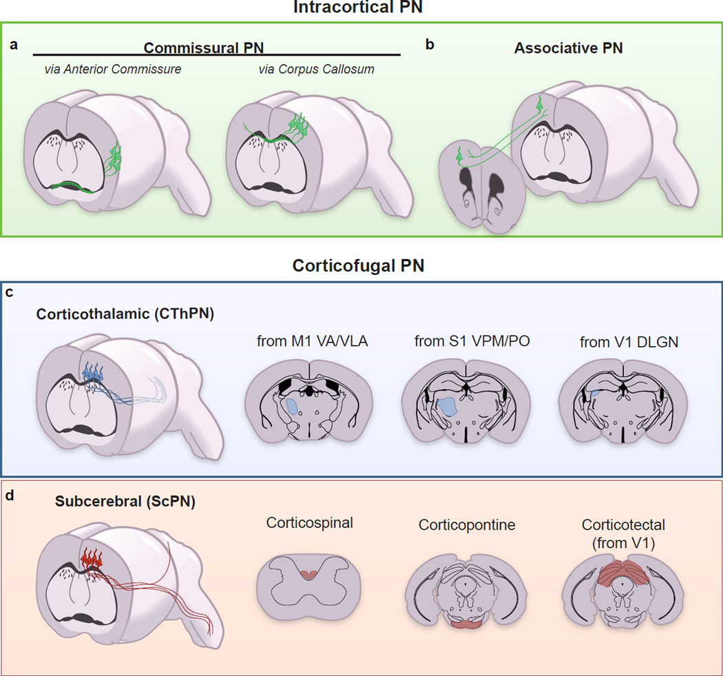
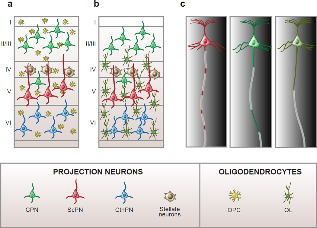
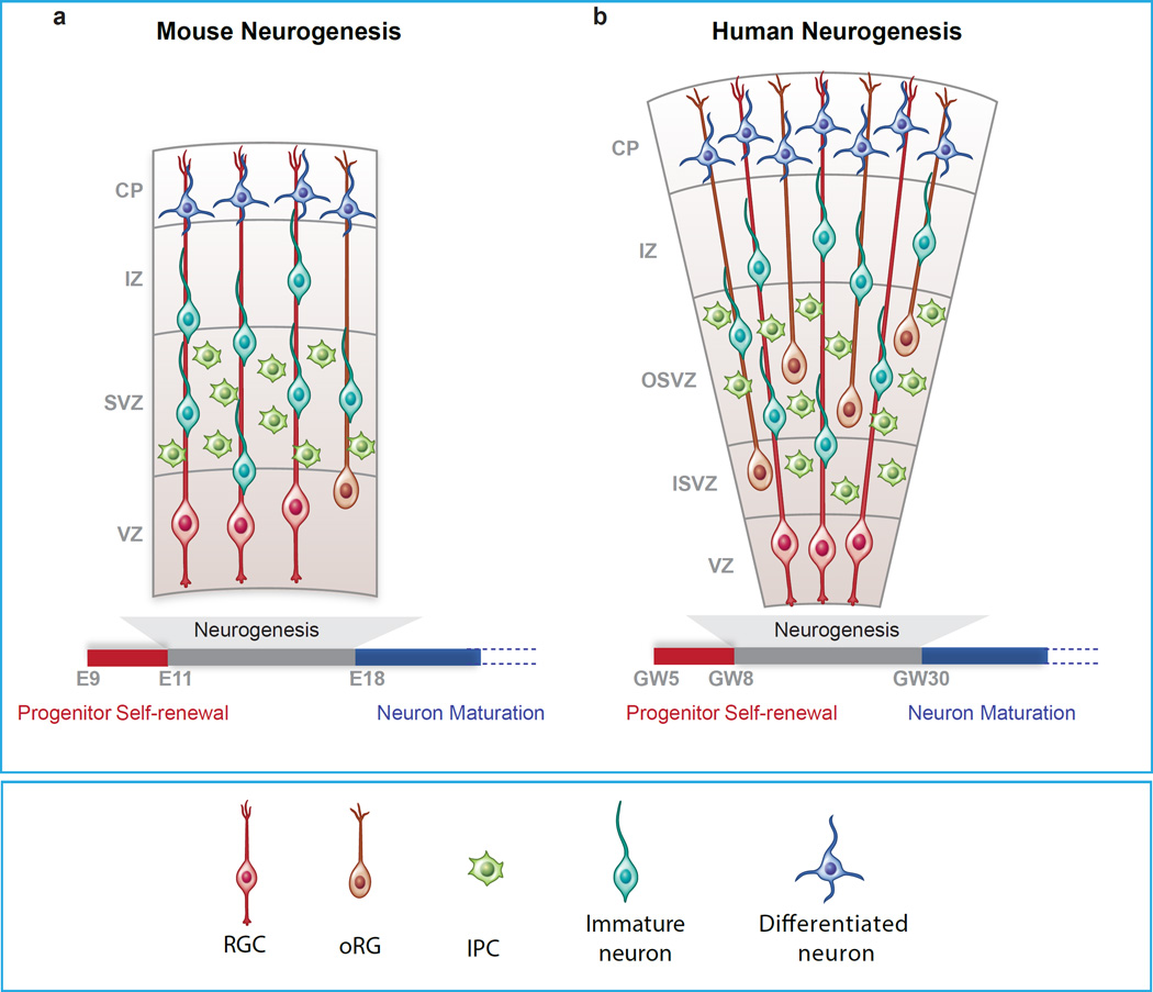
The mammalian cerebral cortex is responsible for the highest levels of associative, cognitive and motor functions. In the central nervous system (CNS) the cortex stands as a prime example of extreme neuronal diversity, broadly classified into excitatory projection neurons (PNs) and inhibitory interneurons (INs). We review here recent progress made in understanding the strategies and mechanisms that shape PN diversity during embryogenesis, and discuss how PN classes may be maintained, postnatally, for the life of the organism. In addition, we consider the intriguing possibility that PNs may be amenable to directed reprogramming of their class-specific features to allow enhanced cortical plasticity in the adult.


Figures

Figure 1
Figure 1. Cortical Projection Neuron Classification by Connectivity
PNs are broadly divided into two groups: Intracortical PNs and Corticofugal PNs. Intracortical PNs are further subdivided into Commissural PNs (a), which project to the contralateral hemisphere, and Associative PNs (b), which project to cortical areas within the same hemisphere (e.g. ipsilateral forward and backward projecting neurons). Some commissural PN connect through the corpus callosum (Callosal PNs, CPNs) while others, residing within the lateral cortex project via the anterior commissure (a). Corticofugal PNs project to subcortical targets and are further divided into Corticothalamic PNs (CThPNs) (c) and Subcerebral PN (ScPNs) (d). CThPNs are located in L6 and project to various nuclei of the thalamus in an area-dependent manner (c). From the primary motor cortex (M1), the majority of CThPNs project to the Ventral Anterior (VA) and Anterior Ventral Lateral (VLA) nuclei. From the somatosensory cortex (S1), the majority of CthPNs project to the Ventral Posterior Medial nucleus (VPM) and the Posterior nucleus (PO). From the visual cortex (V1), the majority of CFuPNs project to the Dorsal Lateral Geniculate Nucleus (dLGN). ScPNs are also further divided based on their axonal targets (d). Corticospinal motor neurons send primary axons to the spinal cord. Corticopontine neurons extend axons to the pontine nuclei within the brainstem, and Corticotectal neurons have axon projections to the optic tectum in the midbrain.
Figure 2
Figure 2. Projection neurons have distinct profiles of longitudinal myelination
In the mammalian neocortex oligodendrocyte progenitors (OPCs) are evenly distributed across all layers (a), but oligodendrocytes (OLs) show preferential distribution in the deep layers (b), reflecting higher levels of myelin in L5 and L6. Cortical PNs display diverse myelination patterns along their axons. At least three types of myelination profiles exist in the mouse neocortex (in addition to axons that are not myelinated) (c). Some projection neurons have axons that are myelinated throughout their entire length with short un-myelinated nodes of Ranvier, others display myelinated segments intercalated with myelinated tracts of different lengths (intermittent myelin). Finally, selected neurons have axons with a long unmyelinated tract between the axon hillock and the first internode (c). The two latter patterns of myelin distribution are found preferentially in PNs of the upper layers, suggesting that myelination patterns may be an integral feature of neuronal class-specific identity. Abbreviations: CPN, callosal PN; CthPN, corticothalamic PN; ScPN, subcerebral PN.
Figure I (Box1)
Figure I (Box1). Progenitors of the cerebral cortex in mice and humans
Different types of progenitors are depicted for mouse (a) and human cortex (b). Time scale of neurogenesis is measured in embryonic days (E) for mice and in gestational week (GW) for humans. Abbreviations: VZ, Ventricular zone; SVZ, Sub-ventricular zone; OSVZ, Outer Sub-ventricular zone; ISVZ, Inner Sub-ventricular zone; IZ, Intermediate zone; CP, Cortical plate; RGC, Radial glial cell; oRG, Outer radial glial cell; IPC, Intermediate precursor cell. The images are not to scale.

References

    1. Golgi C. Sulla Fina Anatomia Degli Organi Centrali del Sistema Nervoso. 1886
    1. Cajal SRy. Histologie du Systéme Nerveaux de l’Homme et des Vertebres. 1911
    1. No RLd. Cerebral Cortex: Architecture, Intracortical Connections, Motor Projections. Oxford university press; 1949.
    1. Greig LC, et al. Molecular logic of neocortical projection neuron specification, development and diversity. Nature reviews. Neuroscience. 2013;14:755–769. - PMC - PubMed
    1. Kepecs A, Fishell G. Interneuron cell types are fit to function. Nature. 2014;505:318–326. - PMC - PubMed

Publication types

LinkOut - more resources