. 2015 May;61(5):1591-602.
doi: 10.1002/hep.27665. Epub 2015 Mar 20.

CXCR4 inhibition in tumor microenvironment facilitates anti-programmed death receptor-1 immunotherapy in sorafenib-treated hepatocellular carcinoma in mice

Affiliations

CXCR4 inhibition in tumor microenvironment facilitates anti-programmed death receptor-1 immunotherapy in sorafenib-treated hepatocellular carcinoma in mice

Yunching Chen et al. Hepatology. 2015 May.

Abstract

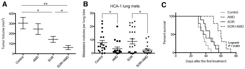
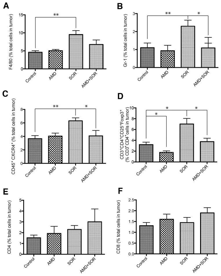
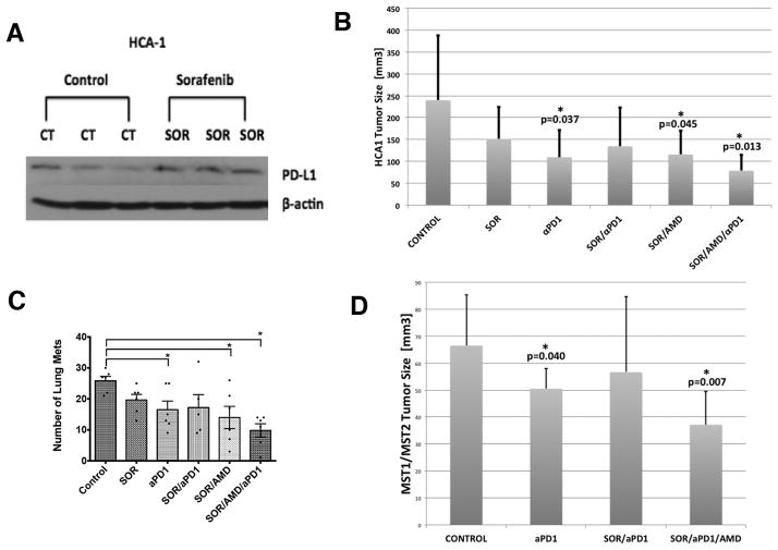
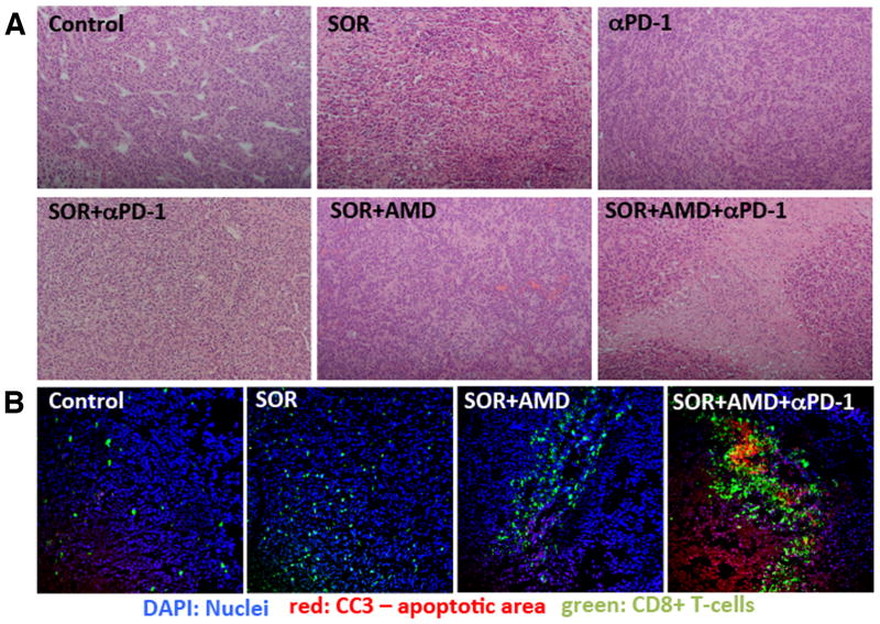
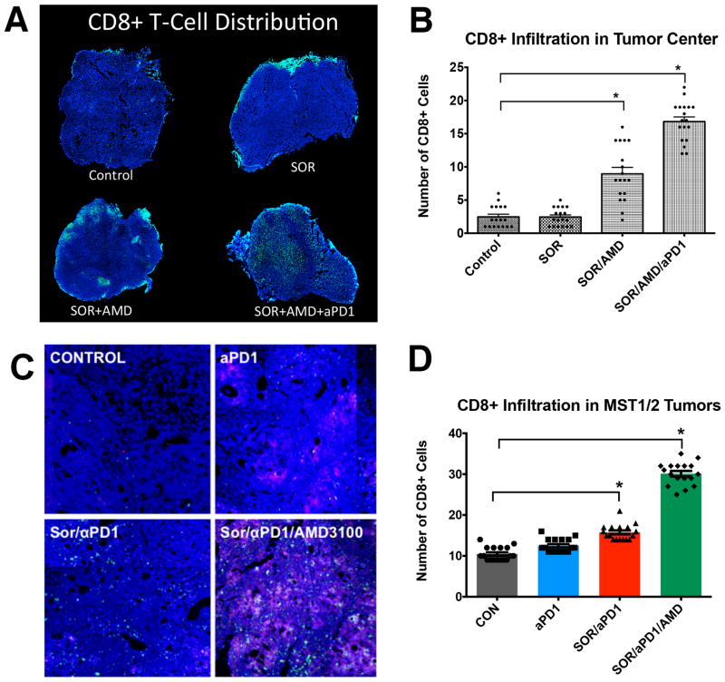
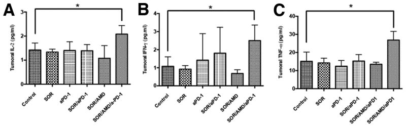
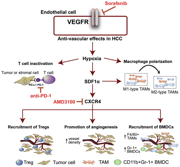
Sorafenib, a broad tyrosine kinase inhibitor, is the only approved systemic therapy for advanced hepatocellular carcinoma (HCC) but provides limited survival benefits. Recently, immunotherapy has emerged as a promising treatment strategy, but its role remains unclear in HCCs, which are associated with decreased cytotoxic CD8(+) T-lymphocyte infiltration in both murine and human tumors. Moreover, in mouse models after sorafenib treatment intratumoral hypoxia is increased and may fuel evasive resistance. Using orthotopic HCC models, we now show that increased hypoxia after sorafenib treatment promotes immunosuppression, characterized by increased intratumoral expression of the immune checkpoint inhibitor programmed death ligand-1 and accumulation of T-regulatory cells and M2-type macrophages. We also show that the recruitment of immunosuppressive cells is mediated in part by hypoxia-induced up-regulation of stromal cell-derived 1 alpha. Inhibition of the stromal cell-derived 1 alpha receptor (C-X-C receptor type 4 or CXCR4) using AMD3100 prevented the polarization toward an immunosuppressive microenvironment after sorafenib treatment, inhibited tumor growth, reduced lung metastasis, and improved survival. However, the combination of AMD3100 and sorafenib did not significantly change cytotoxic CD8(+) T-lymphocyte infiltration into HCC tumors and did not modify their activation status. In separate experiments, antibody blockade of the programmed death ligand-1 receptor programmed death receptor-1 (PD-1) showed antitumor effects in treatment-naive tumors in orthotopic (grafted and genetically engineered) models of HCC. However, anti-PD-1 antibody treatment had additional antitumor activity only when combined with sorafenib and AMD3100 and not when combined with sorafenib alone.

Conclusion: Anti-PD-1 treatment can boost antitumor immune responses in HCC models; when used in combination with sorafenib, anti-PD-1 immunotherapy shows efficacy only with concomitant targeting of the hypoxic and immunosuppressive microenvironment with agents such as CXCR4 inhibitors.


Figures

Figure 1
Figure 1. Treatment with the SDF1α/CXCR4 inhibitor AMD3100 plus sorafenib inhibits primary tumor growth, incidence of lung metastasis formation and improves overall survival in orthotopic HCC models
(A) While sorafenib (SOR) treatment alone marginally delays HCC growth, the addition of AMD3100 (AMD) to SOR – but not AMD alone – induces an additional significant delay in tumor growth (n=8; *p<0.05, **P<0.01). (B) The number of lung metastatic nodules is significantly reduced in AMD-treated mice. (C) Overall survival is significantly prolonged only in orthotopic HCC-bearing mice treated with SOR and AMD. Data are representative of at least two independent experiments and are presented as mean±SEM (n=10). *P<0.05; **P<0.01.
Figure 2
Figure 2. Sorafenib treatment induces a polarization towards a pro-immunosuppressive environment in orthotopic HCA-1 tumors, which is prevented by CXCR4 inhibition in the face of persistent hypoxia
(A–D) Changes in viable tumor-infiltrating immune cells in HCA-1 tumors from mice treated with sorafenib with or without AMD3100 versus control analyzed by flow cytometry. The number of 7AAD–CD45+F4/80+ tumor-associated macrophages (A), 7AAD–CD11b+Gr1+ monocytes (B), 7AAD–CD45+CXCR4+ cells (C), and 7AAD–CD4+CD25+FoxP3+ T regulatory (Treg) cells (D) significantly increased in sorafenib treated HCCs. Combining AMD3100 treatment with sorafenib prevents these effects. (E–F) The number of 7AAD–CD4+CD3+ (E) and 7AAD–CD8+CD3+ (F) T lymphocytes was not significantly different between the four treatment groups in HCA-1 HCCs. *p<0.05; Data are shown as mean±SEM.
Figure 3
Figure 3. PD-1 blockade is active against HCC in grafted and genetically-engineered models and is facilitated by sorafenib when combined with CXCR4 inhibition
(A) Orthotopic HCA-1 tumors were treated when the tumor reached a size of 14mm3 with sorafenib for 28 days and then the tumors were collected. Western blot analysis shows that PD-L1 expression is increased in HCC tissue after sorafenib treatment compared to control treated tumors. (B) PD-1 blockade alone or the combination of sorafenib/AMD3100 significantly delays tumor growth (both p<0.05 vs. control). The most effective tumor growth delay is achieved by the triple combination therapy of sorafenib/AMD3100/anti-PD-1 (p=0.013 vs. control). (C) While the number of lung metastases is significantly reduced by anti-PD-1 treatment or the combination of SOR/AMD3100, the triple therapy of SOR/AMD3100/anti-PD-1 results in most pronounced decrease in lung metastases incidence. (D) PD-1 blockade alone (p=0.04 vs. control) but not in combination with SOR significantly delays HCC growth in the Mst-mutant mouse model (not significant). The most effective tumor growth delay and regression is achieved by the triple combination therapy of sorafenib/AMD3100/anti-PD-1 (p=0.007 vs. control). Data are presented as mean±SEM (N=6 per group). *P<0.05, **P<0.005.
Figure 4
Figure 4. Anti-PD-1 treatment combined with sorafenib and AMD3100 enhances anti-tumor immune responses in HCC
(A) Representative H&E staining showing rare areas of necrosis in untreated tumors, and some necrotic areas in sorafenib or anti-PD-1 antibody only treated HCA1 tumors. Sorafenib plus AMD3100 treatment results in further enhancement of tumor necrosis, and triple combination treatment results in extensive necrosis, with lymphocytic immune cell infiltration in the necrotic areas (H&E Staining, 10× magnification). (B) Representative immunoflorescence staining for CD8+ T cells (FITC, green) and cleaved caspase-3 (Cy5, red) in frozen HCA1 tumor sections. Counterstaining of nuclei by DAPI (blue). Tumor infiltrating cytotoxic CD8+ cells co-localized with areas of cell apoptosis only in the triple combination treatment group.
Figure 5
Figure 5. Anti-PD-1 treatment combined with sorafenib and AMD3100 increases intratumoral CD8+ T lymphocyte distribution in HCC
A–D, The number of cytotoxic T lymphocytes infiltrating the tumor proper is increased only in the SOR+AMD3100+αPD1 treatment group. Representative confocal microscopy of immunofluorescence for CD8+ T lymphocytes (FITC, green) in HCC tissue sections (nuclei by DAPI, in blue) from HCA-1 grafted tumors (A) and representative regions of HCC tumors in the Mst GEM model. Red areas in the MST tumors indicate apoptotic regions of MST HCC tumors (staining for cleaved-caspase 3, Cy5 in red) (C). (B,D) Addition of anti-PD-1 antibody changes the distribution of CD8+ T lymphocytes in the tumors: Triple combination treatment results in significantly higher numbers of cytotoxic T lymphocytes in tumor center. Data are mean±SEM (n =5–6 per group) *P<0.05 vs. control.
Figure 6
Figure 6. Addition of anti-PD-1 antibody to sorafenib and CXCR4 inhibition increases the expression of biomarkers of T lymphocyte activation in HCC
(A–C) PD-1 blockade combined with sorafenib and AMD3100 induces a significant increase of T lymphocyte activation biomarkers IL-2 (A), TNF-α (B), and IFN-γ (C) in HCA-1 tumors versus HCCs in other treatment groups. Data are presented as mean±SEM (N =5 per group) *P<0.05.
Figure 7
Figure 7
Sorafenib treatment induces SDF1α/CXCR4 axis-mediated immunosuppression in HCC microenvironment.

References

    1. Cheng AL, Kang YK, Chen Z, Tsao CJ, Qin S, Kim JS, Luo R, et al. Efficacy and safety of sorafenib in patients in the Asia-Pacific region with advanced hepatocellular carcinoma: a phase III randomised, double-blind, placebo-controlled trial. Lancet Oncol. 2009;10:25–34. - PubMed
    1. Llovet JM, Ricci S, Mazzaferro V, Hilgard P, Gane E, Blanc JF, de Oliveira AC, et al. Sorafenib in advanced hepatocellular carcinoma. N Engl J Med. 2008;359:378–390. - PubMed
    1. Carmeliet P, Jain RK. Molecular mechanisms and clinical applications of angiogenesis. Nature. 2011;473:298–307. - PMC - PubMed
    1. Jain RK. Antiangiogenesis strategies revisited: From starving tumors to alleviating hypoxia. Cancer Cell. 2014;26 doi: 10.1016/j.ccell.2014.10.006. ePub on November 10, 2014. - DOI - PMC - PubMed
    1. Semenza GL. Hypoxia-inducible factors in physiology and medicine. Cell. 2012;148:399–408. - PMC - PubMed

Publication types

MeSH terms