. 2015 Mar;56(3):546-561.
doi: 10.1194/jlr.M055152. Epub 2014 Dec 21.

Lipid-induced NOX2 activation inhibits autophagic flux by impairing lysosomal enzyme activity

Affiliations

Lipid-induced NOX2 activation inhibits autophagic flux by impairing lysosomal enzyme activity

Bharat Jaishy et al. J Lipid Res. 2015 Mar.

Abstract

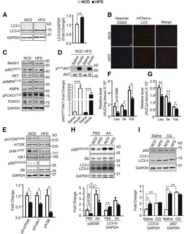
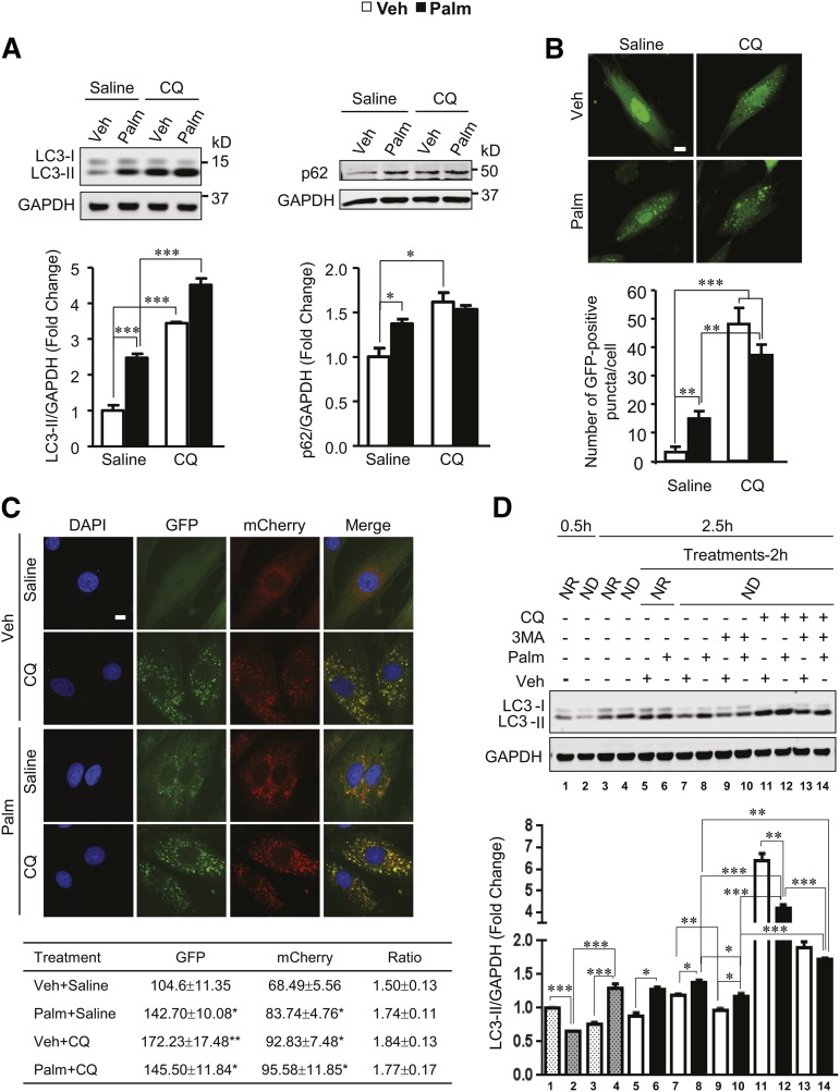
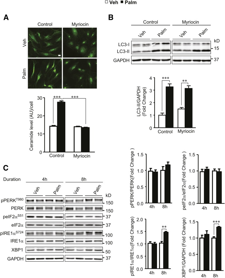
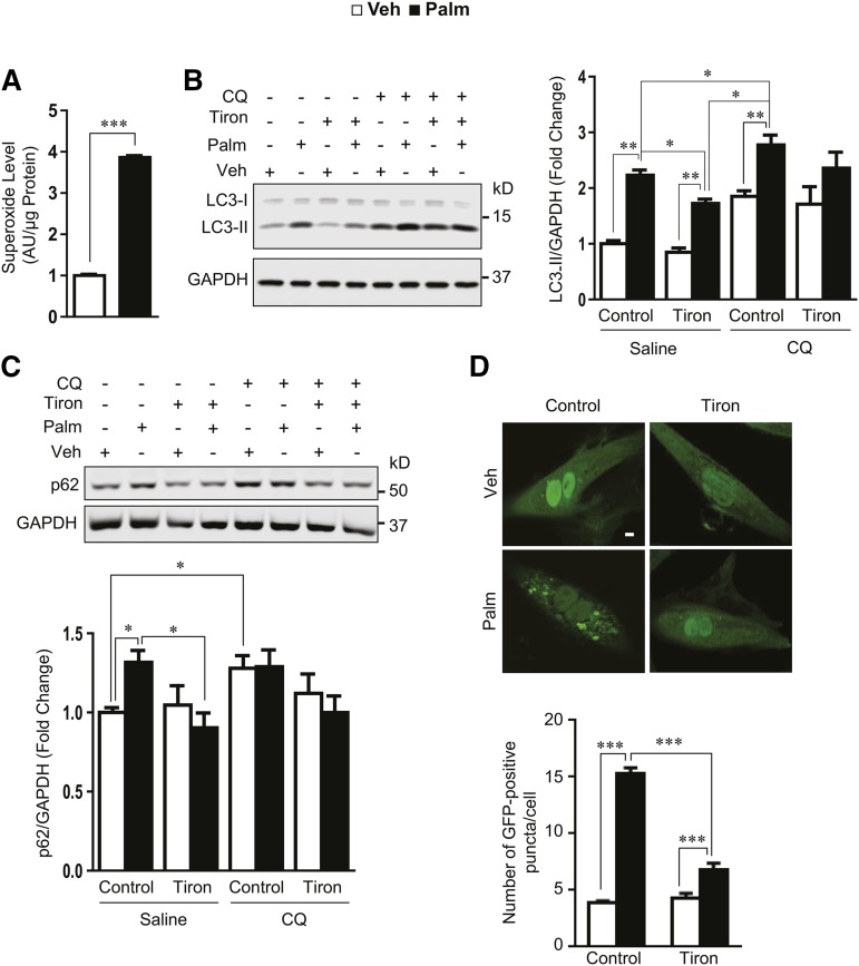
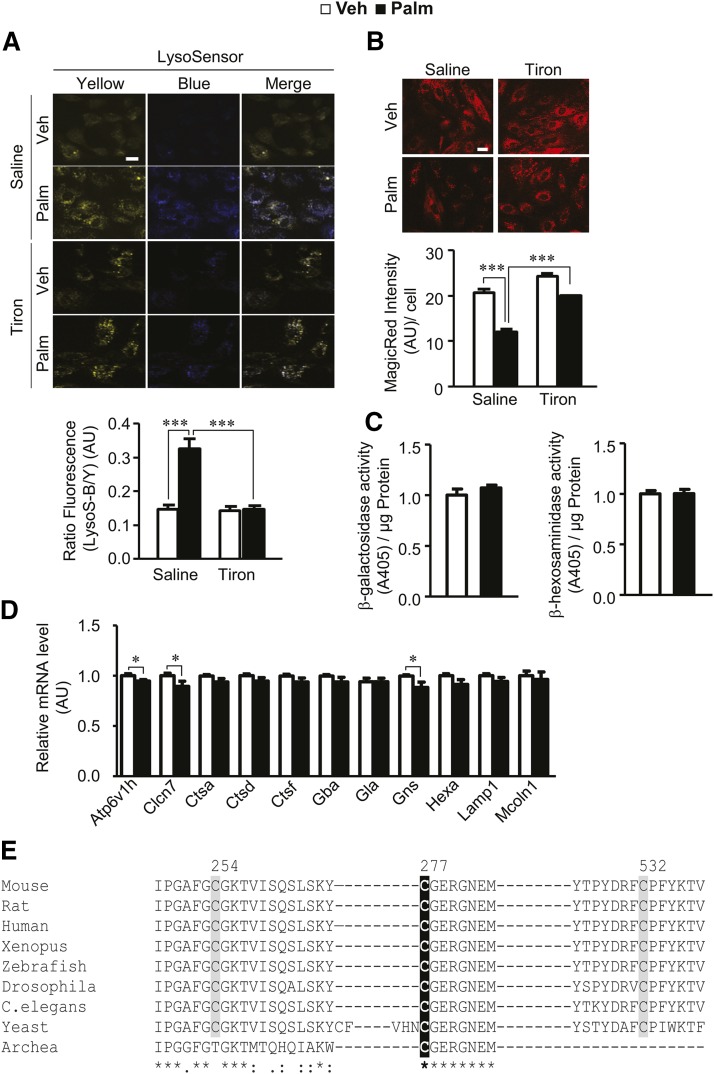
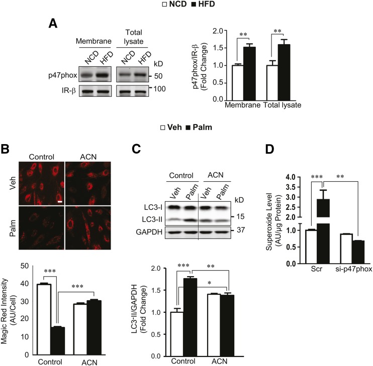
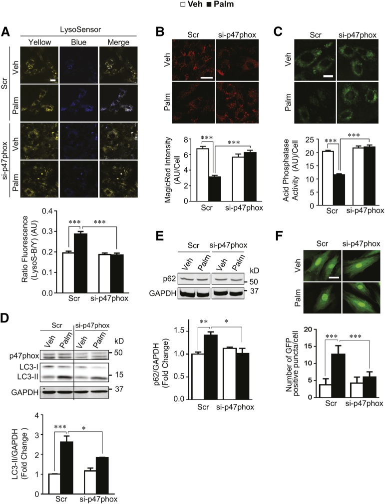
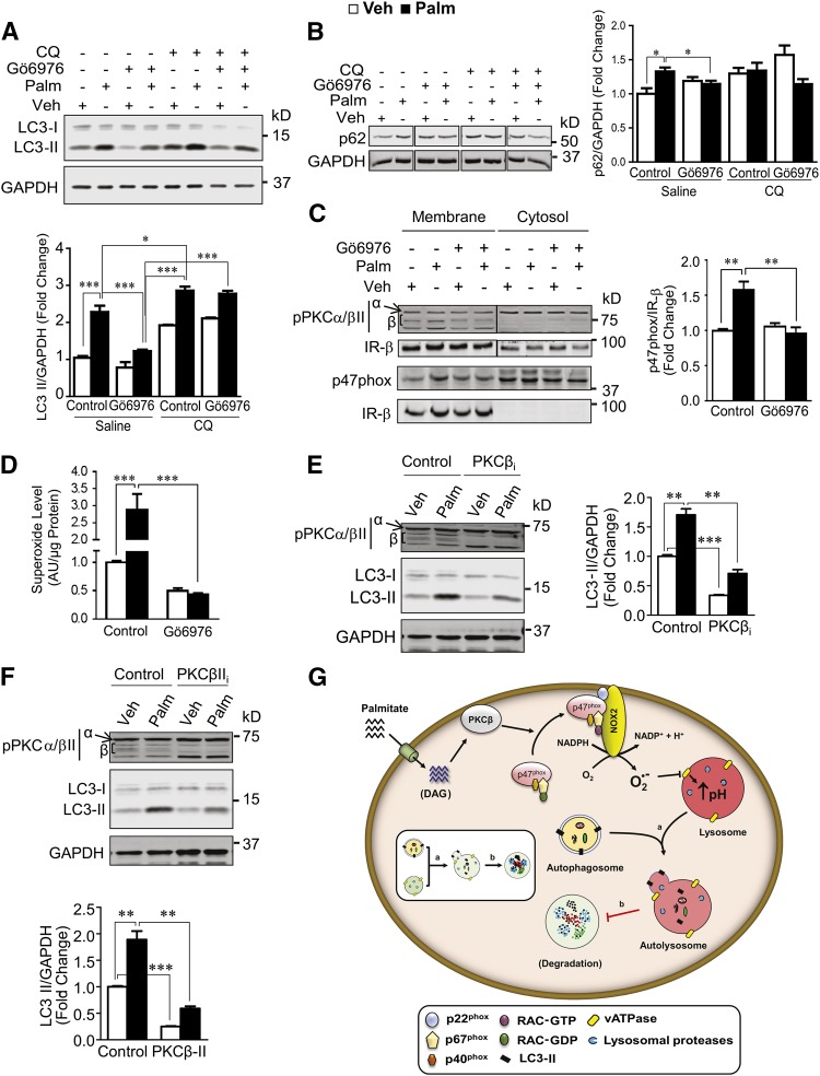
Autophagy is a catabolic process involved in maintaining energy and organelle homeostasis. The relationship between obesity and the regulation of autophagy is cell type specific. Despite adverse consequences of obesity on cardiac structure and function, the contribution of altered cardiac autophagy in response to fatty acid overload is incompletely understood. Here, we report the suppression of autophagosome clearance and the activation of NADPH oxidase (Nox)2 in both high fat-fed murine hearts and palmitate-treated H9C2 cardiomyocytes (CMs). Defective autophagosome clearance is secondary to superoxide-dependent impairment of lysosomal acidification and enzyme activity in palmitate-treated CMs. Inhibition of Nox2 prevented superoxide overproduction, restored lysosome acidification and enzyme activity, and reduced autophagosome accumulation in palmitate-treated CMs. Palmitate-induced Nox2 activation was dependent on the activation of classical protein kinase Cs (PKCs), specifically PKCβII. These findings reveal a novel mechanism linking lipotoxicity with a PKCβ-Nox2-mediated impairment in pH-dependent lysosomal enzyme activity that diminishes autophagic turnover in CMs.

Keywords: NADPH oxidase 2; autophagy; cardiomyocytes; fatty acids; lysosomes; protein kinase Cβ.


Figures

Fig. 1.
Fig. 1.
High fat feeding increases autophagosome abundance in adult murine hearts independently of changes in autophagy induction pathways. A,C, E: Hearts from mice fed with either NCD or HFD for 12 weeks (from 8 to 20 weeks of age) were analyzed for the indicated proteins involved in autophagy by immunoblotting (n = 6). B: Confocal microscopy of transverse cardiac sections from NCD- or HFD-fed mice expressing cardiac-specific mCLC3 (n = 5). Scale bar, 50 μm. D: Mice on NCD or HFD for 12 weeks were administered saline or 0.01 U insulin via ivc injection. Insulin signaling through Akt was assessed in the heart by immunoblotting (n = 4 for NCD-saline and n = 5 for all other groups). F, G: Levels of BCAAs in heart tissue (n = 6) (F) and serum (n = 12) (G) of mice fed with NCD or HFD for 12 weeks. H: Mice on NCD or HFD were injected daily with a BCAA solution (AA) ip during the last 2 weeks of feeding, and heart lysates were analyzed for the indicated proteins by immunoblotting (n = 3, 3, 5, and 3 for NCD-PBS, HFD-PBS, NCD-AA, and HFD-AA, respectively). I: Mice on NCD or HFD were injected ip with saline or CQ and analyzed for LC3-II levels in heart lysates (n = 3). *P < 0.05; **P < 0.01; ***P < 0.001.
Fig. 2.
Fig. 2.
Palmitate promotes autophagosome accumulation in H9C2 CMs by suppressing autophagic turnover. A: Immunoblot analysis of autophagy proteins [LC3 (left) and p62 (right)] in H9C2 CMs incubated with either vehicle (Veh) (1% BSA) or 500 μM palmitate (Palm) for 4 h and in the presence or absence of 80 μM CQ for the last 1 h (n = 3). B, C: H9C2 CMs expressing adenoviral GFP-LC3 (B) or stably expressing retroviral mCherry-GFP-LC3 (C) were incubated with vehicle or palmitate in the presence or absence of CQ as described in (A). B: Confocal images (top) showing the distribution of adenoviral GFP-LC3 (green). Graph (bottom) represents the average number of GFP-positive puncta/CMs ± SEM (n = 50 CMs). C: Confocal images (top) showing GFP (green) and mCherry (red) fluorescence of mCherry-GFP-LC3. DAPI (blue) stains the nuclei. Table (bottom) shows mean fluorescence intensity ± SEM of GFP and mCherry signals from images in (C) and the ratio of GFP to mCherry fluorescence in response to different treatments (n = 21 CMs). Scale bar, 10 μm. *P < 0.05; **P < 0.01 versus vehicle + saline. D: LC3 immunoblot (top) from CMs incubated in ND medium for 0.5 h and treated with vehicle or palmitate in the presence or absence of 3MA and CQ for an additional 2 h in ND medium. CMs incubated with nutrient-rich (NR) or ND medium initially for 0.5 h and then for 2.5 h with vehicle (Veh) or palmitate (Palm) in NR medium for the final 2 h were used as baseline controls (n = 3). Immunoblot lane numbers, correspond with x axis label of densitometry. For all panels except (C), *P < 0.05; **P < 0.01; ***P < 0.001.
Fig. 3.
Fig. 3.
Autophagic response to palmitate is independent of ceramide accumulation and ER stress. A: Levels of ceramide assessed by indirect immunofluorescence in H9C2 CMs incubated with either vehicle (Veh) or palmitate (Palm) for 4 h in the presence or absence of 50 μM myriocin (n = 64 CMs for palmitate-myriocin and 50 CMs for other groups). Scale bar, 50 μm. B: Levels of LC3-II assessed in CMs treated with vehicle or palmitate for 4 h in the presence or absence of myriocin (n = 3). C: Western blot analysis of proteins involved in ER stress response in CMs incubated with either vehicle or 500 μM palmitate for 4 or 8 h (n = 3–4). **P < 0.01; ***P < 0.001.
Fig. 4.
Fig. 4.
Palmitate regulates autophagy in a superoxide-dependent manner. A: Superoxide levels measured by ESR in H9C2 CMs incubated with either vehicle or palmitate for 4 h using CMH as a superoxide spin trap probe (n = 4 samples per group from two independent experiments). B, C: Immunoblot analysis of LC3 (B) and p62 (C) in CMs incubated with vehicle (Veh) or palmitate (Palm) in the presence or absence of 10 mM tiron for 4 h and with or without 80 μM CQ for the last 1 h (n = 3). D: Confocal images of GFP-LC3 in adenoviral GFP-LC3 expressing H9C2 CMs incubated with vehicle or palmitate in the presence or absence of 10 mM tiron (n = 23 CMs). Scale bar, 10 μm. *P < 0.05; **P < 0.01; ***P < 0.001.
Fig. 5.
Fig. 5.
Palmitate impairs lysosomal acidification and enzyme activity in a superoxide dependent manner. A: CMs were incubated with vehicle (Veh) or palmitate (Palm) in the presence or absence of 10 mM tiron for 4 h and subsequently incubated with LysoSensor yellow/blue dye during the last 10 min. Graphs represent the ratio of LysoSensor blue to yellow fluorescence ± SEM quantified from the confocal fluorescence micrographs (n = 40 CMs). B: CMs treated with vehicle or palmitate and with or without tiron were incubated with MR-CatL substrate during the last 1 h of treatment and visualized by confocal microscopy. Graphs represent MR-fluorescence intensity per CM ± SEM (n = 80, 69, 51, and 72 CMs for vehicle-saline, palmitate-saline, vehicle-tiron, and palmitate-tiron, respectively). Scale bar, 20 μm. C: In vitro enzymatic activities of lysosomal enzymes, β-galactosidase and β-hexosaminidase, measured in crude lysosomal preparations from vehicle- or palmitate-treated H9C2 CMs as described in the Materials and Methods (n = 3 independent samples per group). D: H9C2 CMs incubated with vehicle or palmitate were analyzed by quantitative real-time PCR for mRNA levels of genes involved in lysosomal biogenesis, acidification, and enzyme activity (n = 5). E: H9C2 CMs treated with vehicle or palmitate were analyzed for cysteine S-nitrosylation of proteins in the lysosomal fraction by mass spectrometry. Amino acid sequence alignment of ATP6V1A1 subunits from mouse (GenBank accession number: NP_031534), rat (NP_001101788), human (NP_001681), xenopus (NP_001089571), zebrafish (NP_957429), Drosophila (AGB92961), Caenorhabditis elegans (NP_506559), yeast (NP_010096), and archea (YP_007823039), respectively, showing the sequence of interest. The cysteine residues characterized previously are shaded gray whereas the S-nitrosylated cysteine residues identified in this study are shaded black. The numbers represent the position of the cysteine residues on human ATP6V1A1. For panel E, “*”, identical; “:”, conserved; and “.”, semi-conserved amino acid substitution. For other panels, *P < 0.05; ***P < 0.001.
Fig. 6.
Fig. 6.
Nox2-derived superoxides mediate palmitate-induced impairment in lysosomal enzyme activity and autophagosome turnover. A: Immunoblots of p47phox in the plasma membrane fraction and total lysate of hearts from mice on NCD or HFD for 12 weeks (n = 3 for NCD, n = 4 for HFD). B: H9C2 CMs treated with vehicle (Veh) or 500 μM palmitate (Palm) for 4 h in the presence or absence of 10 mM apocynin (ACN) were incubated with MR-CatL substrate for the last 1 h of treatment and analyzed by confocal microscopy as described in Fig. 5B (n = 63, 92, 62, and 88 CMs for vehicle-control, palmitate-control, vehicle-ACN, and palmitate-ACN, respectively). Scale bar, 20 μm. C: Immunoblot analysis of LC3 in CMs treated with vehicle or palmitate as described in (B) (n = 3). D: Superoxide production in H9C2 CMs transfected with scramble (Scr) or p47phox siRNA (si-p47phox) and treated with vehicle or palmitate as described in (B) was measured by ESR using CMH as a spin trap probe (n = 3). *P < 0.05; **P < 0.01; ***P < 0.001.
Fig. 7.
Fig. 7.
Inhibition of Nox2 reverses lipid overload-induced impairment in lysosomal enzyme activity and autophagosome turnover. A: H9C2 CMs were transfected with scramble (Scr) or p47phox siRNA (si-p47phox). CMs treated with vehicle (Veh) or palmitate (Palm) for 4 h were stained with LysoSensor yellow/blue dye (1 μM) for the last 10 min. Graphs represent blue to yellow fluorescence ratio ± SEM (n = 144, 148, 152, and 167 CMs for vehicle-scramble, palmitate-scramble, vehicle-si-p47phox, and palmitate-si-p47phox, respectively). Scale bar, 20 μm. B, C: H9C2 CMs described in (A) were either incubated with MR-CatL substrate (B) or with LysoLive Phosgreen-LAP substrate (C) for the last 1 h of vehicle or palmitate treatment. Graphs represent mean MR-fluorescence intensity ± SEM [n = 28, 51, 28, and 76 CMs for vehicle-scramble, palmitate-scramble, vehicle-si-p47phox, and palmitate-si-p47phox, respectively (B)] or mean LysoLive Phosgreen fluorescence intensity ± SEM [n = 96 CMs (C)]. Scale bar, 20 μm. D, E: Immunoblots of p47phox, LC3 (D) and p62 (E) in H9C2 CMs transfected with scramble (Scr) or p47phox siRNA (si-p47phox) and treated with vehicle or palmitate for 4 h (n = 3). F: Confocal images of GFP-LC3 in adenoviral GFP-LC3 expressing CMs transfected with scramble or p47phox siRNA and incubated with vehicle or palmitate for 4 h (n = 27 CMs for vehicle-scramble, palmitate-scramble, and vehicle-si-p47phox, and 25 CMs for palmitate-si-p47phox). Scale bar, 20 μm. *P < 0.05; **P < 0.01; ***P < 0.001.
Fig. 8.
Fig. 8.
Classical PKCs mediate palmitate-induced activation of Nox2. A, B: Analysis of LC3-II (A) and p62 (B) levels in H9C2 CMs treated with vehicle (Veh) or palmitate (Palm) for 4 h in the presence or absence of 2 μM Gö6976 (n = 3). C: Protein levels of phosphorylated PKCα/βII (pPKCα/βII) (S638/641) and p47phox assessed by Western blot in the plasma membrane and cytosolic fractions isolated from H9C2 CMs described in (A) (n = 3). D: Levels of superoxide in total cell lysates from H9C2 CMs described in (A) (n = 5, 5, 3, and 3 samples for vehicle-control, palmitate-control, vehicle-Gö6976, and palmitate-Gö6976, respectively, from two independent experiments). E: H9C2 CMs were pretreated with control or 10 μM pan-PKCβ pseudosubstrate (PKCβi) for 90 min and subsequently incubated with vehicle or palmitate for 4 h in the presence or absence of the pseudosubstrate. Immunoblots showing pPKCα/βII (S638/641) and LC3 levels in CMs. F: H9C2 CMs were pretreated with control or 2 μM PKCβII peptide inhibitor (PKCβIIi) and subsequently with vehicle or palmitate as described in (E). Immunoblots of pPKCα/βII (S638/641) and LC3 are shown. G: A model for lipid overload-induced impairment in autophagic flux. Lipid overload by palmitate increases intracellular DAG levels that activate PKCβ. Activated PKCβ promotes membrane translocation of the p47phox-containing regulatory subcomplex that induces Nox2 complex activity. Nox2-derived superoxide overproduction leads to oxidative modification of lysosomal vATPases, which inhibits their ability to acidify lysosomal vesicles. Under basal conditions (inset), autolysosome formation (step a) and autophagic substrate degradation (step b) occurs at a normal rate. When lysosomal pH is increased, although autolysosome formation is unaffected, lysosomal enzyme activity to degrade autophagosome cargo is significantly impaired. This leads to the accumulation of autolysosomes in lipid-overloaded CMs indicating impaired autophagic flux. *P < 0.05; **P < 0.01; ***P < 0.001.

Comment in

  • A new pathway regulating autophagy.
    Trent CM, Goldberg IJ. Trent CM, et al. J Lipid Res. 2015 Mar;56(3):485-486. doi: 10.1194/jlr.C057190. Epub 2014 Dec 31. J Lipid Res. 2015. PMID: 25552477 Free PMC article. No abstract available.

References

    1. Hubert H. B., Feinleib M., McNamara P. M., Castelli W. P. 1983. Obesity as an independent risk factor for cardiovascular disease: a 26-year follow-up of participants in the Framingham Heart Study. Circulation. 67: 968–977. - PubMed
    1. Wende A. R., Abel E. D. 2010. Lipotoxicity in the heart. Biochim. Biophys. Acta. 1801: 311–319. - PMC - PubMed
    1. Chiu H. C., Kovacs A., Ford D. A., Hsu F. F., Garcia R., Herrero P., Saffitz J. E., Schaffer J. E. 2001. A novel mouse model of lipotoxic cardiomyopathy. J. Clin. Invest. 107: 813–822. - PMC - PubMed
    1. Yagyu H., Chen G., Yokoyama M., Hirata K., Augustus A., Kako Y., Seo T., Hu Y., Lutz E. P., Merkel M., et al. 2003. Lipoprotein lipase (LpL) on the surface of cardiomyocytes increases lipid uptake and produces a cardiomyopathy. J. Clin. Invest. 111: 419–426. - PMC - PubMed
    1. Chiu H. C., Kovacs A., Blanton R. M., Han X., Courtois M., Weinheimer C. J., Yamada K. A., Brunet S., Xu H., Nerbonne J. M., et al. 2005. Transgenic expression of fatty acid transport protein 1 in the heart causes lipotoxic cardiomyopathy. Circ. Res. 96: 225–233. - PubMed

Publication types

MeSH terms