. 2015 Mar;46(3):963-72.
doi: 10.3892/ijo.2014.2804. Epub 2014 Dec 19.

Shikonin, dually functions as a proteasome inhibitor and a necroptosis inducer in multiple myeloma cells

Affiliations

Shikonin, dually functions as a proteasome inhibitor and a necroptosis inducer in multiple myeloma cells

Naoko Wada et al. Int J Oncol. 2015 Mar.

Abstract

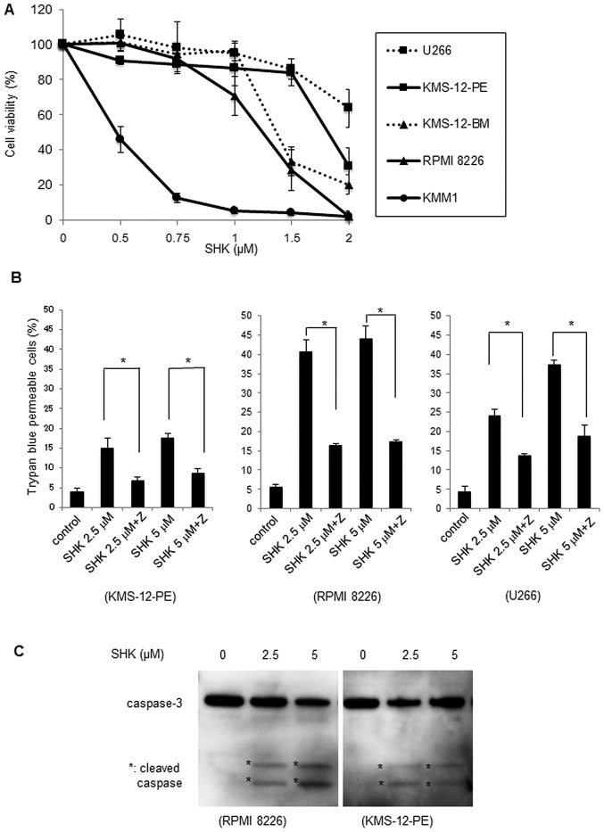
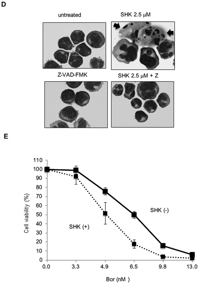
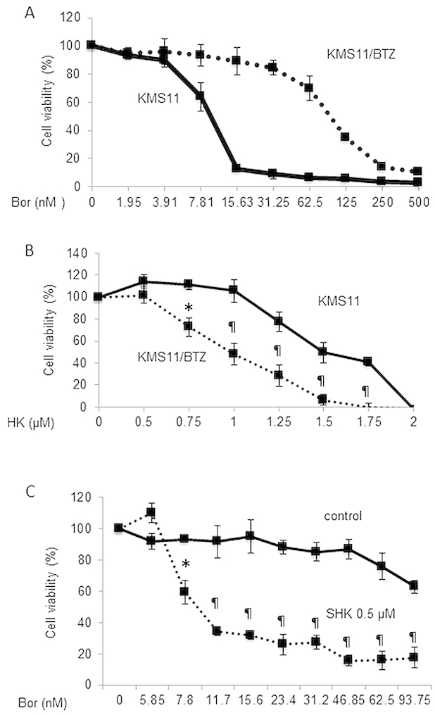
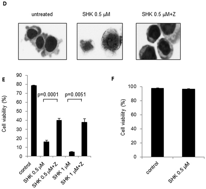
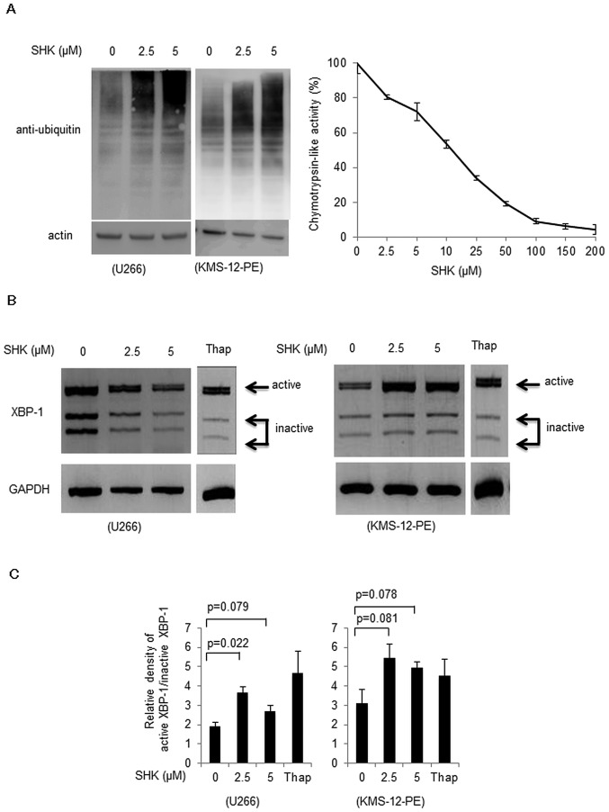
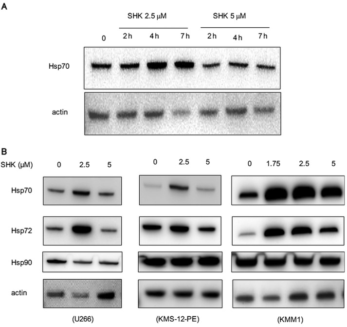
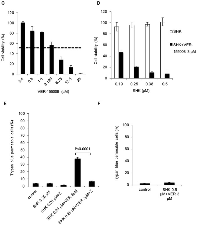
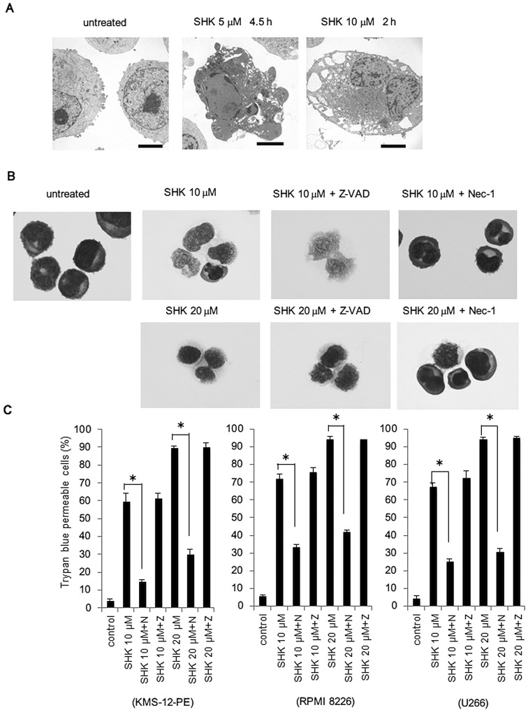
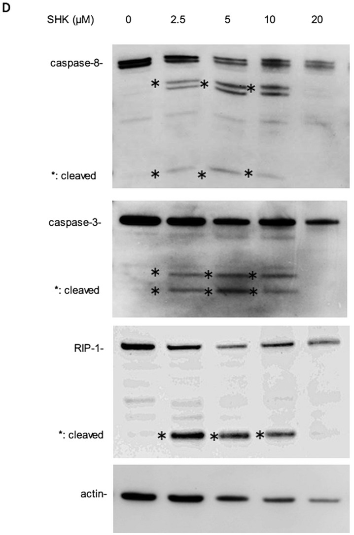
Shikonin (SHK), a natural small agent (MW 288.3), reportedly induces cell death in various tumor cells. We have found that SHK also exerts potent cytocidal effects on human multiple myeloma (MM) cells, but its anticancer mechanism in MM cells remains to be elucidated. SHK at 2.5-5 µM induced apoptosis in seven MM cell lines, including the bortezomib-resistant cell line KMS11/BTZ. The IC50 value of SHK against KMS11/BTZ was comparable to that of a parental cell line KMS11 (1.1 and 1.56 µM, respectively). SHK induces accumulation of ubiquitinated proteins and activates XBP-1 in MM cells, suggesting that SHK functions as a proteasome inhibitor, eventually inducing ER stress-associated apoptosis. SHK increases levels of HSP70/72, which protects cells from apoptosis, and exerts greater cytocidal effects in combination with the HSP70/72 inhibitor VER-155008. At higher concentrations (10-20 µM), SHK induced cell death, which was completely inhibited by a necroptosis inhibitor, necrostatin-1 (Nec-1), while the cytocidal activity was unaffected by Z-VAD-FMK, strongly suggesting that cell death is induced by SHK at high concentrations through necroptosis. The present data show for the first time that SHK induces cell death in MM cells. SHK efficiently induces apoptosis and combination of heat shock protein inhibitor with low dose SHK enhances apoptosis, while high dose SHK induces necroptosis in MM cells. These findings together support the use of SHK as a potential therapeutic agent for MM.


Figures

Figure 1
Figure 1
Induction of apoptosis in MM cells by low concentrations of SHK. (A) Five human MM cell lines (U266, KMS-12-PE, KMS-12-BM, RPMI-8226 and KMM1) were cultured for 24 h in the presence of various concentrations of SHK and analyzed by WST-8 assay. All MM cells tested showed dose-dependent cytotoxic effects of SHK. (B) Three representative MM cell lines (KMS-12-PE, RPMI-8226 and U266) were incubated with SHK at 2.5 or 5 μM for 7 h with or without 20 min pre-treatment with Z-VAD-FMK (indicated as Z) and then analyzed by trypan blue dye exclusion assay. SHK-induced cell death was partly inhibited by Z-VAD-FMK. *P<0.01. (C) Western blot analyses of caspase-3. SHK activated caspase-3 at concentrations of 2.5 and 5 μM for 7 h. (D) Morphological changes of MM cells after incubation with SHK. KMS-12-PE cells were treated with 2.5 μM SHK for 5 h either with or without pre-treatment of Z-VAD-FMK and evaluated by cytospin analysis. Cells were stained with May-Giemsa staining solution. SHK clearly induced apoptotic morphological changes, such as fragmented nucleus (arrows), and apoptosis was inhibited by Z-VAD-FMK. (E) Enhanced cytotoxic effects of bortezomib by SHK. KMS-12-PE cells were incubated with various concentrations of bortezomib either with (dotted line) or without 0.5 μM SHK (solid line). Cell viability was analyzed by WST-8 assay. Marked sensitization of MM cells to bortezomib by SHK was observed.
Figure 1
Figure 1
Induction of apoptosis in MM cells by low concentrations of SHK. (A) Five human MM cell lines (U266, KMS-12-PE, KMS-12-BM, RPMI-8226 and KMM1) were cultured for 24 h in the presence of various concentrations of SHK and analyzed by WST-8 assay. All MM cells tested showed dose-dependent cytotoxic effects of SHK. (B) Three representative MM cell lines (KMS-12-PE, RPMI-8226 and U266) were incubated with SHK at 2.5 or 5 μM for 7 h with or without 20 min pre-treatment with Z-VAD-FMK (indicated as Z) and then analyzed by trypan blue dye exclusion assay. SHK-induced cell death was partly inhibited by Z-VAD-FMK. *P<0.01. (C) Western blot analyses of caspase-3. SHK activated caspase-3 at concentrations of 2.5 and 5 μM for 7 h. (D) Morphological changes of MM cells after incubation with SHK. KMS-12-PE cells were treated with 2.5 μM SHK for 5 h either with or without pre-treatment of Z-VAD-FMK and evaluated by cytospin analysis. Cells were stained with May-Giemsa staining solution. SHK clearly induced apoptotic morphological changes, such as fragmented nucleus (arrows), and apoptosis was inhibited by Z-VAD-FMK. (E) Enhanced cytotoxic effects of bortezomib by SHK. KMS-12-PE cells were incubated with various concentrations of bortezomib either with (dotted line) or without 0.5 μM SHK (solid line). Cell viability was analyzed by WST-8 assay. Marked sensitization of MM cells to bortezomib by SHK was observed.
Figure 2
Figure 2
Antitumor effect of SHK on bortezomib resistant cells, primary MM cells and peripheral blood mononuclear cells. The bortezomib resistant MM cell line KMS-11/BTZ, and the parental cell line KMS11, were cultured with various concentrations of bortezomib (A), SHK (B) or in combination (C) for 24 h and cell viabilities were analyzed by WST-8 assay. (A) The bortezomib resistant MM cell line KMS-11/BTZ (dotted line), and the parental cell line, KMS11 (solid line), were treated with bortezomib for 24 h and subsequently analyzed by WST8 assay. The IC50 values of the KMS11 and KMS11/BTZ cells were 9.9 and 98.5 nM, respectively. (B) The IC50 value of SHK for KMS11/BTZ cells (dotted line) was even lower than that of KMS11 cells (solid line) (1.1 vs. 1.56 μM, respectively). *P<0.05, P<0.01. (C) Treatment of KMS11/BTZ with (dotted line) or without (solid line) low concentration of SHK (0.5 μM), which alone does not show cytotoxic effects, increased the sensitivity to bortezomib. *P<0.005, P<0.0001. (D and E) MM cells from primary bone marrow sample were incubated with 0.5 μM SHK for 16 h and then either evaluated by cytospin analysis (D) or WST-8 assay (E). Both analyses revealed marked increase of dead cells in response to treatment with SHK and inhibition by Z-VAD-FMK. (F) Lack of cytotoxic effect of SHK in normal PBMCs. PBMCs were cultured with 0.5 μM SHK for 24 h and evaluated by trypan blue dye exclusion assay. There was no increase of dead cells by SHK.
Figure 2
Figure 2
Antitumor effect of SHK on bortezomib resistant cells, primary MM cells and peripheral blood mononuclear cells. The bortezomib resistant MM cell line KMS-11/BTZ, and the parental cell line KMS11, were cultured with various concentrations of bortezomib (A), SHK (B) or in combination (C) for 24 h and cell viabilities were analyzed by WST-8 assay. (A) The bortezomib resistant MM cell line KMS-11/BTZ (dotted line), and the parental cell line, KMS11 (solid line), were treated with bortezomib for 24 h and subsequently analyzed by WST8 assay. The IC50 values of the KMS11 and KMS11/BTZ cells were 9.9 and 98.5 nM, respectively. (B) The IC50 value of SHK for KMS11/BTZ cells (dotted line) was even lower than that of KMS11 cells (solid line) (1.1 vs. 1.56 μM, respectively). *P<0.05, P<0.01. (C) Treatment of KMS11/BTZ with (dotted line) or without (solid line) low concentration of SHK (0.5 μM), which alone does not show cytotoxic effects, increased the sensitivity to bortezomib. *P<0.005, P<0.0001. (D and E) MM cells from primary bone marrow sample were incubated with 0.5 μM SHK for 16 h and then either evaluated by cytospin analysis (D) or WST-8 assay (E). Both analyses revealed marked increase of dead cells in response to treatment with SHK and inhibition by Z-VAD-FMK. (F) Lack of cytotoxic effect of SHK in normal PBMCs. PBMCs were cultured with 0.5 μM SHK for 24 h and evaluated by trypan blue dye exclusion assay. There was no increase of dead cells by SHK.
Figure 3
Figure 3
Accumulation of ubiquitinated proteins and activation of XBP-1 by SHK. (A) Left panel, western blot analyses of ubiquitinated proteins. U266 and KMS-12-PE cells were incubated with 2.5 or 5 μM SHK for 7 h. SHK induced an accumulation of ubiquitinated proteins in a dose-dependent manner. Right panel, inhibition of 20S chymotrypsin-like activity by SHK. SHK decreased 20S chymotrypsin-like activity at a dose-dependent manner. (B) Activation of XBP-1 by SHK. XBP-1 mRNA from U266 and KMS-12-PE cells treated with SHK was converted to cDNA by RT-PCR and then subsequently digested with ApaLI to distinguish inactive and active XBP-1. Thapsigargin (Thap) was used as an endoplasmic reticulum stress inducer at 100 nM for 8 h. SHK was used at concentrations of 2.5–5 μM for 2 h. The longer fragment derived from the active form of XBP-1 mRNA (upper band) and two shorter bands derived from the inactive form (middle and lower bands) were detected. A decrease of the inactive bands was found in U266 (left panel) cells, while increase of active band was noted in KMS-12-PE cells (right panel) by treatment with SHK. (C) Relative density of active XBP-1 compared with inactive XBP-1, as shown in (B) was calculated. A significant increase of active XBP-1 was found by treatment with SHK in U266 cells. KMS-12-PE cells showed a similar trend, although the difference was not statistically significant.
Figure 4
Figure 4
Increase of HSP70/72 by SHK and synergistic cytotoxic effects of SHK in combination with HSP70/72 inhibitor. (A) Western blot analyses of HSP70. SHK at a concentration of 2.5 μM transiently increased HSP70 in KMS-12-PE cells in a time-dependent manner, while this was less evident at 5 μM. (B) Western blot analyses of HSP70, HSP72, and HSP90. U266, KMS-12-PE and KMM1 were treated with 2.5 and 5 μM SHK for 7 h. Induction of HSP70 and HSP72 by SHK was observed in all cell lines and maximized at 2.5 μM. There was no change in the expression of HSP90. (C) Cytotoxic effect of the HSP70/72 inhibitor VER-155008 in KMS-12-PE cells. Cells were cultured with various concentrations of VER-155008 for 24 h and evaluated by WST-8 analysis. VER-155008 alone induced cytotoxic effects in MM cells. Note that VER-155008 at ~3 μM showed 55% growth inhibition (dotted line). (D) Combination effects of VER-155008 and SHK. KMS-12-PE cells were treated with SHK at concentrations varying from 0.19 to 0.5 μM either with 3 μM VER-155008 (solid bars) or SHK alone (blank bars) for 24 h. Combinations of SHK and VER-155008 showed significant synergistic effects in induction of cytotoxicity (CI=0.72). (E) The populations of dead cells induced by the combination of SHK and VER-155008 (VER) were partly inhibited by Z-VAD-FMK (P<0.0001). (F) Combination of SHK and VER-155008 did not show toxic effects in normal PBMCs. PBMCs from a normal donor were cultured with SHK and VER-155008 (VER) at 0.5 and 3 μM, respectively, for 24 h and evaluated by trypan blue dye exclusion analysis. No cytotoxic effect was observed.
Figure 4
Figure 4
Increase of HSP70/72 by SHK and synergistic cytotoxic effects of SHK in combination with HSP70/72 inhibitor. (A) Western blot analyses of HSP70. SHK at a concentration of 2.5 μM transiently increased HSP70 in KMS-12-PE cells in a time-dependent manner, while this was less evident at 5 μM. (B) Western blot analyses of HSP70, HSP72, and HSP90. U266, KMS-12-PE and KMM1 were treated with 2.5 and 5 μM SHK for 7 h. Induction of HSP70 and HSP72 by SHK was observed in all cell lines and maximized at 2.5 μM. There was no change in the expression of HSP90. (C) Cytotoxic effect of the HSP70/72 inhibitor VER-155008 in KMS-12-PE cells. Cells were cultured with various concentrations of VER-155008 for 24 h and evaluated by WST-8 analysis. VER-155008 alone induced cytotoxic effects in MM cells. Note that VER-155008 at ~3 μM showed 55% growth inhibition (dotted line). (D) Combination effects of VER-155008 and SHK. KMS-12-PE cells were treated with SHK at concentrations varying from 0.19 to 0.5 μM either with 3 μM VER-155008 (solid bars) or SHK alone (blank bars) for 24 h. Combinations of SHK and VER-155008 showed significant synergistic effects in induction of cytotoxicity (CI=0.72). (E) The populations of dead cells induced by the combination of SHK and VER-155008 (VER) were partly inhibited by Z-VAD-FMK (P<0.0001). (F) Combination of SHK and VER-155008 did not show toxic effects in normal PBMCs. PBMCs from a normal donor were cultured with SHK and VER-155008 (VER) at 0.5 and 3 μM, respectively, for 24 h and evaluated by trypan blue dye exclusion analysis. No cytotoxic effect was observed.
Figure 5
Figure 5
Induction of necroptosis in MM cells by SHK. (A) Electron microscopic examination of KMS-12-PE cells treated with 5 μM SHK for 4 h showed typical apoptotic changes, such as fragmented and condensed nuclei (middle panel). In contrast, treatment of 10 μM SHK for 2 h induced typical necrotic changes, such as translucent cytoplasm and swelling of cell membranes (right panel). Scale bar, 5 μm. (B) Morphological changes of KMS-12-PE cells after incubation with 10 or 20 μM SHK for 5 h. SHK induced ghost cells (left panel) and this was inhibited by treatment with Nec-1 (right panel). No apparent inhibition of cell death was found by Z-VAD-FMK treatment (middle panel). (C) Nec-1 inhibited cell death induced by SHK. KMS-12-PE, RPMI-8226 and U266 cells were incubated with 10 or 20 μM SHK in the presence or absence of Nec-1 (indicated as N) or Z-VAD-FMK (indicated as Z) for 7 h and then analyzed by trypan blue dye exclusion assay. SHK-induced cell death was significantly inhibited by Nec-1 (P<0.01) and not affected by Z-VAD-FMK (*P<0.01). (D) Western blot analyses of caspase-8, -3, and RIP-1. RPMI-8226 cells were treated with SHK at concentrations from 2.5 to 20 μM. SHK activated caspase-8 and -3 at concentrations <10 μM while no changes were detected at 20 μM SHK. RIP-1 was cleaved by SHK <10 μM and remained intact at 20 μM.
Figure 5
Figure 5
Induction of necroptosis in MM cells by SHK. (A) Electron microscopic examination of KMS-12-PE cells treated with 5 μM SHK for 4 h showed typical apoptotic changes, such as fragmented and condensed nuclei (middle panel). In contrast, treatment of 10 μM SHK for 2 h induced typical necrotic changes, such as translucent cytoplasm and swelling of cell membranes (right panel). Scale bar, 5 μm. (B) Morphological changes of KMS-12-PE cells after incubation with 10 or 20 μM SHK for 5 h. SHK induced ghost cells (left panel) and this was inhibited by treatment with Nec-1 (right panel). No apparent inhibition of cell death was found by Z-VAD-FMK treatment (middle panel). (C) Nec-1 inhibited cell death induced by SHK. KMS-12-PE, RPMI-8226 and U266 cells were incubated with 10 or 20 μM SHK in the presence or absence of Nec-1 (indicated as N) or Z-VAD-FMK (indicated as Z) for 7 h and then analyzed by trypan blue dye exclusion assay. SHK-induced cell death was significantly inhibited by Nec-1 (P<0.01) and not affected by Z-VAD-FMK (*P<0.01). (D) Western blot analyses of caspase-8, -3, and RIP-1. RPMI-8226 cells were treated with SHK at concentrations from 2.5 to 20 μM. SHK activated caspase-8 and -3 at concentrations <10 μM while no changes were detected at 20 μM SHK. RIP-1 was cleaved by SHK <10 μM and remained intact at 20 μM.

References

    1. Palumbo A, Anderson K. Multiple myeloma. N Engl J Med. 2011;364:1046–1060. doi: 10.1056/NEJMra1011442. - DOI - PubMed
    1. He S, Liao TT, Chen YT, Kuo HM, Lin YL. Glutathione-S-transferase enhances proliferation-migration and protects against shikonin-induced cell death in breast cancer cells. Kaohsiung J Med Sci. 2011;27:477–484. doi: 10.1016/j.kjms.2011.06.010. - DOI - PMC - PubMed
    1. Duan D, Zhang B, Yao J, Liu Y, Fang J. Shikonin targets cytosolic thioredoxin reductase to induce ROS-mediated apoptosis in human promyelocytic leukemia HL-60 cells. Free Radic Biol Med. 2014;70:182–193. doi: 10.1016/j.freeradbiomed.2014.02.016. - DOI - PubMed
    1. Yoon Y, Kim YO, Lim NY, Jeon WK, Sung HJ. Shikonin, an ingredient of Lithospermum erythrorhizon induced apoptosis in HL60 human premyelocytic leukemia cell line. Planta Med. 1999;65:532–535. doi: 10.1055/s-1999-14010. - DOI - PubMed
    1. Mao X, Yu CR, Li WH, Li WX. Induction of apoptosis by shikonin through a ROS/JNK-mediated process in Bcr/Abl-positive chronic myelogenous leukemia (CML) cells. Cell Res. 2008;18:879–888. doi: 10.1038/cr.2008.86. - DOI - PubMed

Publication types

MeSH terms