. 2015 Jul;25(7):813-26.
doi: 10.1002/hipo.22405. Epub 2015 Jan 8.

Human neural stem cells improve cognition and promote synaptic growth in two complementary transgenic models of Alzheimer's disease and neuronal loss

Affiliations

Human neural stem cells improve cognition and promote synaptic growth in two complementary transgenic models of Alzheimer's disease and neuronal loss

Rahasson R Ager et al. Hippocampus. 2015 Jul.

Abstract

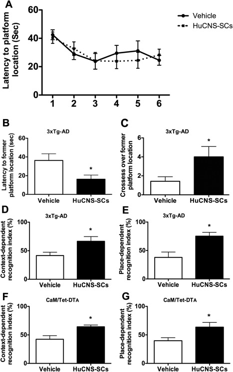
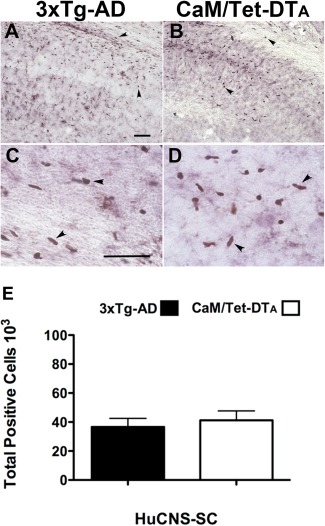
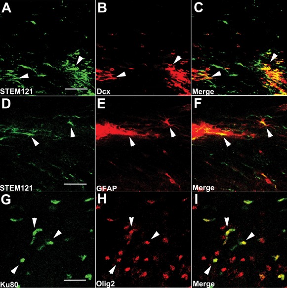
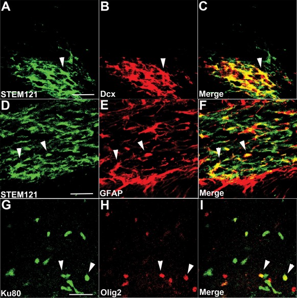
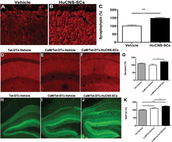
Alzheimer's disease (AD) is the most prevalent age-related neurodegenerative disorder, affecting over 35 million people worldwide. Pathologically, AD is characterized by the progressive accumulation of β-amyloid (Aβ) plaques and neurofibrillary tangles within the brain. Together, these pathologies lead to marked neuronal and synaptic loss and corresponding impairments in cognition. Current treatments, and recent clinical trials, have failed to modify the clinical course of AD; thus, the development of novel and innovative therapies is urgently needed. Over the last decade, the potential use of stem cells to treat cognitive impairment has received growing attention. Specifically, neural stem cell transplantation as a treatment for AD offers a novel approach with tremendous therapeutic potential. We previously reported that intrahippocampal transplantation of murine neural stem cells (mNSCs) can enhance synaptogenesis and improve cognition in 3xTg-AD mice and the CaM/Tet-DT(A) model of hippocampal neuronal loss. These promising findings prompted us to examine a human neural stem cell population, HuCNS-SC, which has already been clinically tested for other neurodegenerative disorders. In this study, we provide the first evidence that transplantation of research grade HuCNS-SCs can improve cognition in two complementary models of neurodegeneration. We also demonstrate that HuCNS-SC cells can migrate and differentiate into immature neurons and glia and significantly increase synaptic and growth-associated markers in both 3xTg-AD and CaM/Tet-DTA mice. Interestingly, improvements in aged 3xTg-AD mice were not associated with altered Aβ or tau pathology. Rather, our findings suggest that human NSC transplantation improves cognition by enhancing endogenous synaptogenesis. Taken together, our data provide the first preclinical evidence that human NSC transplantation could be a safe and effective therapeutic approach for treating AD.

Keywords: Alzheimer's disease; immune suppression; neural stem cells; therapeutic; transplantation.


Figures

Figure 1
Figure 1
Transplantation of HuCNS‐SC improves learning and memory in two complementary models of AD pathogenesis and hippocampal neuronal loss. One‐month after transplantation 3xTg‐AD mice were tested on three independent cognitive tasks. Although no differences were detected in Morris water maze (MWM) acquisition (A), HuCNS‐SC transplantation (black boxes) significantly improved spatial memory as evidenced by decreased probe trail latencies (B) and increased platform crosses (C) vs. vehicle‐treated (white boxes) transgenics (ANOVA P < 0.05, Fisher's PLSD P = 0.034 and P = 0.05). (D) Performance in context‐recognition and (E) place recognition was also significantly improved by HuCNS‐SC transplantation (ANOVA P < 0.05, Fisher's PLSD P = 0.029 and P = 0.01). Beginning 1‐month after transplantation, CaM/Tet‐DTA mice were also tested in context‐ (F) and place‐recognition (G) tasks. HuCNS‐SC transplantation significantly improved performance of lesioned mice vs. vehicle‐injected lesioned mice in both tasks (context: recognition index = 64.3 vs. 42.26, ANOVA P < 0.05, Fisher's PLSD P = 0.04; place: recognition index = 58.87 vs. 39.8, ANOVA P < 0.05, Fisher's PLSD P = 0.04). Data presented as mean ± SEM.
Figure 2
Figure 2
Both novel and traditional immune suppression paradigms allow robust survival and engraftment of human Neural Stem Cells. Given the reported effects of calcineurin inhibitors on AD pathogenesis, we used a novel paradigm that targets leukocyte costimulatory molecules to achieve xenotransplantation in 3xTg‐AD mice. (A) 6 weeks after transplantation, engrafted HuCNS‐SC (human nuclear antigen), were located within the hippocampus and overlying corpus collosum. (B) Immunosuppression with FK506 and anti‐CD4 provided a similar degree of HuCNS‐SC engraftment in CaM/Tet‐DTA mice. (C, D) High power images from subfields of A and B reveal HuCNS‐SC positive for human nuclear antigen (brown). (E) Unbiased stereological assessment of HuCNS‐SC engraftment demonstrates that approximately 40,000 transplanted cells survive per injection site in both 3xTg‐AD and CaM/Tet‐DTA models (optical fractionator probe, N = 6). Data presented as mean ± SEM. Scale bars = 50 μm. [Color figure can be viewed in the online issue, which is available at wileyonlinelibrary.com.]
Figure 3
Figure 3
HuCNS‐SC show initial signs of differentiation in 3xTg‐AD mice. (A–C) HuCNS‐SC (green) injected into the hippocampus express doublecortin (red), a marker of immature neurons, 6 weeks after transplantation. (D–F) Examples of engrafted HuCNS‐SC (green) that coexpress the astrocytic marker GFAP (red) were also observed. (G–I) HuCNS‐SC expressing the nuclear marker Ku80 (green) also coexpress the immature oligodendrocyte marker olig‐2 (red). Scale bars, 25 μm. [Color figure can be viewed in the online issue, which is available at wileyonlinelibrary.com.]
Figure 4
Figure 4
HuCNS‐SCs transplanted into CaM/Tet‐DTA mice also exhibit evidence of neural and glial differentiation. (A–C) HuCNS‐SCs (green) injected into lesioned CaM/Tet‐DTA mice often co‐expressed the early neuronal marker doublecortin (red). (D–F) Examples of HuCNS‐SCs (green) that colocalized with GFAP (red) were also detected. (G–I) In accordance with our findings in 3xTg‐AD mice, HuCNS‐SCs (green) were also found to coexpress the oligodendroglial marker olig‐2 (red). Scale bars, 25 μm. [Color figure can be viewed in the online issue, which is available at wileyonlinelibrary.com.]
Figure 5
Figure 5
HuCNS‐SC transplantation has no effect on Aβ or tau pathology. ELISA analysis reveals no significant difference in either soluble (A) or insoluble (B) Aβ following HuCNS‐SC transplantation. (C–E) Confocal analysis of total human tau also reveals no difference between vehicle‐ and HuCNS‐SC‐transplanted 3xTg‐AD mice. (F–H) Likewise, levels of hyperphosphorylated PHF‐1 positive tau are unaltered by HuCNS‐SC transplantation. (N = 5, data presented as mean ± SEM). Scale bars = 50 μm. [Color figure can be viewed in the online issue, which is available at wileyonlinelibrary.com.]
Figure 6
Figure 6
HuCNS‐SC transplantation increases presynaptic markers and axonal sprouting. (A) Immunolabeling of synaptophysin in vehicle injected 3xTg‐AD mice. (B) HuCNS‐SC injection results in a 47% increase in synaptophysin levels in the stratum radiatum of CA1, quantified in (C, N = 5, t‐test P < 0.008). (D) Synapsin in control unlesioned mice reveals a typical pattern of presynaptic innervation within the stratum radiatum of CA1. (E) Surprisingly, synapsin levels are only slightly diminished in lesioned mice. (F) In contrast, HuCNS‐SC transplantation significantly increases the density of presynaptic innervation, quantified in (G, N = 8–12, ANOVA P < 0.05, Fishers PLSD P = 0.016). (H) Compared to control unlesioned mice, we also detected a significant increase in GAP‐43 immunoreactivity within the dentate gyrus of lesioned CaM/Tet‐DTA mice with vehicle injections (I). More importantly, we observed a further enhancement of GAP‐43 expression in lesioned mice transplanted with HuCNS‐SC (J), quantified in (K). (N = 10, ANOVA P < 0.05, Fishers PLSD P = 0.028, P = 0.006, P = 0.0001). Data presented as mean ± SEM. [Color figure can be viewed in the online issue, which is available at wileyonlinelibrary.com.]

References

    1. Aboody KS, Brown A, Rainov NG, Bower KA, Liu S, Yang W, Small JE, Herrlinger U, Ourednik V, Black PM, XO Breakefield, EY Snyder. 2000. Neural stem cells display extensive tropism for pathology in adult brain: Evidence from intracranial gliomas. Proc Natl Acad Sci USA 97:12846–12851. - PMC - PubMed
    1. Anderson AJ, Haus DL, Hooshmand MJ, Perez H, Sontag CJ, Cummings, B.J. 2011. Achieving stable human stem cell engraftment and survival in the CNS: Is the future of regenerative medicine immunodeficient? Regen Med 6:367–406. - PMC - PubMed
    1. Baglietto‐Vargas D, Moreno‐Gonzalez I, Sanchez‐Varo R, Jimenez S, Trujillo‐Estrada L, Sanchez‐Mejias E, Torres M, Romero‐Acebal M, Ruano D, Vizuete M, J Vitorica, A Gutierrez. 2010. Calretinin interneurons are early targets of extracellular amyloid‐beta pathology in PS1/AbetaPP Alzheimer mice hippocampus. J Alzheimer's Dis 21:119–132. - PubMed
    1. Bakondi B, Shimada IS, Perry A, Munoz JR, Ylostalo J, Howard AB, Gregory CA, Spees JL. 2009. CD133 identifies a human bone marrow stem/progenitor cell sub‐population with a repertoire of secreted factors that protect against stroke. Mol Ther 17:1938–1947. - PMC - PubMed
    1. Basi GS, Jacobson RD, Virag I, Schilling J, Skene JH. 1987. Primary structure and transcriptional regulation of GAP‐43, a protein associated with nerve growth. Cell 49:785–791. - PubMed

Publication types

MeSH terms

Substances