

Shiga Toxin (Stx) Classification, Structure, and Function
- PMID: 25530917
- PMCID: PMC4270005
- DOI: 10.1128/microbiolspec.EHEC-0024-2013
Shiga Toxin (Stx) Classification, Structure, and Function
Abstract
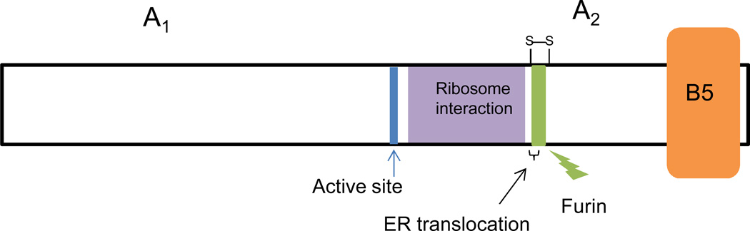
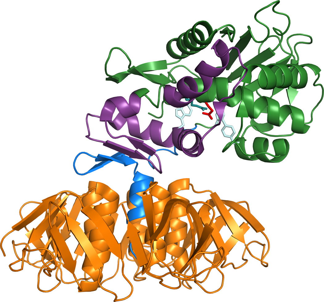
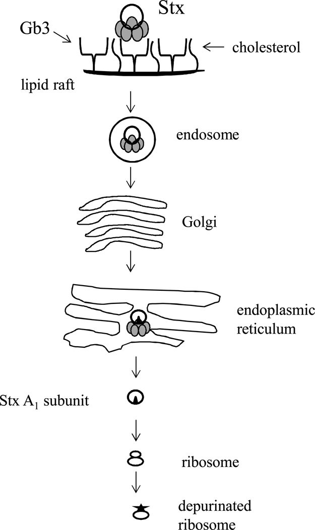
Shiga toxin (Stx) is one of the most potent bacterial toxins known. Stx is found in Shigella dysenteriae 1 and in some serogroups of Escherichia coli (called Stx1 in E. coli). In addition to or instead of Stx1, some E. coli strains produce a second type of Stx, Stx2, that has the same mode of action as Stx/Stx1 but is antigenically distinct. Because subtypes of each toxin have been identified, the prototype toxin for each group is now designated Stx1a or Stx2a. The Stxs consist of two major subunits, an A subunit that joins noncovalently to a pentamer of five identical B subunits. The A subunit of the toxin injures the eukaryotic ribosome and halts protein synthesis in target cells. The function of the B pentamer is to bind to the cellular receptor, globotriaosylceramide, Gb3, found primarily on endothelial cells. The Stxs traffic in a retrograde manner within the cell, such that the A subunit of the toxin reaches the cytosol only after the toxin moves from the endosome to the Golgi and then to the endoplasmic reticulum. In humans infected with Stx-producing E. coli, the most serious manifestation of the disease, hemolytic-uremic syndrome, is more often associated with strains that produce Stx2a rather than Stx1a, and that relative toxicity is replicated in mice and baboons. Stx1a and Stx2a also exhibit differences in cytotoxicity to various cell types, bind dissimilarly to receptor analogs or mimics, induce differential chemokine responses, and have several distinctive structural characteristics.
Figures
References
-
- Trofa AF, Ueno-Olsen H, Oiwa R, Yoshikawa M. Dr. Kiyoshi Shiga: discoverer of the dysentery bacillus. Clin Infect Dis. 1999;29:1303–1306. - PubMed
-
- Conradi H. Uber Iosliche, durch asptische Autolyse erhaltene Giftstoffe vonRuhr- und Typhus-Bazillen. Dtsch. Med. Wochenschr. 1903;29:26–28.
-
- Karmali MA, Steele BT, Petric M, Lim C. Sporadic cases of haemolytic-uraemic syndrome associated with faecal cytotoxin and cytotoxin-producing Escherichia coli in stools. Lancet. 1983;1:619–620. - PubMed
-
- O'Brien AO, Lively TA, Chen ME, Rothman SW, Formal SB. Escherichia coli O157:H7 strains associated with haemorrhagic colitis in the United States produce a Shigella dysenteriae 1 (SHIGA) like cytotoxin. Lancet. 1983;1:702. - PubMed
Publication types
MeSH terms
Substances
Grants and funding
LinkOut - more resources
Full Text Sources
Other Literature Sources
