. 2015 Sep 24;34(39):5025-36.
doi: 10.1038/onc.2014.428. Epub 2014 Dec 22.

LARP1 post-transcriptionally regulates mTOR and contributes to cancer progression

Affiliations

LARP1 post-transcriptionally regulates mTOR and contributes to cancer progression

M Mura et al. Oncogene. .

Abstract

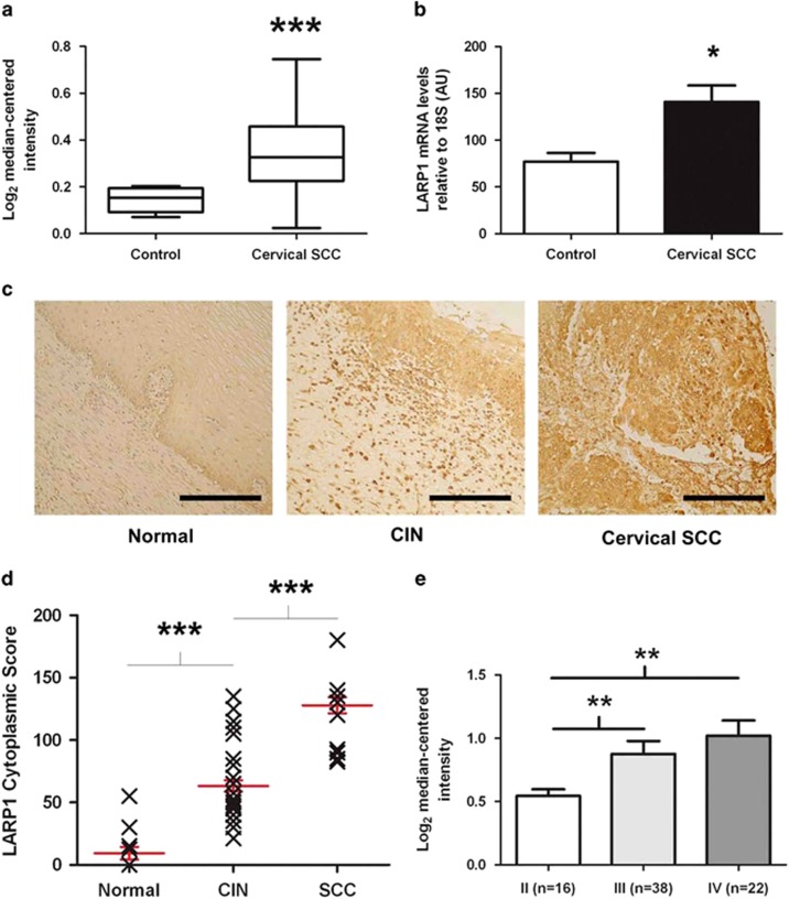
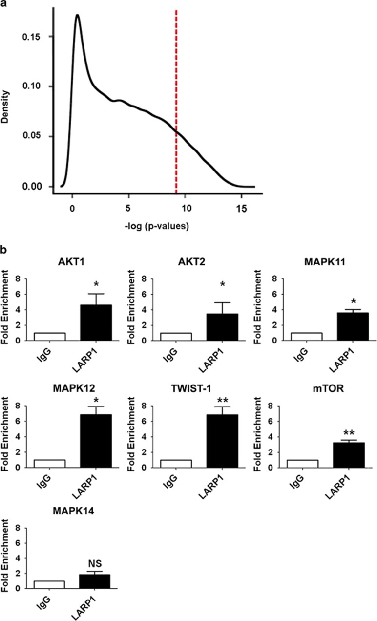
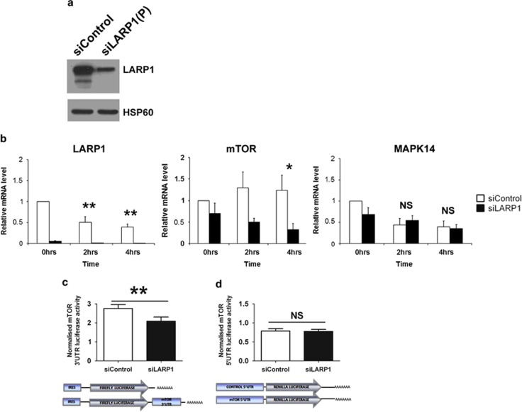
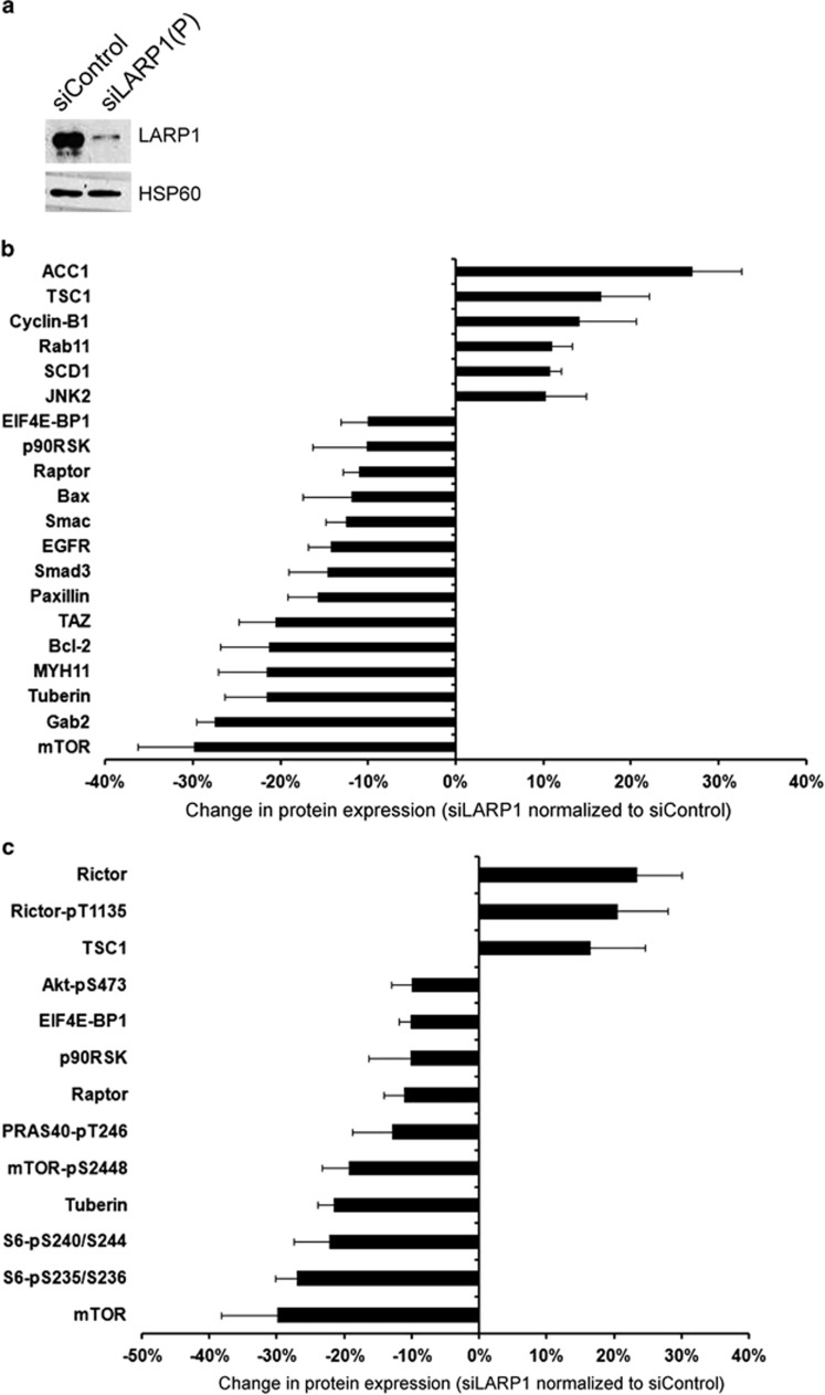
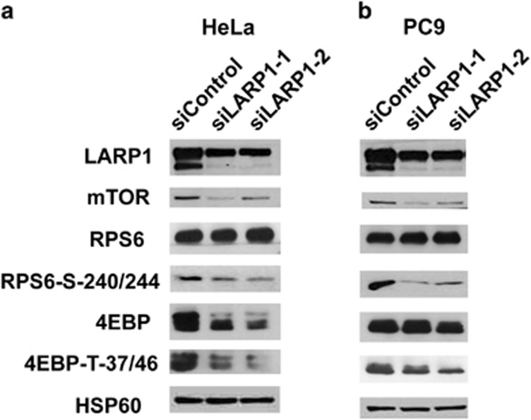
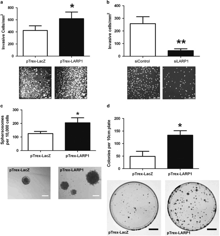
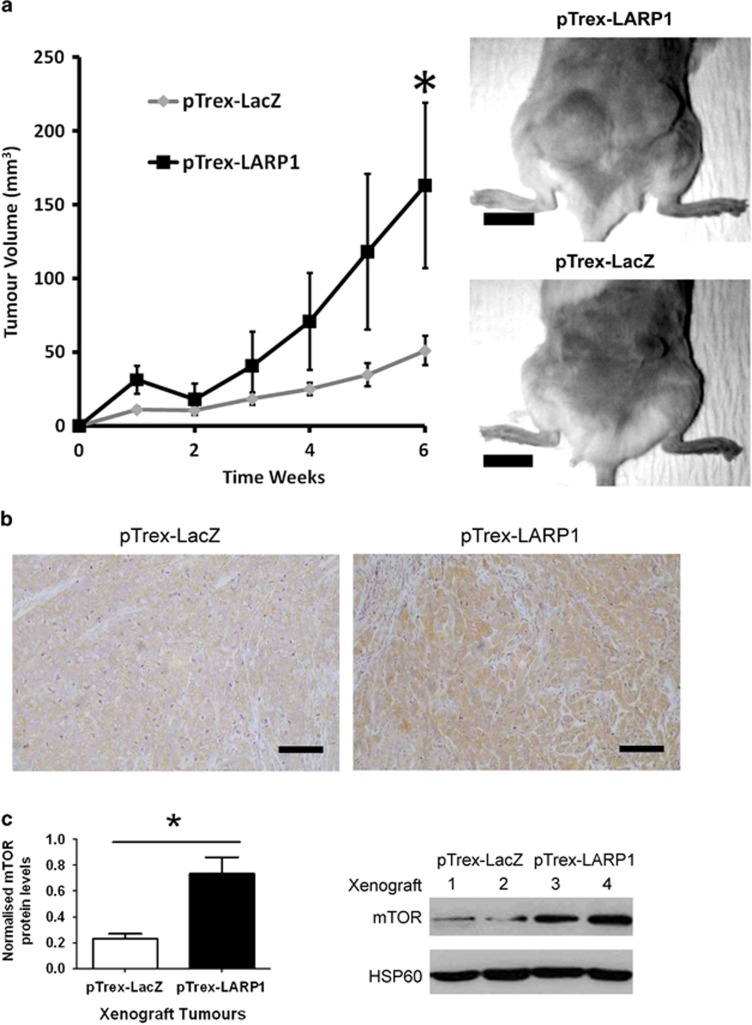
RNA-binding proteins (RBPs) bind to and post-transcriptionally regulate the stability of mRNAs. La-related protein 1 (LARP1) is a conserved RBP that interacts with poly-A-binding protein and is known to regulate 5'-terminal oligopyrimidine tract (TOP) mRNA translation. Here, we show that LARP1 is complexed to 3000 mRNAs enriched for cancer pathways. A prominent member of the LARP1 interactome is mTOR whose mRNA transcript is stabilized by LARP1. At a functional level, we show that LARP1 promotes cell migration, invasion, anchorage-independent growth and in vivo tumorigenesis. Furthermore, we show that LARP1 expression is elevated in epithelial cancers such as cervical and non-small cell lung cancers, where its expression correlates with disease progression and adverse prognosis, respectively. We therefore conclude that, through the post-transcriptional regulation of genes such as mTOR within cancer pathways, LARP1 contributes to cancer progression.


Figures

Figure 1
Figure 1
LARP1 correlates with clinical variables in cervical cancer. (a) Expression dataset comparing LARP1-mRNA levels in cervical squamous cell carcinoma (SCC) and non-cancer tissue. (b) Real-time PCR on RNA extracted from non-cancer cervical tissue (n=4) and cervical SCC (n=7). LARP1-mRNA levels relative to 18S ribosomal RNA (P=0.012, Student's t-test). (c) LARP1 immunostaining (brown) of normal cervical epithelium (N12), cervical intraepithelial neoplasia (CIN; N35) and invasive cervical SCC (N36), counterstained with haematoxylin. (d) LARP1 cytoplasmic scores for CIN compared to normal samples (P<0.0001) and in invasive cervical SCC compared to CIN samples (P<0.0001). (e) Expression dataset showing LARP1 expression in cervical SCC, stratified according to stage. *P⩽0.05, **P⩽ 0.001 and ***P⩽0.0001.
Figure 2
Figure 2
LARP1 exists in complex with mRNAs. (a) Histogram demonstrating relative transcript density across significance thresholds for LARP1-IP-enriched mRNAs (red line, P=1 × 10⁻⁴). (b) RIP-Chip validation by qRT–PCR showing fold enrichment of selected target mRNAs immunoprecipitated with LARP1 antibody, normalized to immunoprecipitation with IgG isotype control. Experiments were performed ⩾3 times. Data are mean±s.e.m. *P⩽0.05 and **P⩽0.001.
Figure 3
Figure 3
LARP1 regulate the stability of mTOR mRNA. (a) Western blot showing LARP1 knockdown with pooled siRNA. HSP60 was used as loading control. (b) Real-time PCR following transcription inhibition by actinomycin-D treatment in HeLa cells transfected with two pooled LARP1 siRNA or control siRNA. Fold changes relative to 18S, normalized to untreated control. (c) Luciferase production in HeLa cells transfected with firefly-luciferase reporter construct containing mTOR 3′UTR sequence normalized to the relative control plasmid (bottom panel: representation of the constructs used) in HeLa cells previously transfected with pooled siRNA to LARP1 or non-targeting siRNA. (d) Luciferase production in HeLa cells transfected with a renilla-luciferase reporter construct containing mTOR 5′UTR sequence normalized to the relative control plasmid (bottom panel: representation of constructs used) in HeLa cells previously transfected with pooled siRNA to LARP1 or non-targeting siRNA. Experiments were performed ⩾3 times. Data are mean±s.e.m. *P⩽0.05 and **P⩽0.001.
Figure 4
Figure 4
Depletion of LARP1 alters protein expression of interactome components, with wider effects on mTOR signaling pathway. (a) Level of LARP1 in HeLa cells transfected with control siRNA and two independent pooled siRNA sequences (siLARP1-1 and siLARP1-2). HSP60 was used as loading control. (b) Reverse-phase protein array after LARP1 knockdown (data normalized to control siRNA) shows altered expression of protein products of corresponding mRNAs identified as existing in complex with LARP1. (c) Change in protein expression and phosphorylation of mTOR signaling pathway components after LARP1 knockdown (data normalized to control siRNA). Experiments were performed ⩾3 times. Data are mean±s.e.m.
Figure 5
Figure 5
LARP1 knockdown decreases mTOR protein level and its downstream targets P-RPS6 and P-4EBP in HeLa and PC9 cell lines. Western blotting analysis of LARP1 in HeLa (a) and PC9 (b) cell lines after LARP1 knockdown using two independent siRNA oligos showing decrease in expression of mTOR, P-4EBP and P-RPS6, but not total RPS6 ribosomal protein. Total 4EBP showed a moderate decrease in HeLa cells. HSP60 was used as a loading control.
Figure 6
Figure 6
LARP1 promotes invasion and anchorage-independent growth. (a) Invasion assay in HeLa cells stably transfected with pTrex-LARP1 and pTrex-LacZ cells or (b) with pooled siRNA to LARP1 and non-targeting control. Graphs are counts of the number of invasive cells per cm2. Representative images of DAPI-stained invasive cells are shown (scale bars, 250 μm). (c) Anchorage-independent growth assay using ultra-low attachment plates in HeLa cells stably transfected with pTrex-LARP1 and pTrex-LacZ. Representative images of spherosomes (scale bars, 200 μm). (d) Number of colonies generated by dissociated spherosomes plated in adherent conditions. Scale bars, 2 cm. Experiments were performed ⩾3 times; data are mean±s.e.m. *P⩽0.05 and **P⩽0.001.
Figure 7
Figure 7
LARP1 promotes tumor progression in vivo and is associated with increased mTOR expression in xenograft tumors. (a) Two million HeLa pTrex-LacZ or pTrex-LARP1 cells were injected subcutaneously into the posterior flank of non-obese diabetic-severe combined immunodeficiency mice and tumor volume was monitored by caliper measurements. Representative mice from each cohort are shown (scale bars, 1 cm). (b) Immunohistochemistry analysis of mTOR protein level in xenograft tumors (scale bars, 200 μm). (c) Relative quantification of mTOR protein levels in xenograft tumors generated from pTrex-LacZ and pTrex-LARP1 cells with a corresponding western blot shown (sequential numbers represent tumors from different mice). Data are means±s.e.m. *P⩽0.05.

References

    1. 1Wu X, Brewer G. The regulation of mRNA stability in mammalian cells: 2.0. Gene 2012; 500: 10–21. - PMC - PubMed
    1. 2Sugiura R, Satoh R, Ishiwata S, Umeda N, Kita A. Role of RNA-binding proteins in MAPK signal transduction pathway. J Signal Transduct 2011; 2011: 109746. - PMC - PubMed
    1. 3Thapar R, Denmon AP. Signaling pathways that control mRNA turnover. Cell Signal 2013; 25: 1699–1710. - PMC - PubMed
    1. 4Keene JD, Lager PJ. Post-transcriptional operons and regulons co-ordinating gene expression. Chromosome Res 2005; 13: 327–337. - PubMed
    1. 5Blagden SP, Willis AE. The biological and therapeutic relevance of mRNA translation in cancer. Nat Rev Clin Oncol 2011; 8: 280–291. - PubMed

Publication types