. 2015 Feb;28(1):19-31.
doi: 10.1089/vim.2014.0108.

Multiple circulating infections can mimic the early stages of viral hemorrhagic fevers and possible human exposure to filoviruses in Sierra Leone prior to the 2014 outbreak

Affiliations

Multiple circulating infections can mimic the early stages of viral hemorrhagic fevers and possible human exposure to filoviruses in Sierra Leone prior to the 2014 outbreak

Matthew L Boisen et al. Viral Immunol. 2015 Feb.

Abstract

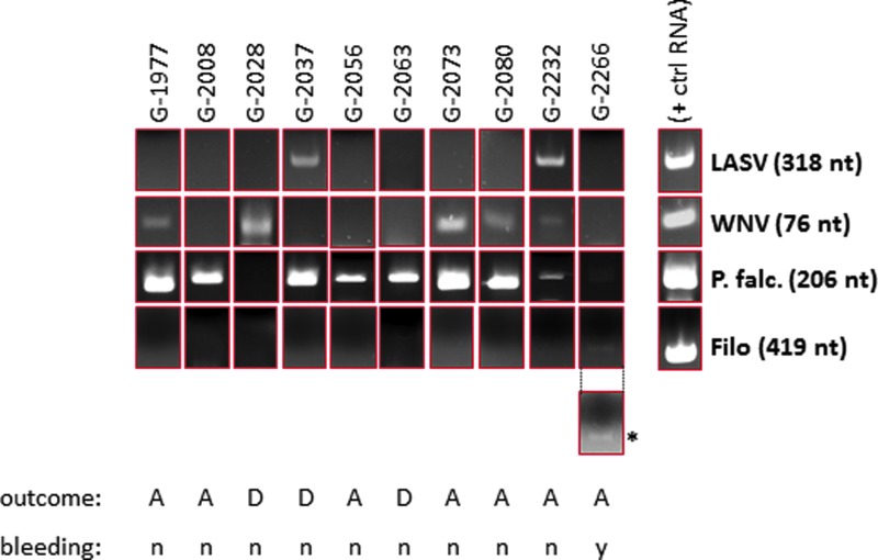
Lassa fever (LF) is a severe viral hemorrhagic fever caused by Lassa virus (LASV). The LF program at the Kenema Government Hospital (KGH) in Eastern Sierra Leone currently provides diagnostic services and clinical care for more than 500 suspected LF cases per year. Nearly two-thirds of suspected LF patients presenting to the LF Ward test negative for either LASV antigen or anti-LASV immunoglobulin M (IgM), and therefore are considered to have a non-Lassa febrile illness (NLFI). The NLFI patients in this study were generally severely ill, which accounts for their high case fatality rate of 36%. The current studies were aimed at determining possible causes of severe febrile illnesses in non-LF cases presenting to the KGH, including possible involvement of filoviruses. A seroprevalence survey employing commercial enzyme-linked immunosorbent assay tests revealed significant IgM and IgG reactivity against dengue virus, chikungunya virus, West Nile virus (WNV), Leptospira, and typhus. A polymerase chain reaction-based survey using sera from subjects with acute LF, evidence of prior LASV exposure, or NLFI revealed widespread infection with Plasmodium falciparum malaria in febrile patients. WNV RNA was detected in a subset of patients, and a 419 nt amplicon specific to filoviral L segment RNA was detected at low levels in a single patient. However, 22% of the patients presenting at the KGH between 2011 and 2014 who were included in this survey registered anti-Ebola virus (EBOV) IgG or IgM, suggesting prior exposure to this agent. The 2014 Ebola virus disease (EVD) outbreak is already the deadliest and most widely dispersed outbreak of its kind on record. Serological evidence reported here for possible human exposure to filoviruses in Sierra Leone prior to the current EVD outbreak supports genetic analysis that EBOV may have been present in West Africa for some time prior to the 2014 outbreak.


Figures

<b>FIG. 1.</b>
FIG. 1.
Representative polymerase chain reaction (PCR) panel showing single, dual, and triple infections identified by PCR screening in a subset of FUO patients. A subset of patients in the study tested positive by PCR for non-Lassa febrile illnesses (NLFIs), such as Plasmodium falciparum (G-2008, G-2056, G-2063), P. falciparum and West Nile virus (WNV; G-1977, G-2073, G-2080), or WNV alone (G-2028). One patient tested positive for Lassa virus (LASV) and P. falciparum (G-2037), and another was triple positive for LASV, WNV, and P. falciparum (G-2232). One patient in the study had a very low level of P. falciparum antigen and tested positive in a filoviral reverse transcription (RT)-PCR screen (G-2266). Positive control RNAs were generated by runoff in vitro transcription reactions from a T7 promoter immediately upstream of the corresponding amplicon. The positive control P. falciparum DNA was amplified from a cloned 206 nt 18S RNA gene fragment that generated a distinct amplicon from a P. vivax 18S RNA cDNA. Each subset of PCR reactions included a negative control RNA or DNA isolated from normal, negative serum of United States and Sierra Leone origin, as well as master PCR mixes containing oligonucleotides but without input nucleic acids. For clarity, the panel displaying the single filoviral amplicon (*) was brightness (+40%) and contrast (+40%) enhanced. The original image is shown above the enhanced panel. All other panels are shown as originally captured. The outcome (A=alive; D=deceased) and bleeding (n=no bleeding; y=bleeding) status for each patient is shown below each set of panels. Ten representative PCR screening profiles are shown. A complete profile of the PCR screening is displayed in Supplementary Table S3. FUO, fever of unknown origin. Color images available online at www.liebertpub.com/vim
<b>FIG. 2.</b>
FIG. 2.
Normal quantile plot of the distribution of optical density (OD) 450 nm values in Ebola virus (EBOV) immunoglobulin G (IgG) enzyme-linked immunosorbent assay (ELISA) with sera from patients with FUO collected from 2011 to March 2014 (n=242), and a set of Sierra Leone (n=14) and United States normal (n=13). Color images available online at www.liebertpub.com/vim
<b>FIG. 3.</b>
FIG. 3.
Distribution of OD 450 nm values in EBOV IgG combo ELISA with the EBOV GP-specific KZ52 control monoclonal antibody over a titration range (blue bars), a panel of 14 normal Sierra Leone donors (Kenema Government Hospital [KGH] LF team members, green bars), and 13 normal United States donors (brown bars). (A) Conservative cutoff OD 450 nm=0.360, established on the 80th percentile of normal sera from SL donors, which intersected three times with the mean OD generated by a United States normal sera panel, was used for analysis of the data in this study. (B) ReEBOV IgG ELISA screening of a panel of LF, NLFI, LPE, and FUO patients presenting to the KGH Lassa Ward between 2011 and March 2014 (n=242). The plot displays mean raw data obtained over a titration range with EBOV GP-specific KZ52 antibody (green bars), a negative control serum (orange bar), and a 1:100 dilution of each patient serum (blue bars). The cutoff OD 450 nm=0.360 results in a 20% positive rate for samples registering IgG antibodies specific to EBOV proteins (GP and VP40). A significant correlation between increasing IgG titers and temporal presentation of subjects at the KGH Lassa Ward could not be established (R2=0.0021; data not shown). A temporal distribution of sera analyzed, from mid-September 2011 to late April 2014, and corresponding dry and rainy seasons, and average temperatures (in °C) are graphically represented. In Sierra Leone, dry and rainy seasons correlate with specific agricultural activities: The main planting season is April–July, with harvesting occurring between September and January. Agricultural activities increase the contact between humans and animal species in fields and wooded areas. These studies did not identify an obvious correlation between seasons and the emergence of seroreactivity against EBOV antigens. Color images available online at www.liebertpub.com/vim

References

    1. Baize S, Pannetier D, Oestereich L, et al. . Emergence of Zaire Ebola virus disease in Guinea. N Engl J Med 2014;371:1418–1425 - PubMed
    1. Barzon L, Lavezzo E, Militello V, et al. . Applications of next-generation sequencing technologies to diagnostic virology. Int J Mol Sci 2011;12:7861–7884 - PMC - PubMed
    1. Bausch DG, and Schwarz L. Outbreak of Ebola virus disease in Guinea: where ecology meets economy. PLoS Negl Trop Dis 2014;8:e3056. - PMC - PubMed
    1. Bausch DG, Sesay SS, and Oshin B. On the front lines of Lassa fever. Emerg Infect Dis 2004;10:1889–1890 - PMC - PubMed
    1. Becker S, Feldmann H, Will C, et al. . Evidence for occurrence of filovirus antibodies in humans and imported monkeys: do subclinical filovirus infections occur worldwide? Med Microbiol Immunol 1992;181:43–55 - PubMed

Publication types