. 2014 Dec 22;9(12):e114583.
doi: 10.1371/journal.pone.0114583. eCollection 2014.

PARP inhibition restores extrinsic apoptotic sensitivity in glioblastoma

Affiliations

PARP inhibition restores extrinsic apoptotic sensitivity in glioblastoma

Georg Karpel-Massler et al. PLoS One. .

Abstract

Background: Resistance to apoptosis is a paramount issue in the treatment of Glioblastoma (GBM). We show that targeting PARP by the small molecule inhibitors, Olaparib (AZD-2281) or PJ34, reduces proliferation and lowers the apoptotic threshold of GBM cells in vitro and in vivo.

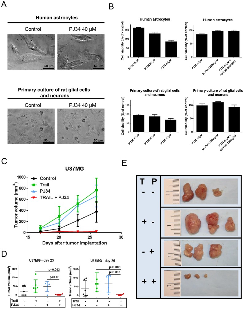
Methods: The sensitizing effects of PARP inhibition on TRAIL-mediated apoptosis and potential toxicity were analyzed using viability assays and flow cytometry in established GBM cell lines, low-passage neurospheres and astrocytes in vitro. Molecular analyses included western blots and gene silencing. In vivo, effects on tumor growth were examined in a murine subcutaneous xenograft model.

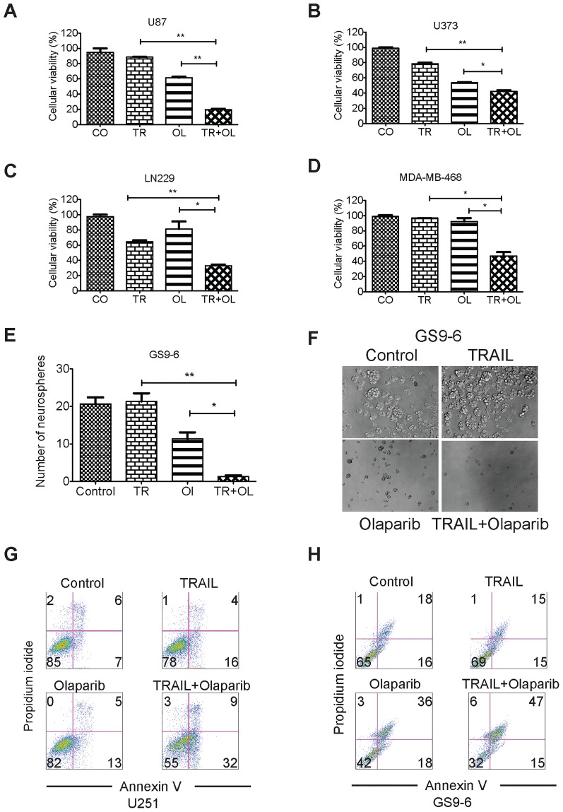
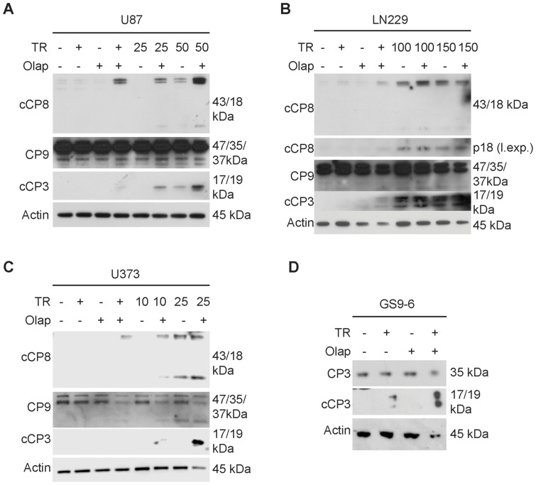
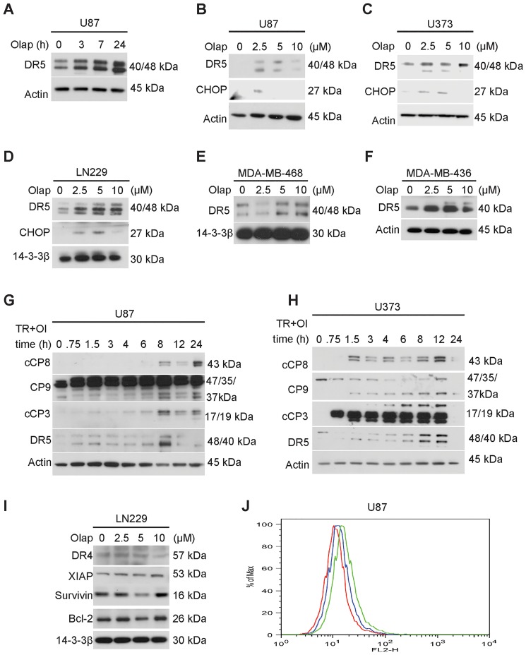
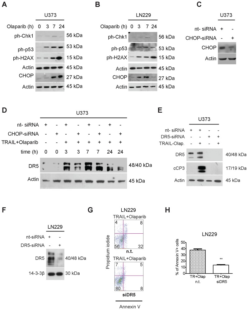
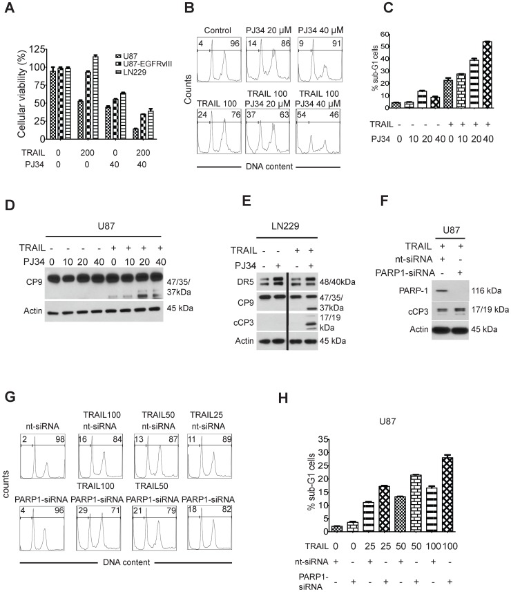
Results: The combination treatment of PARP inhibitors and TRAIL led to an increased cell death with activation of caspases and inhibition of formation of neurospheres when compared to single-agent treatment. Mechanistically, pharmacological PARP inhibition elicited a nuclear stress response with up-regulation of down-stream DNA-stress response proteins, e.g., CCAAT enhancer binding protein (C/EBP) homology protein (CHOP). Furthermore, Olaparib and PJ34 increased protein levels of DR5 in a concentration and time-dependent manner. In turn, siRNA-mediated suppression of DR5 mitigated the effects of TRAIL/PARP inhibitor-mediated apoptosis. In addition, suppression of PARP-1 levels enhanced TRAIL-mediated apoptosis in malignant glioma cells. Treatment of human astrocytes with the combination of TRAIL/PARP inhibitors did not cause toxicity. Finally, the combination treatment of TRAIL and PJ34 significantly reduced tumor growth in vivo when compared to treatment with each agent alone.

Conclusions: PARP inhibition represents a promising avenue to overcome apoptotic resistance in GBM.


Conflict of interest statement

Competing Interests: The authors have declared that no competing interests exist.

Figures

Figure 1
Figure 1. Cooperative cell death induction of the combination treatment with TRAIL and Olaparib.
A–D): U87 (A), U373 (B), LN229 (C) and MDA-MB-468 (D) cells were treated with suboptimal dosages of TRAIL (A: 100 ng/ml, B: 25 ng/ml, C: TRAIL 200 ng/ml, D: 10 ng/ml), Olaparib (A–C: 10 µM, D: 5 µM) or the combination of both reagents for 48 hours. Thereafter, MTT assays were performed to determine cellular viability. E–F) GBM neurosphere culture (GS9-6) was treated with suboptimal dosages of TRAIL, Olaparib or the combination of both reagents and assessed for neurosphere formation 2 weeks after plating. G–H): U251 and GS9-6 (GBM neurosphere culture) cells were treated with suboptimal dosages of TRAIL, Olaparib or the combination of both reagents and stained with Annexin-V (FITC-conjugated) and Propidium iodide 24 hours after treatment. Cells were analyzed by flow cytometry to determine the fraction of apoptotic cells. Values are given as mean ± SEM of representative experiments. The unpaired t-test was used to calculate the p-values. A p-value of less than 0.05 (0.01 to 0.05) is indicated by one star “*”, whereas a p-value of less than 0.01 (0.001 to 0.01) is highlighted by two stars “**”. A p-value less than 0.001 is indicated by a star triplet (***). CO – Control, TR – TRAIL, OL – Olaparib, TR+OL – TRAIL + Olaparib.
Figure 2
Figure 2. The combination therapy, consisting of TRAIL and Olaparib, elicits enhanced activation of initiator- and effector caspases.
A–C) U87 (A), LN229 (B) and U373 (C) GBM cells were treated with TRAIL (ng/ml), Olaparib (10 µM) or the combination of both for 7 hours, subsequently harvested for immunoblotting and analyzed for the expression of cleaved caspase-8 (cCP8), full length caspase-9 (CP9) or effector caspase-3 (cCP3). In B the small fragment of cleaved caspase-8 is exposed longer (separate longer exposure (l. exp.)). Actin serves as a loading control. D) GBM neurosphere culture cells, GS9-6, were incubated with TRAIL, Olaparib or the combination of both reagents for 7 hours, subjected to immunoblotting and analyzed for full length caspase-3 (CP3) and cleaved caspase-3 (cCP3). Actin serves as a loading control. TR – TRAIL, Olap – Olaparib.
Figure 3
Figure 3. Olaparib elicits an increase in TRAIL receptor 2 (DR5) expression in GBM cells.
A) U87 GBM cells were treated with Olaparib (10 µM) for the indicated time points, subjected to immunoblotting and analyzed for the expression of DR5. B–D) U87 (B), U373 (C) and LN229 GBM cells (D) were treated with increasing concentrations of Olaparib (µM) for 7 hours, subjected to immunoblotting and analyzed for the expression of CHOP and DR5. E–F) MDA-MB-468 (E) and MDA-MB-436 (F) were treated with increasing concentrations (µM) of Olaparib for 7 hours, harvested for immunoblotting and analyzed for the expression of DR5. G–H) U87 (G) and U373 (H) GBM cells were treated with the combination of TRAIL/Olaparib (U87: TRAIL 100 ng/ml, Olaparib 10 µM; U373: TRAIL 25 ng/ml Olaparib 10 µM) for a time course analysis, subjected to immunoblotting and analyzed for expression of cleaved caspase-8 (cCP8), full length caspase-9 (CP9), cCP3 (cleaved caspase-3) and DR5. Actin serves as a loading control. I) LN229 GBM cells were treated with increasing concentrations of Olaparib (µM), subjected to immunoblotting and analyzed for the expression of DR4, XIAP, Survivin and Bcl-2. J) U87 glioma cells were treated with Olaparib (5 µM) overnight, stained with a primary PE labeled antibody against DR5 and subjected to flow cytometry. A representative isotype control served as a control. The isotype-control is indicated in red, DR5 expression upon treatment with solvent is indicated in blue and DR5 expression after treatment with Olaparib is indicated in green. Olap – Olaparib, TR+OL – TRAIL + Olaparib.
Figure 4
Figure 4. Olaparib causes a nuclear stress response in a time-dependent manner in GBM cells and regulates DR5 expression through CHOP.
A–B) U373 (A) and LN229 (B) were treated with Olaparib (10 µM) for 3, 7, and 24 h. Cells were harvested for immunoblotting at the indicated timepoints and subjected to analysis for expression of ph-Chk1 (Serine 345), ph-p53 (Serine 15), ph-H2AX (Serine 139) and CHOP. C) U373 GBM cells were transfected with a non-targeting or a CHOP specific siRNA. 72 hours later, cells were harvested, subjected to immunoblotting and analyzed for CHOP expression. D) U373 GBM cells were transfected as indicated with either a non-targeting or a CHOP-specific siRNA. 72 hours later cells were treated with TRAIL/Olaparib and harvested for immunoblotting at the indicated time points. Thereafter, protein expression for DR5 was determined by immunoblotting. E) U373 GBM cells were transfected with a non-targeting or a DR5-specific siRNA. 72 hours after transfection cells were treated with the combination of TRAIL (50 ng/ml) and Olaparib (10 µM) for 7 hours, harvested for immunoblotting and analyzed for the expression of DR5 and cCP3. F) LN229 glioma cells were transfected with a non-targeting siRNA or a DR5-specific siRNA. 72 hours after transfection, cells were harvested for immunoblotting and DR5 expression was determined. G–H) LN229 cells transfected with a non-targeting (n.t.) or a DR5-specific siRNA were treated with TRAIL (200 ng/ml) and Olaparib (10 µM), stained with Annexin V/Propidium iodide and analyzed by flow cytometry. A p-value of less than 0.01 is indicated by two stars “**”. Columns, mean; bars, SEM. TR -TRAIL, Olap – Olaparib.
Figure 5
Figure 5. Inhibition of PARP-1 by the pharmacological inhibitor, PJ34, as well as PARP-1-specific siRNA-mediated suppression enhances TRAIL-mediated apoptosis in GBM cells.
A) U87, U87 EGFRvIII and LN229 were treated with TRAIL (200 ng/ml), PJ34 (40 µM) or the combination of both for 72 hours. Subsequently, cells were analyzed by MTT-assay. Values are given as mean ± SEM. B,C) U87 GBM cells were treated with TRAIL, PJ34 or the combination of both for 24 hours and subsequently analyzed for specific apoptosis (sub-G1 fraction) by flow cytometry. In B, representative plots of these experiments are provided and C shows a quantitative analysis for these experiments. D) U87 GBM cells were treated with PJ34 (µM), TRAIL (100 ng/ml) or the combination of both with the indicated concentrations for 7 hours and subjected to immunoblotting analysis for cleavage of caspase-9. E) LN229 GBM cells were treated with PJ34 (20 µM), TRAIL (200 ng/ml) or the combination of both and analyzed for the expression of DR5, caspase-9 (CP9) and cleaved caspase-3 (cCP3). The vertical line on the immunoblot indicates that the first and second samples were noncontiguous, but run on the same gel simultaneously with the other samples. F) U87 gliobastoma cells were transfected with non-targeting or PARP-1-specific siRNA for 72 hours and subsequently incubated with TRAIL (100 ng/ml). Protein expression of cCP3 and PARP-1 was evaluated by immunoblotting. G–H) U87 cells were transfected with either non-targeting siRNA or with a siRNA specific for PARP-1. 72 hours after transfection cells were incubated with increasing concentrations of TRAIL (concentrations in ng/ml) and subsequently analyzed for apoptosis by flow cytometry (specific apoptosis, sub-G1 fraction). Shown are both representative plots (G) as well as a quantitation of the indicated results (H). Columns, mean; bars, SEM.
Figure 6
Figure 6. The combination of TRAIL and PJ34 is non-toxic to human astrocytes and primary rat neurons and glial cells and exerts stronger anti-proliferative activity against malignant glioma than the respective single treatments in vivo.
A) Representative microphotographs of human astrocytes and primary glial/neurons cells treated with PJ34 for 72 hours. B) Human astrocytes and primary neurons/glial cells were treated with increasing concentrations of PJ34 or in combination with human TRAIL (huTRAIL) or murine TRAIL (muTRAIL) for 72 hours and then analyzed by MTT assay. C) Shown is the tumor growth curve with the four different treatment groups: Control (DMSO), TRAIL, PJ34, TRAIL/PJ34 (each n = 6 tumors). Tumors and treatment groups were established as described in the material and methods section. D) Quantification and statistical analysis of treatment groups after 23 and 26 days, respectively. The Mann-Whitney test was used for statistical analysis and a p-value of less than 0.05 was deemed statistically significant. E) Gross images of representative tumors from different treatment groups. T – TRAIL, P – PJ34.

References

    1. Stupp R, Mason WP, van den Bent MJ, Weller M, Fisher B, et al. (2005) Radiotherapy plus concomitant and adjuvant temozolomide for glioblastoma. N Engl J Med 352:987–996. - PubMed
    1. von Minckwitz G, Muller BM, Loibl S, Budczies J, Hanusch C, et al. (2011) Cytoplasmic poly(adenosine diphosphate-ribose) polymerase expression is predictive and prognostic in patients with breast cancer treated with neoadjuvant chemotherapy. J Clin Oncol 29:2150–2157. - PubMed
    1. Gan A, Green AR, Nolan CC, Martin S, Deen S (2013) Poly(adenosine diphosphate-ribose) polymerase expression in BRCA-proficient ovarian high-grade serous carcinoma; association with patient survival. Hum Pathol 44:1638–1647. - PubMed
    1. Murai J, Zhang Y, Morris J, Ji J, Takeda S, et al. (2014) Rationale for Poly(ADP-ribose) Polymerase (PARP) inhibitors in combination therapy with camptothecins or temozolomide based on PARP trapping versus catalytic inhibition. J Pharmacol Exp Ther 349:408–416. - PMC - PubMed
    1. Russo AL, Kwon HC, Burgan WE, Carter D, Beam K, et al. (2009) In vitro and in vivo radiosensitization of glioblastoma cells by the poly (ADP-ribose) polymerase inhibitor E7016. Clin Cancer Res 15:607–612. - PMC - PubMed

Publication types

MeSH terms