. 2015 Feb;16(2):161-9.
doi: 10.1038/ni.3078. Epub 2014 Dec 22.

IL-25-responsive, lineage-negative KLRG1(hi) cells are multipotential 'inflammatory' type 2 innate lymphoid cells

Affiliations

IL-25-responsive, lineage-negative KLRG1(hi) cells are multipotential 'inflammatory' type 2 innate lymphoid cells

Yuefeng Huang et al. Nat Immunol. 2015 Feb.

Abstract

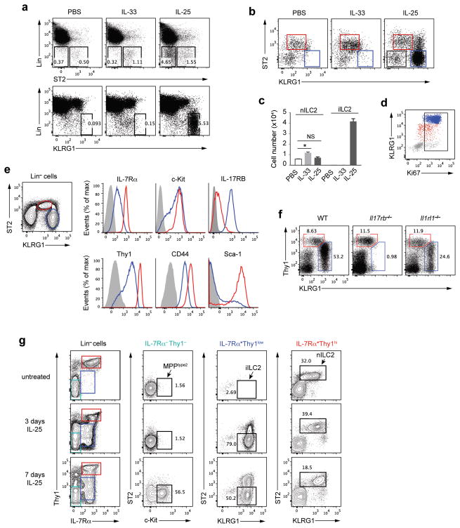
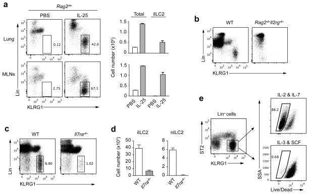
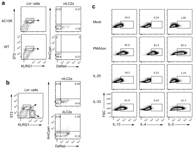
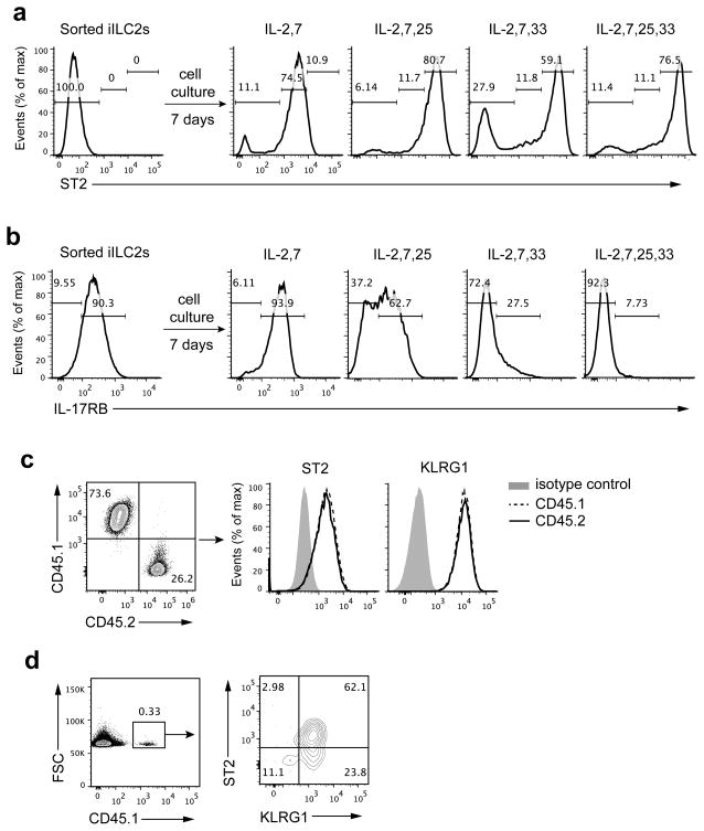
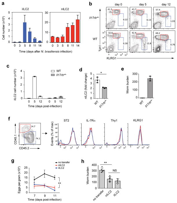
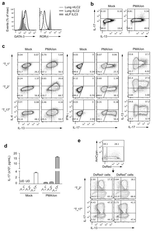
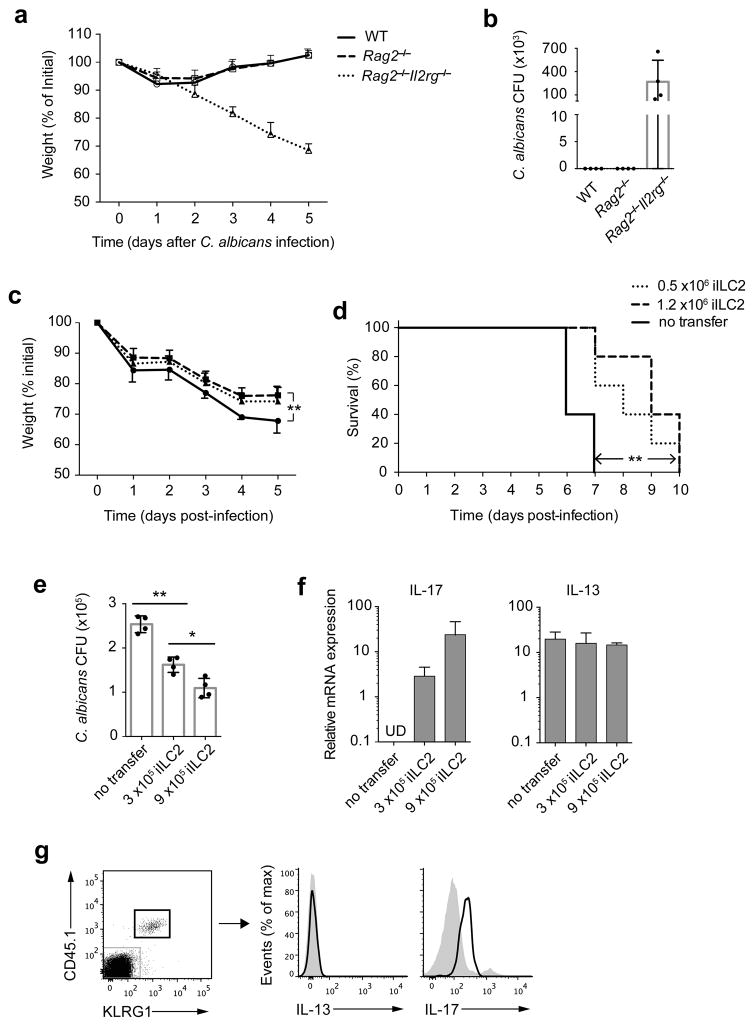
Innate lymphoid cells (ILCs) are lymphocyte-like cells that lack T cell or B cell antigen receptors and mediate protective and repair functions through cytokine secretion. Among these, type 2 ILCs (ILC2 cells) are able to produce type 2 cytokines. We report the existence of an inflammatory ILC2 (iILC2) population responsive to interleukin 25 (IL-25) that complemented IL-33-responsive natural ILC2 (nILC2) cells. iILC2 cells developed into nILC2-like cells in vitro and in vivo and contributed to the expulsion of Nippostrongylus brasiliensis. They also acquired IL-17-producing ability and provided partial protection against Candida albicans. We propose that iILC2 cells are transient progenitors of ILCs mobilized by inflammation and infection that develop into nILC2-like cells or ILC3-like cells and contribute to immunity to both helminths and fungi.


Conflict of interest statement

COMPETING FINANCIAL INTERESTS

The authors declare no competing financial interest.

Figures

Figure 1
Figure 1
IL-25 induces a LinST2KLRG1hi cell population distinct from nILC2 or MPPtype2. (a) Wild-type C57BL/6 (B6) mice were treated i.p. with PBS, IL-33 or IL-25 (200ng per mouse per day for each cytokine) daily for 3 days. Leukocytes in the lung were isolated and analyzed by flow cytometry for ST2, KLRG1 and “lineage” expression. Lineage (Lin) includes the antibodies to CD3ε, CD5, CD19, B220, TCRγδ, NK1.1, CD11b, CD11c, Gr-1, FcεR1 and TER119. (b) KLRG1 and ST2 expression on Lin cells in the lungs of the mice treated as in a. Red gate, nILC2; blue gate, iILC2. (c) Cell numbers of nILC2s or iILC2s in the lungs of the mice treated as in a. (d) Ki67 expression on lung leukocytes from IL-25-treated mice as in the right of b. Red (nILC2) dots were gated on LinST2+KLRG1int cells, blue (iILC2) dots were gated on LinST2KLRG1hi cells and gray dots were gated on LinST2KLRG1 cells. (e) Expression of ILC2 markers on Lin cells from the lung of IL-25-treated mice. Red line, nILC2; Blue line, iILC2; gray shadow, negative control (e.g., Lin+IL-7Rα cells were gated as negative control for IL-7Rα expression). (f) Wild-type (WT), Il17rb−/− or Il1rl1−/− mice were treated with IL-25 for 3 days and lung leukocytes were analyzed by flow cytometry for lineage, Thy1 and KLRG1 expression. Cells were gated on Lin. (g) Wild-type B6 mice were untreated or treated with IL-25 for 3 or 7 days. Lung leukocytes were analyzed by flow cytometry for ILC markers expression. MPPtype2 cells were gated on LinThy1IL-7RαST2c-Kit+, iILC2s were gated on LinThy1lowIL-7Rα+ST2KLRG1hi, and nILC2s were gated on LinThy1hiIL-7Rα+ST2+KLRG1int. c, mean ± s.e.m.; NS, not significant; *P≤0.05 (unpaired two-tailed t test). Data are representative of three independent experiments (ae) or representative of two independent experiments (f,g). a,b,c, n=3 mice for each group in each experiment; d,e, n=2 mice in each experiment; f,g, n=2 mice for each group in each experiment.
Figure 2
Figure 2
iILC2 development depends on γc chain and IL-7Rα. (a) Left, Rag2−/− mice were treated with PBS or IL-25 for 3 days and leukocytes from lungs or MLNs were analyzed by flow cytometry for lineage and KLRG1 expression. iILC2s were gated as LinKLRG1hi. Right, cell numbers of total lymphocytes and iILC2s in the lungs (upper) or MLNs (lower). (b) Lung leukocytes from IL-25-treated wild-type or Rag2−/−Il2rg−/− mice were analyzed by flow cytometry for lineage and KLRG1 expression. (c) Wild-type and Il7ra−/− mice were treated and analyzed as in b. (d) Cell numbers of iILC2s and nILC2s in c. (e) LinST2 KLRG1hi cells were purified by cell sorting from the lungs of IL-25-treated wild-type mice and were cultured either in IL-2 plus IL-7 or in IL-3 plus SCF (10ng/ml for each cytokine). 3 days later, cell viability was determined by Dead/Live staining. Live cells were gated as Dead/Live. a,d, mean ± s.e.m.. Data are representative of more than five independent experiments (a), three independent experiments (bd) or two independent experiments (e). ad, n=3 mice for each group in each experiment; e, n=2 cell culture wells for each condition in each experiment.
Figure 3
Figure 3
iILC2 cells produce type 2 cytokines. (a) Lung leukocytes of naive 4C13R or non-transgenic B6 mice were isolated and analyzed by flow cytometry for lineage, KLRG1, ST2, AmCyan (IL-4) and DsRed (IL-13) expression. nILC2s were gated on LinST2+KLRG1int. (b) 4C13R mice were treated i.p. with IL-25 for 3 days, and lung leukocytes were analyzed as in a. nILC2s were gated on LinST2+KLRG1int and iILC2s were gated on LinST2KLRG1hi. (c) iILC2s, sorted from IL-25-treated wild-type B6 mice, were cultured with IL-2, IL-7 and IL-25 (10ng/ml for each cytokine) for 3 days and then unstimulated (Mock) or stimulated with PMA plus ionomycin, IL-25 (50ng/ml) or IL-33 (50ng/ml) for 6 hours. IL-13, IL-4 and IL-5 production was measured by intracellular cytokine staining followed by flow cytometry. Data are representative of three independent experiments (ac) (n=2~5 mice for each group in each experiment of a; n=2 mice for each experiment of b; n=3 cell culture wells for each stimulus in each experiment of c).
Figure 4
Figure 4
iILC2 cells develop into ST2+ nILC2-like cells both in vitro and in vivo. (a,b) iILC2s sorted from IL-25-treated Rag2−/− mice were cultured in different cytokine combinations as indicated. ST2 and IL-17RB expression were measured on day 7 by flow cytometry. (c) iILC2s, sorted from IL-25-treated CD45.1 Rag1−/− mice, and nILC2s, sorted from naive CD45.2 Rag1−/− mice, were mixed at a ratio of 2.7:1 iILC2s to nILC2s and cultured with IL-2, IL-7 and IL-25. The ratio of CD45.1: CD45.2 and expression of ST2 and KLRG1 were measured on day 7 by flow cytometry. (d) iILC2s, sorted from IL-25-treated CD45.1 Rag1−/− mice, were intravenously (i.v.) transferred (5 ×10⁵ cells per mouse) into Rag2−/−Il2rg−/− mice. 8 days later, transferred cells in the lung of recipient mice were analyzed for KLRG1 and ST2 expression by flow cytometry. Data are representative of two independent experiments (ad) (n=3 cell culture wells for each condition in each experiment of a, b; n=2 repeats in each experiment of c; n=2 recipient mice in each experiment of d).
Figure 5
Figure 5
iILC2 cells regulate anti-helminth immunity. (a) Wild-type B6 mice were infected with N. brasiliensis for various days and leukocytes in the lungs were counted and analyzed by flow cytometry. nILC2s were gated as Lin Thy1hi KLRG1int and iILC2s were gated as LinThy1low KLRG1hi. (b) Wild-type or Il17rb−/− mice were infected with N. brasiliensis for various days and leukocytes in the lungs were analyzed as described in a. (c) Absolute cell numbers of iILC2s in b. (d) nILC2 expansion on day 12 relative to day 0 in b. (e) Worm burden in the intestine on day 12. (f) 2.5 ×10⁴ nILC2s (sorted from naive CD45.1 Rag1−/−) and 20 ×104 iILC2s (sorted from IL-25-treated CD45.2 Rag1−/− mice) were mixed and i.v. transferred into Rag2−/−Il2rg−/− mice, which were infected with N. brasiliensis on the same day. 14 days later, transferred cells in the lung were analyzed for the ratio of CD45.1: CD45.2 and for ILC2 surface markers by flow cytometry. (g) 2 ×10⁵ sorted nILC2s or iILC2s were i.v. transferred into Rag2−/−Il2rg−/− mice that were infected with N. brasiliensis. Worm eggs in the feces were counted on day 7, day 9 and day 11 after infection. (h) Worm burden in intestine of the mice in g were counted on day 14 post infection. a,c,d,e,g,h, mean ± s.e.m.; NS, not significant; *P≤0.05, **P≤0.01 (unpaired two-tailed t test). Data are representative of two independent experiments (a,f,g,h) or represent one experiment (be) (n=3 mice for each group in each experiment of a; n=3 mice in be; n=2 recipient mice in each experiment of f; n=5 mice for each group in each experiment of g,h).
Figure 6
Figure 6
iILC2 cells express RORγt and have the capacity to develop into IL-17 producers. (a) Flow cytometry analysis of GATA-3 and RORγt expression on nILC2s and iILC2s from lung of IL-25-treated wild-type B6 mice and ILC3s (LinIL-7Rα+RORγthi) from small intestinal lamina propria (siLP) of naïve mice. (b) Freshly sorted iILC2s from IL-25-treated Rag2−/− mice were stimulated with or without PMA plus ionomycin for 6 hours. IL-13 and IL-17 production was measured with intracellular cytokine staining, followed by flow cytometry. (c) iILC2s sorted from IL-25-treated Rag2−/− mice were cultured in various conditions (described in Methods). 7 days after culture, cells were stimulated with or without PMA plus ionomycin for 6 hours and production of IL-4, IL-13, IL-17 and IFN-γ were determined by intracellular staining. (d) iILC2s were cultured as in c for 7 days and secreted IL-17 protein in supernatants of cultured cells was measured with or without PMA plus ionomycin stimulation, by enzyme-linked immunosorbent assay. UD, undetectable. (e) iILC2s from IL-25-treated 4C13R mice were divided into DsRed+ and DsRed populations through cell sorting. The purified populations were cultured in either “TH2” conditions or “TH17” conditions for 7 days. IL-13 and IL-17 production was measured by intracellular staining after 6-hour stimulation with PMA plus ionomycin. d, mean ± s.e.m.. Data are representative of three (ac) or two (d,e) independent experiments (n=2 mice for each group in each experiment of a; n=2 cell culture wells for each condition or stimulus in each experiment of b,c,e; n=3 cell culture wells for each condition in each experiment of d).
Figure 7
Figure 7
iILC2 cells contribute to anti-fungal immunity. (a) Wild-type, Rag2−/− and Rag2−/−Il2rg−/− mice were infected sublingually with C. albicans and weighed daily. (b) Fungal burden in entire tongue of the mice as in a on day 5 post infection. (c,d) 0.5 ×10⁶ or 1.2 ×10⁶ iILC2s sorted from IL-25-treated Rag2−/− mice were i.v. transferred into Rag2−/−Il2rg−/− mice. Recipient mice were infected sublingually with C. albicans on the next day. The mice were observed for survival and weighed daily. (e) 3 ×10⁵ or 9 ×10⁵ iILC2s sorted from IL-25-treated CD45.1 Rag1−/− mice were i.v. transferred into Rag2−/−Il2rg−/− mice, followed by C. albicans sublingual infection on next day. On day 5 post infection, two-thirds of the tongue tissue was analyzed for fungal burden. (f) The other one-third tongue tissue in e was analyzed for IL-17 and IL-13 mRNA expression. (g) Lung leukocytes of the mice receiving 9 ×10⁵ iILC2s in e were analyzed for IL-13 and IL-17 production without stimulation, by intracellular cytokine staining. a,c, mean ± s.e.m.; **P≤ 0.01 (paired two-tailed t test). b,e,f, mean ± s.e.m.; *P≤0.05, **P≤0.01 (unpaired two-tailed t test). d, **P≤0.01 (log-rank (Mantel-Cox) test). Data are representative of three (a) or two (bg) independent experiments (n=3~5 mice for each group in each experiment of ag).

Comment in

References

    1. Walker JA, Barlow JL, McKenzie AN. Innate lymphoid cells--how did we miss them? Nat Rev Immunol. 2013;13:75–87. - PubMed
    1. Spits H, Cupedo T. Innate lymphoid cells: emerging insights in development, lineage relationships, and function. Annu Rev Immunol. 2012;30:647–675. - PubMed
    1. Spits H, et al. Innate lymphoid cells--a proposal for uniform nomenclature. Nat Rev Immunol. 2013;13:145–149. - PubMed
    1. Moro K, et al. Innate production of T(H)2 cytokines by adipose tissue-associated c-Kit(+)Sca-1(+) lymphoid cells. Nature. 2010;463:540–544. - PubMed
    1. Neill DR, et al. Nuocytes represent a new innate effector leukocyte that mediates type-2 immunity. Nature. 2010;464:1367–1370. - PMC - PubMed

Publication types

MeSH terms