. 2014 Dec 22;9(12):e115604.
doi: 10.1371/journal.pone.0115604. eCollection 2014.

Use of echocardiography reveals reestablishment of ventricular pumping efficiency and partial ventricular wall motion recovery upon ventricular cryoinjury in the zebrafish

Affiliations

Use of echocardiography reveals reestablishment of ventricular pumping efficiency and partial ventricular wall motion recovery upon ventricular cryoinjury in the zebrafish

Juan Manuel González-Rosa et al. PLoS One. .

Abstract

Aims: While zebrafish embryos are amenable to in vivo imaging, allowing the study of morphogenetic processes during development, intravital imaging of adults is hampered by their small size and loss of transparency. The use of adult zebrafish as a vertebrate model of cardiac disease and regeneration is increasing at high speed. It is therefore of great importance to establish appropriate and robust methods to measure cardiac function parameters.

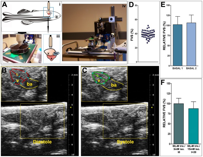
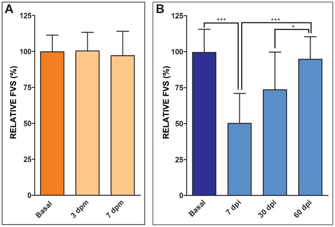
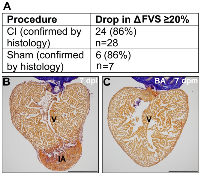
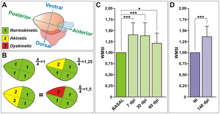
Methods and results: Here we describe the use of 2D-echocardiography to study the fractional volume shortening and segmental wall motion of the ventricle. Our data show that 2D-echocardiography can be used to evaluate cardiac injury and also to study recovery of cardiac function. Interestingly, our results show that while global systolic function recovered following cardiac cryoinjury, ventricular wall motion was only partially restored.

Conclusion: Cryoinjury leads to long-lasting impairment of cardiac contraction, partially mimicking the consequences of myocardial infarction in humans. Functional assessment of heart regeneration by echocardiography allows a deeper understanding of the mechanisms of cardiac regeneration and has the advantage of being easily transferable to other cardiovascular zebrafish disease models.


Conflict of interest statement

Competing Interests: The authors have declared that no competing interests exist.

Figures

Figure 1
Figure 1. Echocardiographic image acquisition and basal fractional volume shortening (FVS) quantification.
(A) Schematic representation of animal positioning for image acquisition and picture of the set up. (i) Animals are positioned ventrally and (ii) are immobilized in the same way as for surgical procedures, in a Petri dish, and are covered with fish water containing anaesthetic solution. This positioning allows for a transducer to be placed directly over the body wall at the level of the heart (iii). The transducer is attached to a holder to allow a stable position during acquisition (iv). (B,C) Details from representative 2D echocardiography images from an uninjured zebrafish heart showing maximal ventricular dilatation (B, diastole) and maximal ventricular contraction (C, systole). The diastolic (red) and systolic (green) ventricular areas are outlined and the length of the apical image long axis is also indicated (L). Red and green lines in B and C highlight ventricular border in diastole and systole, respectively. Yellow lines indicate the bulbus arteriosus (BA). (D) FVS obtained in basal conditions (n = 47, mean ± SD  =  39 ± 5). (E) Comparison of FVS in basal conditions at two different days, with an interval of 7 days. Shown are means ± SD. The relative FVS (RFVS) of BASAL2 versus BASAL1 within the same animal are statistically comparable (p = 0.1099, Wilcoxon matched-pairs signed rank test). (F) FVS measured in basal conditions with different dosages of anesthesia and throughout time in the same animal. Initial anesthesia conditions are the same as for all acquisitions (60 µM tricaine/3 mM isoflurane). The final acquisition was taken 20 minutes later and the final anesthesia dose was 60 µM tricaine/15 mM isoflurane. Differences in the average FVS are not statistically significant (p = 0.1094, Wilcoxon matched-pairs signed rank test). A, atrium; ba, bulbus arteriosus; FVS, fractional volume shortening; L, length of the apical image long axis; RFVS, relative fractional volume shortening; v, ventricle.
Figure 2
Figure 2. Cryoinjury transiently impairs ventricular pumping efficiency.
(A,B) Temporal evolution of changes in the relative FVS in sham (A) and cryoinjured (B) animals in a longitudinal study. Graphs represent relative mean values and SD. (A) The relative FVS (RFVS) is not significantly changed in animals after sham operation at 3 (n = 14) or 7 (n = 21) dpm (p = 0.884; one-way ANOVA). (B) Cryoinjured animals show a temporal decrease in the RFVS of 50%, which is gradually recovered around 60 dpi (*** p<0.001; ** p<0.01; * p<0.05; one-way ANOVA followed by Tukey's honest significant difference test, n = 31). dpi, days postinjury; dpm, days postmanipulation; FVS, fractional volume shortening; RFVS, relative fractional volume shortening.
Figure 3
Figure 3. Correlation between histology of imaged hearts and echocardiographic analysis.
(A,B) Groups of animals in which cryoinjury was confirmed by histological AFOG staining after echocardiography. In 24 out of 28 fish, cryoinjury was diagnosed at 7 dpi by measurement of a drop of the RFVS ≥ 20% compared to the equivalent basal measurement. Only one from 7 sham-operated fish presented a drop in RFVS ≥ 20%. (C) Subsequent histological staining however did not support an alteration in the cardiac morphology or injury in any of the sham operated animals. BA, bulbus arteriosus; IA, injured area; dpi, days postinjury; dpm, days postmanipulation; V, ventricle. Size bars, 200 µm.
Figure 4
Figure 4. Ventricular wall motion is not fully recovered after cryoinjury.
(A) Schematic representation of segmental criteria of the zebrafish ventricle, considering the antero-posterior and dorso-ventral axis. Depending on their motility, segments are scored as normokinetic (“1”), akinetic (“2”) or dyskinetic (“3”). (B) Theoretical representations of the wall motion score index (WMSI) showing ventricles from healthy controls (WMSI = 1) and cryoinjured animals (WMSI>1). (C) Temporal evolution of changes in the WMSI in cryoinjured animals in a longitudinal study. After injury, the WMSI increased and remained elevated even at 60 dpi, indicating that wall motion is affected. Graphs represent mean values and SD (*** p<0.001; * p<0.05; one-way ANOVA followed by Tukey's honest significant difference test, n = 20). (D) The WMSI is not recovered at extended stages of regeneration (n = 17), and it is not affected in siblings (NI) of the same age (n = 3). *** p = 0.00009, Wilcoxon Signed Rank Test comparing to a theoretical mean of 1. dpi, days postinjury; NI, not injured; WMSI, wall motion score index.
Figure 5
Figure 5. Cryoinjury induces local, long-term alterations in myocardial organization.
(A,B) Immunohistochemistry on sagittal sections of control (A,A’) and cryoinjured (B-B”) hearts at 130 dpi from the Tg(myl7:nucDsRed) line. A’–B” are zoomed images of boxed areas in A and B, additionally showing autofluorescence to reveal tissue organization. (A-A’) In control hearts, one or two cells constitute the thickness of the compact myocardium (CM). (B-B”) At 130 dpi, the injured wall (IW) shows an abnormal increase in the number and distribution of cardiomyocytes compared with the contralateral wall (CLW). (C) Quantification of the nuclear density relative to the compact tissue reveals an increase in cardiomyocyte density in the IW compared to the CLW. Graph represents mean values and SD (*** p = 0.006, two tailed Student's t-test; 100–150 cells counted per section, 3 sections per heart, n = 3 animals analyzed). (D) qPCR from ventricular RNA samples reveal induction of the natriuretic peptide encoding gene nppa upon cryoinjury. Graph represents mean values and SD, n  =  4-5 replicates, Expressions levels were normalized to that of ef1α and rps11 and further normalized to that of the uninjured sample. (* p<0.05; one-way ANOVA followed by Tukey's honest significant difference test). (E-H) Sections of cryoinjured hearts at the indicated times post-injury hybridized with a riboprobe for nppa mRNA. Yellow arrows mark areas of strong nppa expression. (E) In control hearts, nppa is highly expressed in the atrium (yellow arrow) and at lower levels in the trabecular myocardium (white arrow). (F-G) Shortly after injury, nppa is strongly upregulated in the ventricular myocardium. (H) At 90 dpi, the levels of nppa expression are similar to those detected in control hearts. Observe the increase in thickness of the compact layer of the injured wall (asterisk) revealed by no expression of nppa. AT, atrium; BA, bulbus arteriosus, CLW, contralateral wall; CM, compact myocardium; hpi, hours postinjury; dpi, days postinjury; IA, injured area; IW, injured wall; V, ventricle. Bars, 200 µm (full views), 50µm (magnifications).

Similar articles

Cited by

References

    1. Laflamme MA, Murry CE (2011) Heart regeneration. Nature 473:326–335. - PMC - PubMed
    1. Sedmera D, Thompson RP (2011) Myocyte proliferation in the developing heart. Dev Dyn 240:1322–1334. - PMC - PubMed
    1. Jennings RB, Murry CE, Steenbergen C Jr, Reimer KA (1990) Development of cell injury in sustained acute ischemia. Circulation 82:II2–12. - PubMed
    1. Choi WY, Poss KD (2012) Cardiac regeneration. Curr Top Dev Biol 100:319–344. - PMC - PubMed
    1. Raya A, Koth CM, Buscher D, Kawakami Y, Itoh T, et al. (2003) Activation of Notch signaling pathway precedes heart regeneration in zebrafish. Proc Natl Acad Sci U S A 100 Suppl 1 11889–11895. - PMC - PubMed

Publication types

MeSH terms