. 2015 Feb;12(2):140-6.
doi: 10.1038/nmeth.3217. Epub 2014 Dec 22.

Simultaneous all-optical manipulation and recording of neural circuit activity with cellular resolution in vivo

Affiliations

Simultaneous all-optical manipulation and recording of neural circuit activity with cellular resolution in vivo

Adam M Packer et al. Nat Methods. 2015 Feb.

Erratum in

  • Nat Methods. 2015 Jul;12(7):692

Abstract

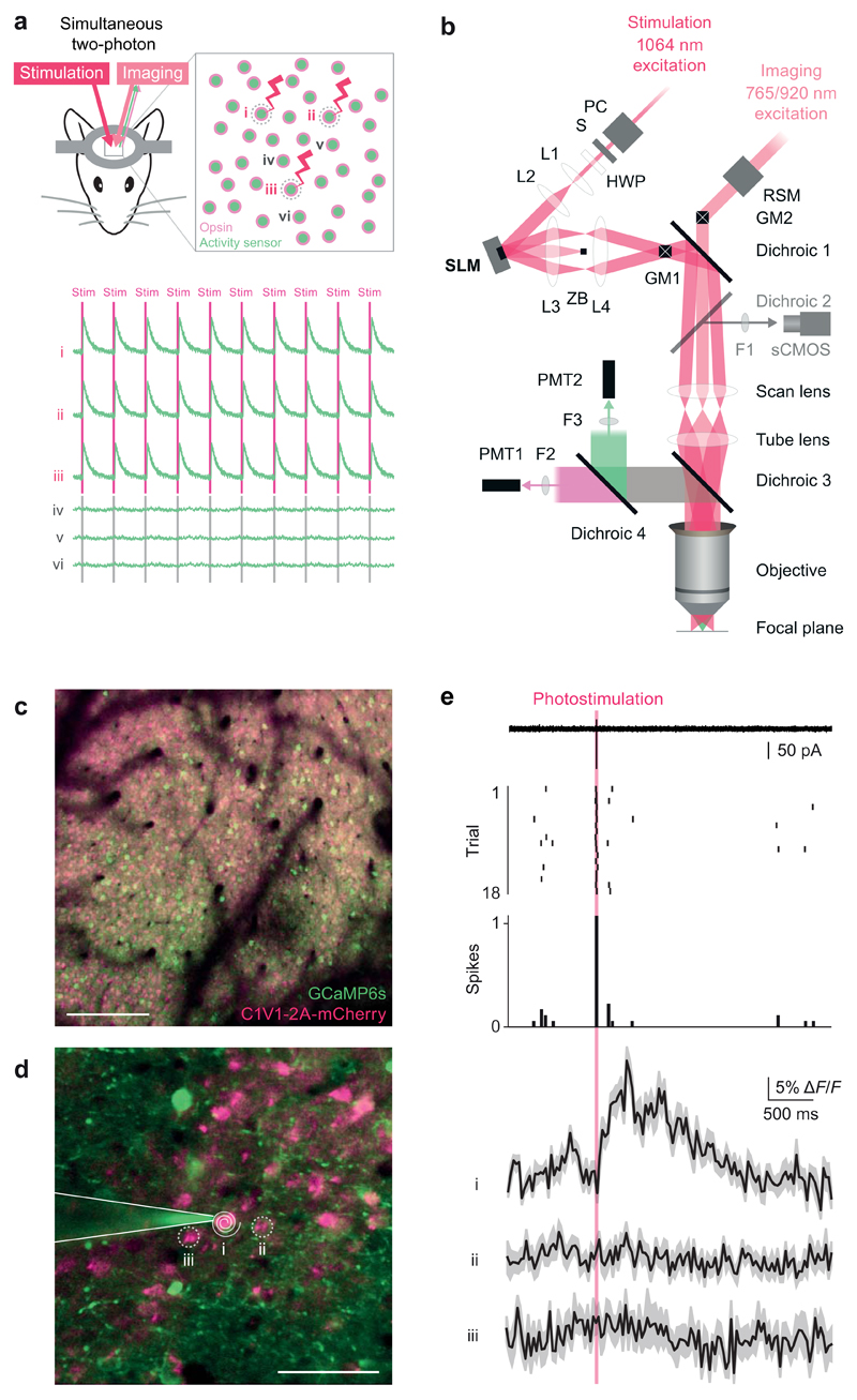
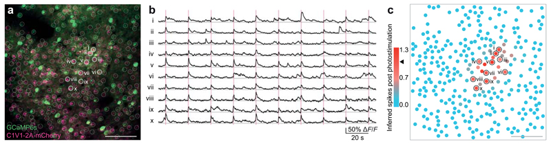
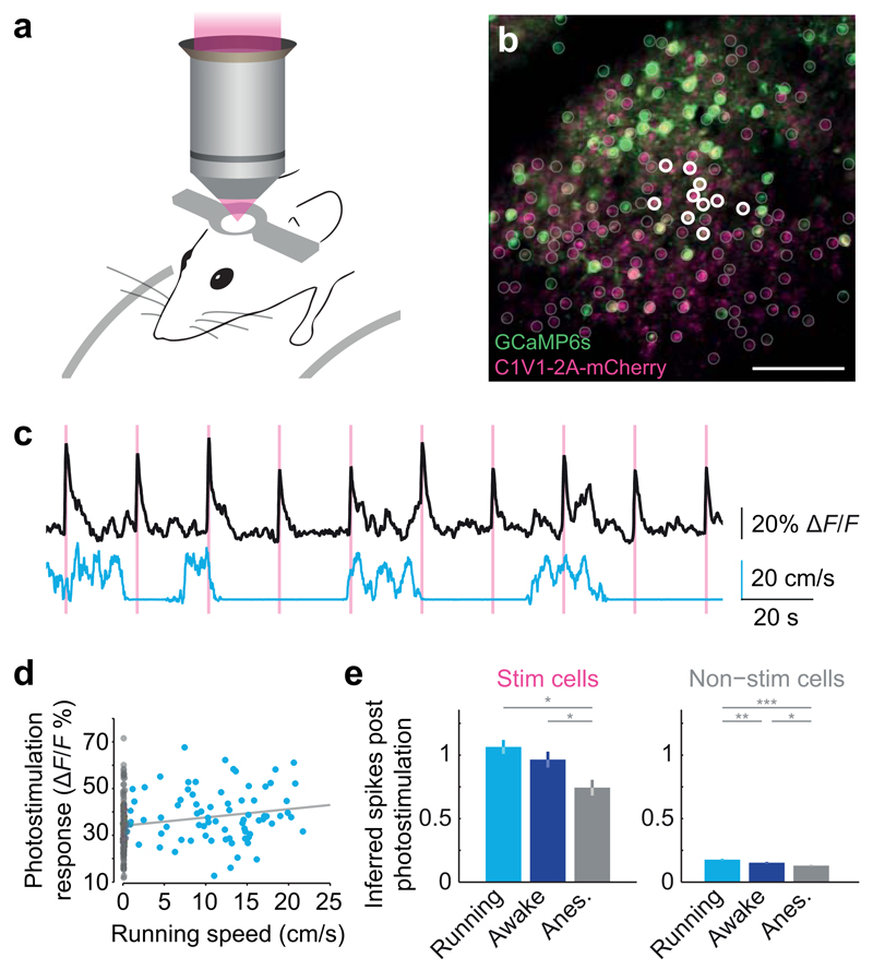
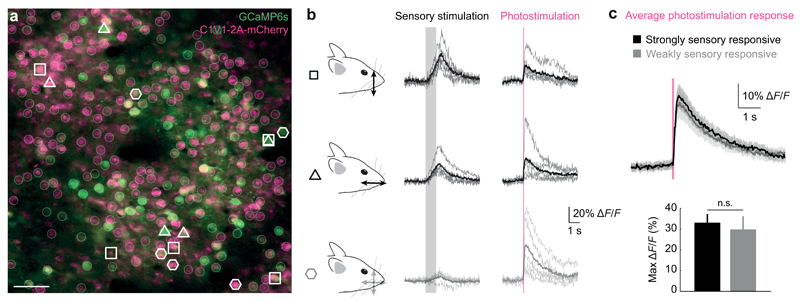
We describe an all-optical strategy for simultaneously manipulating and recording the activity of multiple neurons with cellular resolution in vivo. We performed simultaneous two-photon optogenetic activation and calcium imaging by coexpression of a red-shifted opsin and a genetically encoded calcium indicator. A spatial light modulator allows tens of user-selected neurons to be targeted for spatiotemporally precise concurrent optogenetic activation, while simultaneous fast calcium imaging provides high-resolution network-wide readout of the manipulation with negligible optical cross-talk. Proof-of-principle experiments in mouse barrel cortex demonstrate interrogation of the same neuronal population during different behavioral states and targeting of neuronal ensembles based on their functional signature. This approach extends the optogenetic toolkit beyond the specificity obtained with genetic or viral approaches, enabling high-throughput, flexible and long-term optical interrogation of functionally defined neural circuits with single-cell and single-spike resolution in the mouse brain in vivo.


Conflict of interest statement

Statement of Competing Financial Interests

All authors declare that there are no competing financial interests

Figures

Figure 1
Figure 1. Single-cell two-photon optogenetic photostimulation and single action potential readout in vivo
(a) Schematic illustration of the experimental goal. Successful interrogation of neural circuits at the resolution and precision at which they function requires a method with the specificity to simultaneously manipulate and record individual, user-selected neurons in the awake behaving animal. An all-optical approach to perform such experiments would be beneficial given the chronic and non-invasive nature in addition to the ability to localize neuron’s locations precisely in circuits where physical topology is critical. Top: Our approach utilizes a coexpression strategy to imbue neurons with read and write abilities: a calcium sensor generates an optical readout of activity while an opsin enables photostimulation. Bottom: The experimental goal would realize robust and reliable photostimulation in the user-selected neurons 1, 2, and 3 with sufficient resolution to avoid stimulating the immediately adjacent neurons 4, 5, and 6. (b) Optical layout of the SLM-based (spatial light modulator) two-photon patterned photostimulation, two-photon resonant-scanning, moving in vivo microscope. The photostimulation beam (300 fs, 5 W, 1064 nm pulsed laser, Fianium Ltd or 100 fs, 2.3 W, 1055 nm pulsed laser, Coherent) was controlled by a Pockels’ cell (PC, Conoptics Ltd.) and shutter (S, Vincent Associates), resized using a beam-expanding telescope (L1 [f = 50 mm] and L2 [f = 200 mm], plano-convex lenses, Thorlabs) to fill the SLM active area and polarization maximized for diffraction efficiency from the SLM with a half wave plate (HWP, WPH10M-1064, Thorlabs). SLM: reflective spatial light modulator, 7.68 mm × 7.68 mm active area, 512 × 512 pixels, (Boulder Nonlinear Systems). L3: 1” achromatic doublet, f = 400 or 250 mm. ZB: zero order block. L4: 2” achromatic doublet, f = 150 mm. GM1: Galvanometer mirror pair, 3 or 6 mm (Cambridge Technology, integrated into dual beam Ultima microscope by Bruker Corp. [formerly Prairie Technologies]). GM2: Galvanometer mirror pair, 6mm (Cambridge Technology, integrated by Bruker Corp.). The imaging beam path can also be scanned along the fast axis by a resonant scanning galvanometer mirror (RSM, Cambridge Technology, integrated by Bruker Corp.) relayed onto GM2. Dichroic 1: 1030 nm short pass (T1030SP, Chroma Technology). Dichroic 2: 660 nm long pass (660LP, Chroma). This dichroic is used to image SLM beamlets in widefield mode on the sCMOS camera only when dichroic 3 is removed. F1: 675/67 nm bandpass filter (Semrock). sCMOS camera: ORCA-Flash4.0 (Hamamatsu). Scan lens: f=75 mm. Tube lens f=180 mm. Dichroic 3: 700 nm long pass (T700lpxxr-xxt, Chroma). Dichroic 4: 575LP (HQ575dcxr, Chroma). F2: 525/70 nm bandpass filter (525/70m-2P, Chroma). F3: 607/45 nm bandpass filter (607/45m-2P, Chroma). PMT1: Multi-alkali photomultiplier tube (Hamamatsu). PMT2: GaAsP photomultiplier tube (Hamamatsu). Objective: 16X 0.8 NA (Nikon). (c) A large field of view of neurons co-expressing GCaMP6s and C1V1 (scale bar, 100 μm). (d) Inset from a large field of view (200 × 200 μm) for the experiment shown in (e) (scale bar, 50 μm). A two-photon targeted cell-attached patch clamp recording was obtained from neuron 1, which co-expressed GCaMP6s and C1V1. This neuron was targeted for optogenetic stimulation by a beamspot produced by the SLM that was then driven in a spiral pattern by the galvanometer mirrors (white spiral). (e) Top: electrophysiological recording during photostimulation trials reveals reliable generation of single action potentials during the photostimulation period (red bar) as evidenced in the single sweep (from trial 2), raster plot, and peristimulus time histogram. Bottom: calcium imaging recordings obtained simultaneously showed a transient (mean ± SEM matching the amplitude and kinetics expected for a single action potential with GCaMP6s in the photostimulated neuron, while the nearby neurons showed no detectable transients. Similar results were seen in n = 3 neurons.
Figure 2
Figure 2. Precise photostimulation of multiple identified neurons in vivo
(a) Left: An example field of view of somatosensory cortex layer 2/3 neurons expressing C1V1-2A-YFP (scale bar, 100 μm). Right: Protocol to target spiral photostimulation patterns to multiple locations using the SLM and galvanometer mirrors (scale bars, 100 μm). (b) Left: Magnification of a, showing cell-attached patch-clamp recording configuration in which multiple locations were photostimulated while electrophysiologically confirming action potential generation in one of the targets. Middle: Raster of spike times around stimulus delivery over 10 trials (photostimulation, red bar). Right: Overlaid raw data showing low latency and jitter. (c) Metrics evaluating performance for 10 and 20 spot photostimulation patterns and spiral photostimulations lasting 11, 16, or 34 ms. Error bars are SEM, n = six neurons, four mice.
Figure 3
Figure 3. Simultaneous fast calcium imaging and photostimulation of multiple neurons in vivo
(a) A field of view of neurons in layer 2/3 of somatosensory cortex colabeled with C1V1-2A-mCherry and GCaMP6s (scale bar, 100 μm). Ten neurons were targeted for simultaneous photostimulation (white circles). (b) Calcium transients from the ten photostimulated neurons showing the responses on all individual trials. (c) Color-coded plot of the strength of photostimulation as measured by the sum of inferred spikes immediately post-stimulation. Stimulated neurons are circled in black. 300 total neurons, 120 trials, mean inferred action potentials post photostimulation = 1.0 ± 0.1 (mean ± SD, black arrow in legend).
Figure 4
Figure 4. Behavioral state-dependence of network perturbations
(a) Mice were head-fixed and allowed to run freely on a fixed-axis styrofoam treadmill. (b) A field of view in which ten nearby neurons in layer 2/3 of mouse somatosensory cortex were chosen for photostimulation while they and the surrounding neurons across the field of view were simultaneously observed with high-speed calcium imaging. (c) Top: mean calcium transient of all stimulated neurons in response to simultaneous photostimulation of ten neurons. Bottom: running speed. (d) Correlation between mean perturbation of responses to photostimulation and running speed. (e) Comparison of mean ± SD inferred spike responses to photostimulation during different behavioural states (n = 3 mice, 576 imaged neurons; Tukey’s test for multiple comparisons, * denotes p < 0.05).
Figure 5
Figure 5. Targeting neurons for optogenetic manipulation based on their individual functional signatures in vivo
(a) A field of view (scale bar, 50 µm) showing neurons co-expressing GCaMP6s (green) and C1V1-2A-mCherry (purple) in the C2 barrel of mouse somatosensory cortex. (b) Groups of individually identified neurons were selected for photostimulation based on their response to dorsal-ventral and rostro-caudal whisker stimulation (open symbols). Five neurons that responded differently or not at all to sensory stimulation (gray shading) were simultaneously photostimulated (red line). (c) Simultaneous photostimulation of five neurons responding strongly to a given sensory stimulation or weakly to both stimuli revealed similar responses (mean ± SEM) to photostimulation (n = 1 mouse).

References

    1. Packer AM, Roska B, Hausser M. Targeting neurons and photons for optogenetics. Nat Neurosci. 2013;16:805–815. - PMC - PubMed
    1. Grienberger C, Konnerth A. Imaging calcium in neurons. Neuron. 2012;73:862–885. - PubMed
    1. Rickgauer JP, Deisseroth K, Tank DW. Simultaneous cellular-resolution optical perturbation and imaging of place cell firing fields. Nat Neurosci. 2014 - PMC - PubMed
    1. Huber D, et al. Sparse optical microstimulation in barrel cortex drives learned behaviour in freely moving mice. Nature. 2008;451:61–64. - PMC - PubMed
    1. Fajardo O, Zhu P, Friedrich RW. Control of a specific motor program by a small brain area in zebrafish. Front Neural Circuits. 2013;7:67. - PMC - PubMed

Publication types

MeSH terms