Comparative Study
. 2015 Feb;8(1):174-85.
doi: 10.1161/CIRCEP.114.002195. Epub 2014 Dec 22.

Sympathetic nerve stimulation, not circulating norepinephrine, modulates T-peak to T-end interval by increasing global dispersion of repolarization

Affiliations
Comparative Study

Sympathetic nerve stimulation, not circulating norepinephrine, modulates T-peak to T-end interval by increasing global dispersion of repolarization

Daigo Yagishita et al. Circ Arrhythm Electrophysiol. 2015 Feb.

Abstract

Background: T-peak to T-end interval (Tp-e) is an independent marker of sudden cardiac death. Modulation of Tp-e by sympathetic nerve activation and circulating norepinephrine is not well understood. The purpose of this study was to characterize endocardial and epicardial dispersion of repolarization (DOR) and its effects on Tp-e with sympathetic activation.

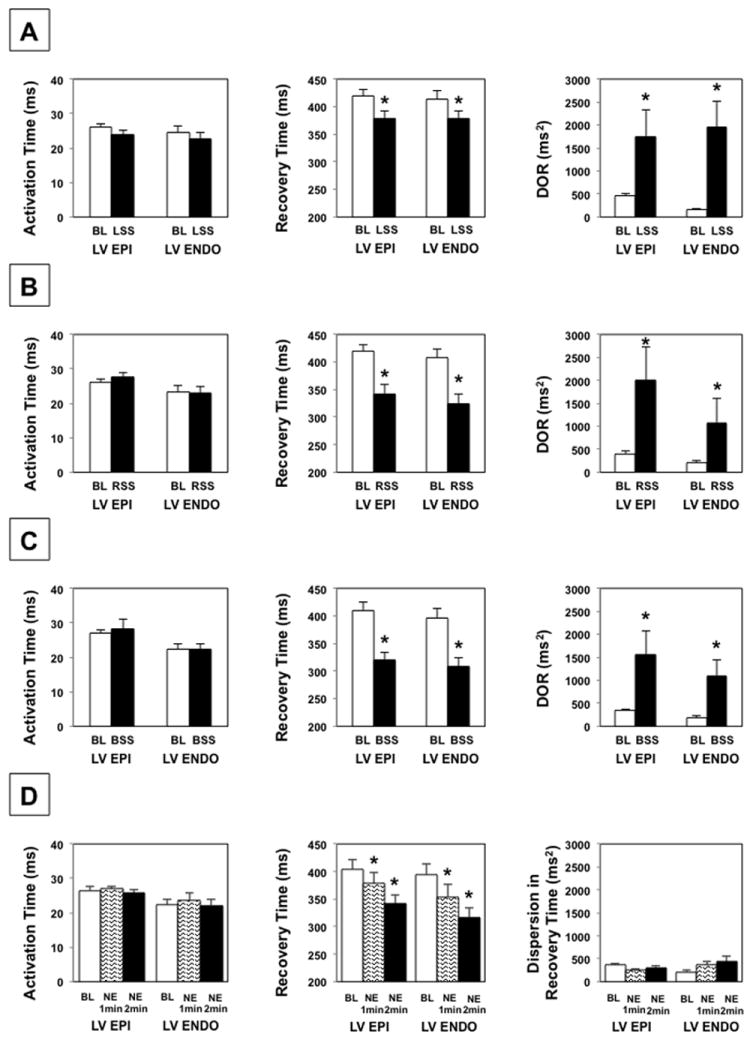
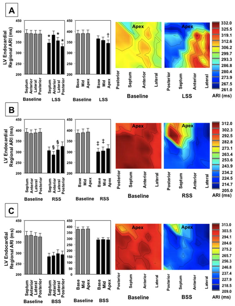
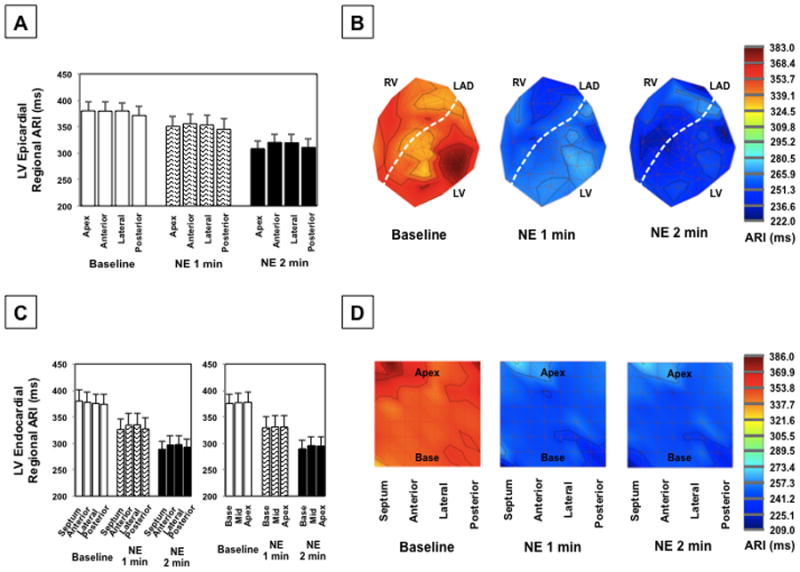
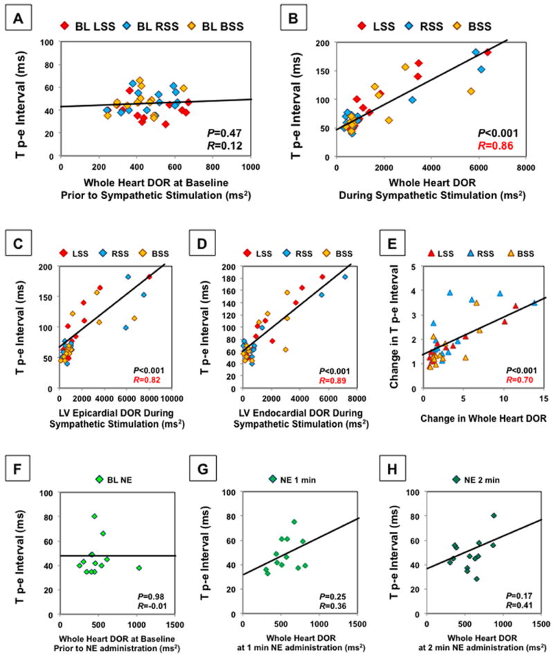
Methods and results: In Yorkshire pigs (n=13), a sternotomy was performed and the heart and bilateral stellate ganglia were exposed. A 56-electrode sock and 64-electrode basket catheter were placed around the epicardium and in the left ventricle (LV), respectively. Activation recovery interval, DOR, defined as variance in repolarization time, and Tp-e were assessed before and after left, right, and bilateral stellate ganglia stimulation and norepinephrine infusion. LV endocardial and epicardial activation recovery intervals significantly decreased, and LV endocardial and epicardial DOR increased during sympathetic nerve stimulation. There were no LV epicardial versus endocardial differences in activation recovery interval during sympathetic stimulation, and regional endocardial activation recovery interval patterns were similar to the epicardium. Tp-e prolonged during left (from 40.4±2.2 ms to 92.4±12.4 ms; P<0.01), right (from 47.7±2.6 ms to 80.7±11.5 ms; P<0.01), and bilateral (from 47.5±2.8 ms to 78.1±9.8 ms; P<0.01) stellate stimulation and strongly correlated with whole heart DOR during stimulation (P<0.001, R=0.86). Of note, norepinephrine infusion did not increase DOR or Tp-e.

Conclusions: Regional patterns of LV endocardial sympathetic innervation are similar to that of LV epicardium. Tp-e correlated with whole heart DOR during sympathetic nerve activation. Circulating norepinephrine did not affect DOR or Tp-e.

Keywords: ECG; T wave; action potential; autonomic nervous system; dispersion; sympathetic.


Conflict of interest statement

Conflict of Interest Disclosures: The University of California, Los Angeles has intellectual property developed by one of the authors (KS) that relate to epicardial interventions.

Figures

Figure 1
Figure 1
(A) A 56-electrode sock is placed over the ventricles for recording of epicardial electrograms. (B) Sock electrode configuration for creation of polar maps is shown. (C) The 64-electrode catheter used for endocardial recordings is shown. (D) The basket catheter splines are placed on a 2-D plaque type configuration for polar map visualization. LA = left atrium, LAD = left anterior descending coronary artery, LV = left ventricle, RV = right ventricle, RVOT = right ventricular outflow tract.
Figure 2
Figure 2
Effects of SG stimulation and NE infusion on whole heart mean ARI (A), dispersion in ARI (B), AT (C), RT (D), DOR (E), and Tp-e interval (F) are shown. Both sympathetic nerve stimulation and NE infusion decrease mean ARI. However, only SG stimulation increased DOR and Tp-e. * P < 0.05 for baseline vs. SG stimulation or NE infusion. † P < 0.01 for baseline vs. SG stimulation or NE infusion. P values obtained using the Wilcoxon signed rank test. BL = baseline, LSS = left stellate stimulation, RSS = right stellate stimulation, BSS = bilateral stellate stimulation, NE = norepinephrine infusion.
Figure 3
Figure 3
Effects of LSS (A), RSS (B), BSS (C), and NE infusion (D) on AT, RT, and DOR of LV epicardium and endocardium. * P < 0.01 for baseline vs. SG stimulation or NE administration. BL = baseline, Epi = epicardium, Endo = endocardium, LSS = left stellate stimulation, RSS = right stellate stimulation, BSS = bilateral stellate stimulation, NE = norepinephrine infusion. P values obtained using the Wilcoxon signed rank test.
Figure 4
Figure 4
Regional epicardial ARI effects of (A) LSS, (B) RSS, and (C) BSS are shown in the left panels, while the right panels demonstrate a polar map from a representative animal during each condition. * P < 0.01 for comparison of mean ARI of the LV anterior wall to other regions. † P < 0.01 for comparison of mean ARI of LV posterior wall to other regions. LSS = left stellate stimulation, RSS = right stellate stimulation, BSS = bilateral stellate stimulation. Regional comparisons performed using the linear mixed effects regression model with heterogeneous variances.
Figure 5
Figure 5
Regional endocardial ARI effects of LSS (A), RSS (B), and BSS (C) are shown in the left panels, while the right panels demonstrate a representative ARI polar maps from a single animal. * P < 0.05 for comparison of mean ARI of the anterior wall to other regions. § P < 0.05 for comparison of mean ARI of the posterior wall to other regions. † P < 0.01 when comparing apical ARI with mid wall or basal ARIs during LSS. ‡ P < 0.05 for comparison of apical with mid wall or basal ARIs during RSS. Regional comparisons performed using the linear mixed effects model with heterogeneous variances.
Figure 6
Figure 6
There is no effect on global or regional LV transmural differences in ARI during LSS (A), RSS (B), and BSS (C). The Wilcoxon signed rank test was used for the comparison of the transmural differences in ARI. The mixed effects model with heterogenous variances was used for comparison of regional transmural differences in ARI.
Figure 7
Figure 7
(A) Regional epicardial ARIs for all animals and (B) representative polar maps at baseline and during NE infusion at 1 min and 2 min. (C) Regional endocardial ARIs and (D) representative polar maps during NE infusion. The mixed effects model with heterogeneous variances was used for comparison of regional differences in ARI.
Figure 8
Figure 8
Tp-e does not correlate with DOR at baseline prior to sympathetic nerve stimulation (A). However, it strongly correlates with the DOR of the entire LV and RV epicardium and LV endocardium during SG stimulation (B). Correlation with LV epicardial DOR (C) and LV endocardial DOR (D) during SG stimulation is shown. The change in Tp-e was strongly correlated with the change in whole heart DOR (E). Tp-e did not significantly correlate with DOR at baseline prior to NE infusion (F) or at 1 and 2 minutes after NE infusion (G,H). For comparison of the correlation between Tp-e and DOR, Pearson product-moment correlation coefficient was used.

References

    1. Vaseghi M, Shivkumar K. The role of the autonomic nervous system in sudden cardiac death. Prog Cardiovasc Dis. 2008;50:404–419. - PMC - PubMed
    1. Zhou S, Jung BC, Tan AY, Trang VQ, Gholmieh G, Han SW, Lin SF, Fishbein MC, Chen PS, Chen LS. Spontaneous stellate ganglion nerve activity and ventricular arrhythmia in a canine model of sudden death. Heart Rhythm. 2008;5:131–139. - PubMed
    1. Zipes DP, Wellens HJ. Sudden cardiac death. Circulation. 1998;98:2334–2351. - PubMed
    1. Bourke T, Vaseghi M, Michowitz Y, Sankhla V, Shah M, Swapna N, Boyle NG, Mahajan A, Narasimhan C, Lokhandwala Y, Shivkumar K. Neuraxial modulation for refractory ventricular arrhythmias: Value of thoracic epidural anesthesia and surgical left cardiac sympathetic denervation. Circulation. 2010;121:2255–2262. - PMC - PubMed
    1. Schwartz PJ, Locati EH, Moss AJ, Crampton RS, Trazzi R, Ruberti U. Left cardiac sympathetic denervation in the therapy of congenital long qt syndrome. A worldwide report. Circulation. 1991;84:503–511. - PubMed

Publication types