Review
. 2014 Dec 19;4(4):1092-116.
doi: 10.3390/life4041092.

Synthetic Biology: A Bridge between Artificial and Natural Cells

Affiliations
Review

Synthetic Biology: A Bridge between Artificial and Natural Cells

Yunfeng Ding et al. Life (Basel). .

Abstract

Artificial cells are simple cell-like entities that possess certain properties of natural cells. In general, artificial cells are constructed using three parts: (1) biological membranes that serve as protective barriers, while allowing communication between the cells and the environment; (2) transcription and translation machinery that synthesize proteins based on genetic sequences; and (3) genetic modules that control the dynamics of the whole cell. Artificial cells are minimal and well-defined systems that can be more easily engineered and controlled when compared to natural cells. Artificial cells can be used as biomimetic systems to study and understand natural dynamics of cells with minimal interference from cellular complexity. However, there remain significant gaps between artificial and natural cells. How much information can we encode into artificial cells? What is the minimal number of factors that are necessary to achieve robust functioning of artificial cells? Can artificial cells communicate with their environments efficiently? Can artificial cells replicate, divide or even evolve? Here, we review synthetic biological methods that could shrink the gaps between artificial and natural cells. The closure of these gaps will lead to advancement in synthetic biology, cellular biology and biomedical applications.


Figures

Figure 1
Figure 1
Bridging gaps between artificial and natural cells using synthetic biology approaches. For artificial cells, significant progress has been made in constructing different genetic circuits, optimizing factors for gene expression, facilitating cell-cell communication and mimicking replication. Despite the progress, there is still a gap between artificial cells (green circle) and natural cells (pink rectangle). Synthetic biology can be exploited to bridge the gap. For example, de novo synthesized genome DNA can be designed to encode artificial cells with more cellular functions. The natural cellular environment can be mimicked inside artificial cells to achieve efficient gene expression and signal transduction. Different membrane proteins can be reconstituted to endow the membrane of artificial cells with complex functions. Division machinery may be implemented to achieve self-replication in artificial cells.
Figure 2
Figure 2
Construction of artificial cells in three steps. First step: genetic circuits are constructed in vivo using synthetic modules. These genetic circuits control information flow in artificial cells. Second step: the constructed circuits are tested in cell-free systems, which provide the transcription and translation engine. The feedback loop between Step 1 and Step 2 illustrates the testing and optimization of newly-constructed genetic circuits. Third Step: the circuits and the cell-free systems are encapsulated inside synthetic liposomes (the shell). The steps can be repeated in cycles to achieve optimal, efficient artificial cells.
Figure 3
Figure 3
(a) Kinetics of α-hemolysin-eGFP expression. The presence of α-hemolysin (filled circles) prolonged the expression of eGFP from ~20 h to days. Filled circles: 0.5 nM pIVEX2.3d-α-hemolysin-eGFP. Filled squares: the expression of eGFP inside liposomes without α-hemolysin. The inset indicates the first 10 h of gene expression. (b) The E. coli extract was encapsulated in vesicles with pIVEX2.3d-α-hemolysin-eGFP surrounded by feeding solution. Expression of α-hemolysin-eGFP was observed in aggregate vesicles (left), single vesicle (middle) and doublet (right) (scale bar, 20 μm) (reprinted with permission from [71], Copyright 2004, The National Academy of Sciences). (c) Schematic diagram of a positive feedback loop (PFL). The T3 RNA polymerase (T3 RNAP) gene was regulated by T3-lacO promoter. The addition of IPTG induced T3 RNAP expression. The T3 RNAP promoted its own transcription and activated GFP expression. (d) Comparison of GFP expression with or without a PFL. DNA fragments (3 nM) shown in (c) were mixed with a cell-free system containing purified LacI and T3 RNAP. GFP expression was measured at 180 min after the addition of IPTG. The signal-to-noise ratio was increased from 75 to 800 with the PFL (reprinted with permission from [83], Copyright 2013, Royal Society of Chemistry.)
Figure 4
Figure 4
The effect of molecular crowding on gene expression. (a) Gene expression rates in environments containing big crowding agent (Dextran-Big). The reporter gene, cyan fluorescent protein (cfp), was under the control of a normal T7 promoter (PT7), a weak T7 promoter (PT7, weak) or a weak ribosome binding site (RBSweak). The black line represents the predicted expression rates of cfp from normal PT7. The grey line represents the predicted expression rates of cfp from weak PT7. Experimental data (open triangles for WT, open squares for RBSweak, filled squares for T7weak) follow the prediction. (b) Perturbation of gene expression rates using different concentrations of potassium glutamate (K+), magnesium acetate (Mg2+), ammonium acetate (NH4+), spermidine (Sp.) and folinic acid (Fol.). Gene expression was less perturbed in highly-crowded environments (black open bars) than that in low crowded environments (grey bars) (reprinted with permission from [66], Copyright, 2013, Nature Publishing Group).
Figure 5
Figure 5
Artificial cells translate chemical signals for E. coli. (a) Theophylline cannot diffuse through the cell membranes of E. coli. Without artificial cells (circles), E. coli cannot sense theophylline. (b) Theophylline can diffuse through artificial cell membranes. Artificial cells sense theophylline and express α-hemolysin to form unspecific pores on their membranes. The entrapped IPTG is released to trigger GFP expression in E. coli (reprinted with permission from [73], Copyright 2014, Nature Publishing Group).
Figure 6
Figure 6
Schematic diagram of reconstituted olfactory receptor. The expression circuits of the olfactory receptor (BmOR1) and its co-receptor (BmOrco) were constructed inside giant vesicles (GVs). Canine pancreatic microsomal membranes (the small vesicle inside the GV) were added inside GVs to promote cell-free synthesis. BmOR1 and BmOrco were expressed and inserted into the GV membranes to form a complex. The olfactory complex was stimulated by its ligand, bombykol. The effect of stimulation can be detected by a voltage clamp (reprinted with permission from [72], Copyright 2014, Royal Society of Chemistry).
Figure 7
Figure 7
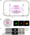
Basic elements and processes for self-replication of artificial cells. (a) Schematic of artificial cells with the capability of lipid-synthesis. Sn-glycerol-3-phosphate acyltransferase (GPAT) catalyzed glycerol-3-phosphate (G3P) to form lysophosphatidic acid (LPA). Lysophosphatidic acid acyltransferase (LPAAT) generated phosphatidic acid (PA) using LPA. GPAT and LPAAT were expressed in artificial cells to sustain PA synthesis (reprinted with permission from [19], Copyright 2009, Elsevier.) (b) Schematic of MreB, YFP-MreB and MreC expression in artificial cells (left). Co-expression of MreB, YFP-MreB and MreC inside artificial cells, which were imaged by a microscope (right). The fluorescence of YFP (green pseudocolor) showed that MreB formed a filamentous structure in the presence of MreC. Rhodamine (red pseudocolor) showed that artificial cells were isolated from the extracellular feeding solution (scale bar, 10 μm) (reprinted with permission from [176], Copyright 2012, American Chemical Society). (c) The tubulin-like protein, FtsZ, formed a Z ring on the artificial cell membrane. (Top) Z rings were first observed to form a constriction on the artificial cell (0 min). (Bottom) A more obvious constriction formed after 6 min (scale bar, 5 μm) (reprinted with permission from [177], Copyright 2008, The American Association of the Advancement of Science).

References

    1. Hooke, R. Micrographia: Or Some Physiological Descriptions of Minute Bodies Made by Magnifying Glasses With Observations and Inquiries Thereupon; John Martyn, printer to the Royal Society, and are to be sold at his shop at the Bell a little without Temple Barr. Martin and Allestry, London, UK, 1665.

    1. Schwann T. Mikroskopische untersuchungen über die übereinstimmung in der struktur und dem wachstum der tiere und pflanzen. In: Jahn I., editor. Klassische Schriften zur Zellenlehre. 2nd ed. Wissenschaftlicher Verlag Harri Deutsch GmbH; Frankfurt, Germany: 2003.
    1. Chang T.M. 1957 Report on “method for preparing artificial hemoglobin corpuscles”. [(accessed on 11 December 2014)]. Available online: http://www.worldscientific.com/doi/pdf/10.1142/9789812770370_bmatter. - DOI
    1. Noireaux V., Maeda Y.T., Libchaber A. Development of an artificial cell, from self-organization to computation and self-reproduction. Proc. Natl. Acad. Sci. USA. 2011;108:3473–3480. doi: 10.1073/pnas.1017075108. - DOI - PMC - PubMed
    1. Zhang Y., Ruder W.C., LeDuc P.R. Artificial cells: Building bioinspired systems using small-scale biology. Trends Biotechnol. 2008;26:14–20. doi: 10.1016/j.tibtech.2007.09.006. - DOI - PubMed