. 2014 Dec 23:4:7564.
doi: 10.1038/srep07564.

G protein-coupled estrogen receptor protects from atherosclerosis

Affiliations

G protein-coupled estrogen receptor protects from atherosclerosis

Matthias R Meyer et al. Sci Rep. .

Erratum in

Abstract

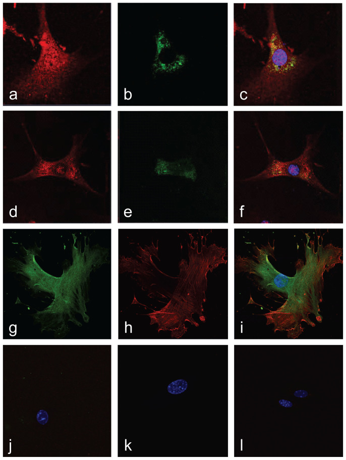
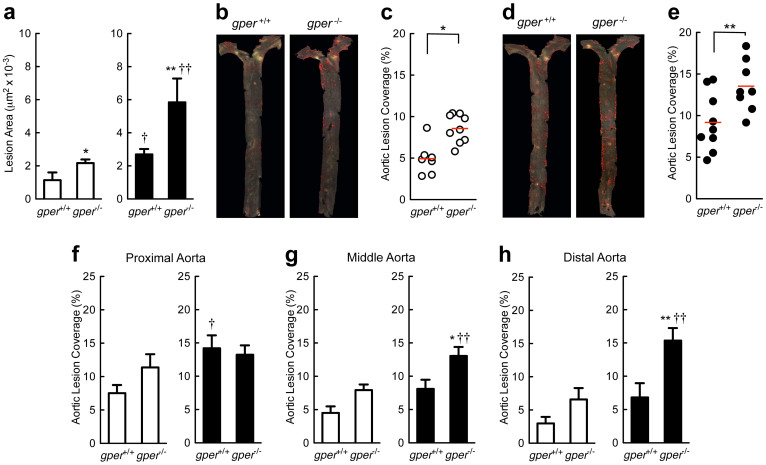
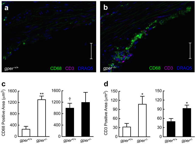
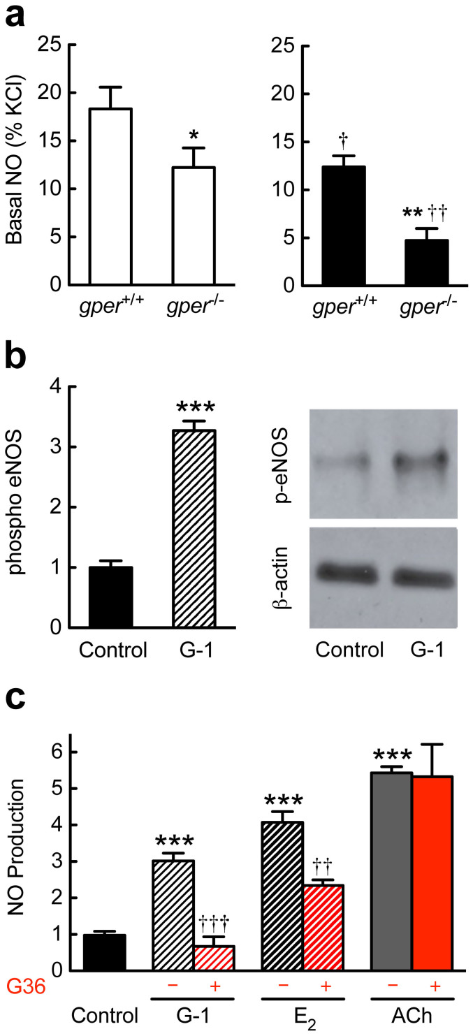
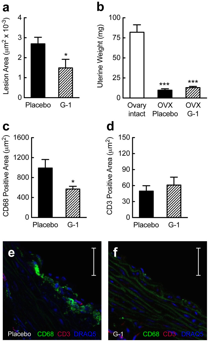
Coronary atherosclerosis and myocardial infarction in postmenopausal women have been linked to inflammation and reduced nitric oxide (NO) formation. Natural estrogen exerts protective effects on both processes, yet also displays uterotrophic activity. Here, we used genetic and pharmacologic approaches to investigate the role of the G protein-coupled estrogen receptor (GPER) in atherosclerosis. In ovary-intact mice, deletion of gper increased atherosclerosis progression, total and LDL cholesterol levels and inflammation while reducing vascular NO bioactivity, effects that were in some cases aggravated by surgical menopause. In human endothelial cells, GPER was expressed on intracellular membranes and mediated eNOS activation and NO formation, partially accounting for estrogen-mediated effects. Chronic treatment with G-1, a synthetic, highly selective small molecule agonist of GPER, reduced postmenopausal atherosclerosis and inflammation without uterotrophic effects. In summary, this study reveals an atheroprotective function of GPER and introduces selective GPER activation as a novel therapeutic approach to inhibit postmenopausal atherosclerosis and inflammation in the absence of uterotrophic activity.


Figures

Figure 1
Figure 1. GPER is an intracellular membrane receptor in vascular endothelial and smooth muscle cells.
(a)–(f), Endothelial cells were stained for GPER (red; a, d) and either endoplasmic reticulum (b) or Golgi apparatus (e), demonstrating colocalization of GPER with both markers in the merged images (c), (f), which include DAPI staining of the nucleus (blue). (g)–(j), Vascular smooth muscle cells were stained for GPER (green; g), employing an antibody targeting an “extracellular” epitope, and α-actin (red; h). merged with GPER staining in (i) under both permeabilizing conditions (g)–(i) and non-permeabilizing conditions (j). Pre-immune anti-GPER serum and negative control IgG were used to test the specificity of the GPER anti-serum and α-actin antibodies under permeabilizing (k) and non-permeabilizing (l) conditions. The cell nucleus is stained with DAPI (blue; i–l). The amino terminus of GPER, expected to be extracellular if the receptor is expressed on the cell surface (plasma membrane), is accessible only upon cell permeabilization, indicating that GPER is expressed predominantly on internal membranes.
Figure 2
Figure 2. Increased atherosclerosis in mice lacking GPER.
Quantification of atherosclerosis in the aortic root (a) and macroscopic atherosclerosis on the aortic surface (b)–(h). Data are shown for ovary intact (open bars/circles) and ovariectomized (filled bars/circles) mice treated with an atherogenic diet. Deletion of gper increased both aortic root atherosclerosis (a) as well as macroscopic atherosclerosis (b)–(e). Both aortic root as well as macroscopic atherosclerosis development were also accelerated after ovariectomy (a), (d), (e). The effect of ovariectomy was further aggravated by deletion of gper (a), (d), (e). The predilection site for atherosclerotic lesions in gper+/+ animals was the proximal aortic segment (f), with lesions intensity decreasing from the middle (g) to the distal (h) segment. This distribution pattern was unaffected by gper deficiency or by ovariectomy alone (f)–(h); however, in ovariectomized gper−/− mice, the distribution pattern changed markedly, revealing equally extensive atherosclerosis in all three aortic segments (f)–(h). *P < 0.05 and **P < 0.01 compared with gper+/+ mice, †P < 0.05 and ††P < 0.01 compared with ovary intact genotype matched mice (ANOVA with Bonferroni post-hoc test). All data (n = 4–9 per group) represent mean ± s.e.m.
Figure 3
Figure 3. Gper deficiency results in vascular accumulation of inflammatory cells.
Quantification of CD68+ cells (macrophages) and CD3+ cells (T cells) in the aortic root using quantitative immunohistochemistry. Data are shown for ovary intact (open bars) and ovariectomized mice (filled bars) treated with an atherogenic diet. Compared with ovary intact gper+/+ mice (a), (c), deletion of gper yielded a pronounced increase in CD68+ cells (b), (c). Ovariectomy alone also increased staining for CD68+ cells in gper+/+ mice, whereas deletion of gper had no further effect on this increase (c). Compared to either ovary intact or ovariectomized gper+/+ mice (a), (d), respectively, deletion of gper yielded a pronounced increase in CD3+ cells (b), (d). Ovariectomy did not further increase CD3+ staining in either gper+/+ or gper−/− mice (d). *P < 0.05 and **P < 0.01 compared with gper+/+ mice, †P < 0.05 compared to ovary intact genotype-matched mice (ANOVA with Bonferroni post-hoc test). All data (n = 3–6 per group) are mean ± s.e.m. Scale bar, 100 μm.
Figure 4
Figure 4. GPER regulates NO synthase function ex vivo and in vitro.
Basal vascular nitric oxide (NO) bioactivity was measured ex vivo (a) in mice treated with an atherogenic diet, with eNOS phosphorylation (b) and NO formation (c) determined in human endothelial cells. (a), Basal vascular NO bioactivity in ovary intact (open bars) and ovariectomized (filled bars) mice. Deletion of gper reduced vascular NO bioactivity to a similar extent (about 30%) as did ovariectomy in gper+/+ mice. However, in ovariectomized gper−/− mice, NO bioactivity was further reduced by more than 70%. *P < 0.05 and **P < 0.01 compared with gper+/+ mice, †P < 0.05 and ††P < 0.01 compared to ovary intact genotype-matched mice (ANOVA with Bonferroni post-hoc test). All data are mean ± s.e.m. (n = 5–7). (b), Stimulation of human endothelial cells with the GPER-selective agonist G-1 increased levels of activated eNOS-phosphorylated on serine1177. ***P < 0.001 compared with control (vehicle only, Student's t-test). Data are mean ± s.e.m. (n = 4). (c), Endothelial NO production was determined through the detection of stable NO metabolites NO2/NO3. Stimulation of GPER in human endothelial cells with the selective (G-1) or non-selective (17β-estradiol, E2) GPER agonist increased NO formation, as did the M3 muscarinic receptor agonist, acetylcholine (ACh). A selective GPER antagonist, G36, completely blocked G-1-stimulated endothelial NO formation, while E2-stimulated NO formation was only partly reduced. G36 had no effect on ACh-stimulated NO formation. ***P < 0.001 compared with control, ††P < 0.01 and †††P < 0.001 compared with no antagonist (Student's t-test). All data (n = 3–9 per group) are mean ± s.e.m.
Figure 5
Figure 5. A small molecule GPER-selective agonist inhibits atherosclerosis and vascular inflammation.
Treatment effects on atherosclerosis (a), uterine wet weight (b) or quantification of CD68+ (c), (e), (f) and CD3+ (d)–(f) staining of macrophages and T cells, respectively, in the aortic root. Data were obtained in ovariectomized (surgically postmenopausal) gper+/+ mice, which display accelerated atherogenesis (cf. Fig 1a–d). Effects are shown in response to treatment with placebo (filled bars) or G-1 (hatched bars), a selective small molecule agonist of GPER. G-1 treatment reduced atherosclerosis by 45% (a). Ovariectomy reduced uterine weight by about 90% compared with ovary-intact animals (b), in which estrogen levels are high. G-1 treatment had no feminizing effect on the uterus similar to placebo treatment (b). G-1 treatment reduced staining for CD68+ cells by 43% (c), (e), (f), but had no effect on CD3+ immunostaining (d)–(f). *P < 0.05 compared with placebo, ***P < 0.001 compared with ovary intact (Student's t-test). All data (n = 5–11 per group) are mean ± s.e.m. Scale bar, 100 μm.

Similar articles

Cited by

References

    1. Barrett-Connor E. Menopause, atherosclerosis, and coronary artery disease. Curr Opin Pharmacol 13, 186–191 (2013). - PMC - PubMed
    1. Turgeon J. L., McDonnell D. P., Martin K. A. & Wise P. M. Hormone therapy: physiological complexity belies therapeutic simplicity. Science 304, 1269–1273 (2004). - PubMed
    1. Hodis H. N. & Mack W. J. Hormone replacement therapy and the association with coronary heart disease and overall mortality: clinical application of the timing hypothesis. J Steroid Biochem Mol Biol 142, 68–75 (2014). - PubMed
    1. Phillips G. B., Pinkernell B. H. & Jing T. Y. Relationship between serum sex hormones and coronary artery disease in postmenopausal women. Arterioscler Thromb Vasc Biol 17, 695–701 (1997). - PubMed
    1. Khalil R. A. Estrogen, vascular estrogen receptor and hormone therapy in postmenopausal vascular disease. Biochem Pharmacol 86, 1627–1642 (2013). - PMC - PubMed

Publication types

MeSH terms

Substances