Review
. 2015 Apr:66:217-39.
doi: 10.1146/annurev-physchem-040214-121637. Epub 2014 Dec 17.

Mechanisms of virus assembly

Affiliations
Review

Mechanisms of virus assembly

Jason D Perlmutter et al. Annu Rev Phys Chem. 2015 Apr.

Abstract

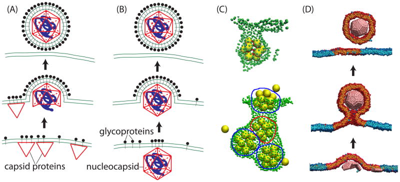
Viruses are nanoscale entities containing a nucleic acid genome encased in a protein shell called a capsid and in some cases are surrounded by a lipid bilayer membrane. This review summarizes the physics that govern the processes by which capsids assemble within their host cells and in vitro. We describe the thermodynamics and kinetics for the assembly of protein subunits into icosahedral capsid shells and how these are modified in cases in which the capsid assembles around a nucleic acid or on a lipid bilayer. We present experimental and theoretical techniques used to characterize capsid assembly, and we highlight aspects of virus assembly that are likely to receive significant attention in the near future.

Keywords: RNA packaging; capsid; kinetics; membrane; simulation; thermodynamics.


Figures

Figure 1
Figure 1
The geometry of icosahedral lattices. Moving h and k steps along each of the ĥ and lattice vectors results in a triangle with area T/4 (for unit spacing between lattice points), where T is the triangulation number defined as T = h2 + hk + k2. The blue and purple triangles correspond to T=1 and T=3 respectively. The resulting icosahedrons are shown in the center and right images, with triangular facets in distinct (quasi-equivalent) environments distinguished by color. The purple triangle from the left image is inscribed on the T=3 icosahedron.
Figure 2
Figure 2
(A) Light scattering measured as a function of time for 5 μM dimer of HBV capsid protein at indicated ionic strengths. The image is reprinted with permission from Ref. (11) Copyright (1999) American Chemical Society. (B) Assembly products at long times for a 20-subunit icosahedral shell as a function of temperature (i.e. inverse of interaction strength) and particle concentration. Representative structures for several regions are shown on the right. Figure adapted with permission from Ref. (43), Copyright (2007) American Chemical Society.
Figure 3
Figure 3
Schematic of the assembly mechanism for cowpea chlorotic mottle virus (CCMV) (12). In the nucleation phase, addition of capsid protein dimers is unfavorable until reaching the critical nucleus. Subsequent additions (the growth phase) are relatively favorable, though still reversible, until the capsid is completed. Subunits must interconvert between different quasi-equivalent conformations to assemble the T=3 icosahedral geometry (Fig. 1); different conformations are distinguished by color. The diameter of the complete CCMV capsid is 28 nm.
Figure 4
Figure 4
Relationship between genome length and capsid charge. (A) Survey of the charge ratio, or number of nucleotides in the genome divided by total positive charge on the inner capsid surface, for ssRNA viruses. (B) The thermodynamic optimum charge ratio predicted from simulations (99) ( formula image symbols) is compared to actual charge ratios ( formula image symbols) for several viruses. Predicted optimal charge ratios in the absence of base-pairing are also shown ( formula image symbols). The thermodynamic optimum charge ratio is defined as the NA length which minimizes the free energy for encapsidating the genome divided by the positive capsid charge.
Figure 5
Figure 5
(A) Crystal structure of the SV40 basic assembly unit (107), which is a homopentamer of the capsid protein capsid subunit, and a coarse-grained model pentameric subunit. The locations of the positively charged ARMs are shown in yellow (most of the ARM residues are not resolved in the crystal structure). (B) The dominant products of assembly around a linear polyelectrolyte as a function of ionic strength and subunit-subunit interaction strength at thermodynamically optimal polyelectrolyte lengths, which vary from 350–575 depending on the ionic strength. (C) Simulation snapshots which exemplify the dominant assembly outcomes. (D) A doublet formed in simulations around a polyelectrolyte with 1200 segments (twice the optimal length). (E) A doublet assembled from CCMV capsid proteins around RNA with 6400 nucleotides (about twice the number of nucleotides encapsidated in native CCMV virions) (108). Image provided by R. Garmann, C. Knobler and W. Gelbart.
Figure 6
Figure 6
Two mechanisms for assembly around a polyelectrolyte (110). (A) Low ionic strength (strong subunit-polyelectrolyte interactions) and weak subunit-subunit interactions lead to the en masse mechanism typified by disordered intermediates. (B) High ionic strength (weak subunit-polymer interactions) and strong subunit-subunit interactions lead to the nucleation-and-growth mechanism in which an ordered nucleus forms on the polymer followed by sequential addition of subunits.
Figure 7
Figure 7
Viral budding pathways. (A),(B) Schematic of the two classes of budding pathways for enveloped viruses. (A) Assembly of capsid proteins (CPs, red) drives budding and recruitment of glycoproteins (e.g. type C retroviruses). (B) Glycoproteins (GPs, black) drive budding of a pre-assembled nucleocapsid core (e.g. alphaviruses). (C) Snapshots from simulations in which patchy sphere icosahedrons assemble on and bud from a triangulated membrane. The top image is reprinted with permission from Ref. (151) Copyright (2012) American Institute of Physics. The bottom image is reprinted from Ref. (152). (D) Model subunits assembling in and budding from a membrane microdomain (153).

References

    1. Ban N, McPherson A. The structure of satellite panicum mosaic virus at 1.9 Å resolution. Nat Struct Mol Biol. 1995;2:882–890. - PubMed
    1. Philippe N, Legendre M, Doutre G, Coute Y, Poirot O, et al. Pandoraviruses: Amoeba Viruses with Genomes Up to 2.5 Mb Reaching That of Parasitic Eukaryotes. Science. 2013;341:281–286. - PubMed
    1. Fraenkel-Conrat H, Williams RC. Reconstitution of Active Tobacco Mosaic Virus from Its Inactive Protein and Nucleic Acid Components. Proc Natl Acad Sci U S A. 1955;41:690–698. - PMC - PubMed
    1. Mateu MG. Assembly, stability and dynamics of virus capsids. Arch Biochem Biophys. 2013;531:65–79. - PubMed
    1. Hagan MF. Modeling Viral Capsid Assembly. Adv Chem Phys. 2014;155:1–68. - PMC - PubMed

Publication types

MeSH terms