. 2015 Feb 25;175(2-4):157-66.
doi: 10.1016/j.vetmic.2014.10.025. Epub 2014 Nov 1.

Levels of feline infectious peritonitis virus in blood, effusions, and various tissues and the role of lymphopenia in disease outcome following experimental infection

Affiliations

Levels of feline infectious peritonitis virus in blood, effusions, and various tissues and the role of lymphopenia in disease outcome following experimental infection

Niels C Pedersen et al. Vet Microbiol. .

Abstract

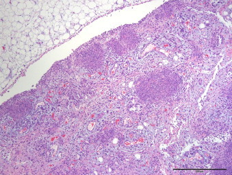
Twenty specific pathogen free cats were experimentally infected with a virulent cat-passaged type I field strain of FIPV. Eighteen cats succumbed within 2-4 weeks to effusive abdominal FIP, one survived for 6 weeks, and one seroconverted without outward signs of disease. A profound drop in the absolute count of blood lymphocytes occurred around 2 weeks post-infection (p.i.) in cats with rapid disease, while the decrease was delayed in the one cat that survived for 6 weeks. The absolute lymphocyte count of the surviving cat remained within normal range. Serum antibodies as measured by indirect immunofluorescence appeared after 2 weeks p.i. and correlated with the onset of disease signs. Viral genomic RNA was either not detectable by reverse transcription quantitative real-time PCR (RT-qPCR) or detectable only at very low levels in terminal tissues not involved directly in the infection, including hepatic and renal parenchyma, cardiac muscle, lung or popliteal lymph node. High tissue virus loads were measured in severely affected tissues such as the omentum, mesenteric lymph nodes and spleen. High levels of viral genomic RNA were also detected in whole ascitic fluid, with the cellular fraction containing 10-1000 times more viral RNA than the supernatant. Replicating virus was strongly associated with macrophages by immunohistochemistry. Virus was usually detected at relatively low levels in feces and there was no evidence of enterocyte infection. Viral genomic RNA was not detected at the level of test sensitivity in whole blood, plasma, or the white cell fraction in terminal samples from the 19 cats that succumbed or in the single survivor. These studies reconfirmed the effect of lymphopenia on disease outcome. FIPV genomic RNA was also found to be highly macrophage associated within diseased tissues and effusions as determined by RT-qPCR and immunohistochemistry but was not present in blood.

Keywords: Experimental; FIP virus; Immunohistochemistry; Macrophages; RT-qPCR; Viremia.


Figures

None
Graphical abstract
Fig. 1
Fig. 1
Sensitivity of the RT-qPCR used in this study to detect FIPV genomes.
Fig. 2
Fig. 2
Febrile responses of cats that were experimentally infected with FIPV.
Fig. 3
Fig. 3
Anti-feline coronavirus titers in plasma of cats experimentally infected with FIPV.
Fig. 4
Fig. 4
Absolute total white cell and lymphocyte counts of 18 cats that died within 2–4 weeks following experimental infection with FIPV.
Fig. 5
Fig. 5
Absolute total white blood cell and lymphocyte counts of two cats experimentally infected with FIP, one (p26) of which resisted disease and one (p31) that died at 6 weeks post-infection.
Fig. 6
Fig. 6
Gross appearance of the abdomen in a cat with experimentally induced effusive FIP. The omentum is edematous, omental vessels prominent and reddened, the surface of the spleen is covered with whitish plaques (pyogranulomas) and fibrin, and the abdomen is filled with a yellowish exudate.
Fig. 7
Fig. 7
Omentum (Hematoxylin and Eosin). The omentum is greatly expanded by pyogranulomatous inflammatory nodules and peritoneal inflammation. Blood vessels are prominent and there is multifocal serous atrophy of fat.
Fig. 8
Fig. 8
Omentum stained for FIPV antigen (Immunoperoxidase). FIP antigen is present in inflammatory nodules and on the peritoneal surface.
Fig. 9
Fig. 9
Omentum (Immunoperoxidase). High magnification of an omental pyogranuloma demonstrates a predominance of strongly CD18 positive macrophages (400×).
Fig. 10
Fig. 10
Omentum (Immunoperoxidase). Immunoreactivity for CD3 demonstrates few T lymphocytes present in omental pyogranulomas (400×).
Fig. 11
Fig. 11
Omentum (Immunoperoxidase): High magnification of an omental pyogranuloma shows a predominance of mononuclear cells. Immunopositivity for FIPV antigen is present in a population of large foamy macrophages (400×).
Fig. 12
Fig. 12
Terminal ileum (Immunoperoxidase). FIP antigen is present within macrophages on the inflamed serosal surface, but is not observed in enterocytes.
Fig. 13
Fig. 13
Ascites fluid (Immunoperoxidase). Cytospin concentrated CD11b positive cells from FIP infected cat ascites fluid is comprised prodominantly of macrophages and lesser numbers of neutrophils. Macrophages demonstrate abundant membranous and variable cytoplasmic immunoreactivity for FIPV antigen by immunocytochemistry. Neutrophils are diffusely negative for FIPV.
Fig. 14
Fig. 14
Levels of FIPV genomic RNA in various tissues from cats experimentally infected with FIPV.
Fig. 15
Fig. 15
Levels of FIPV genomic RNA in ascitic fluid from cats experimentally infected with FIPV.

References

    1. Can-Sahna K., Soydal Ataseven V., Pinar D., Oğuzoğlu T.C. The detection of feline coronaviruses in blood samples from cats by genomic RNA RT-PCR. J. Feline Med. Surg. 2007;9:369–437. - PMC - PubMed
    1. Chang H.W., de Groot R.J., Egberink H.F., Rottier P.J. Feline infectious peritonitis: Insights into feline coronavirus pathobiogenesis and epidemiology based on genetic analysis of the viral 3c gene. J. Gen. Virol. 2010;91:415–420. - PubMed
    1. Chang H.W., Egberink H., Halpin R., Spiro D.J., Rottier P.J. Spike protein fusion peptide and feline coronavirus virulence. Emerg. Infect. Dis. 2012;18:1089–1095. - PMC - PubMed
    1. de Groot-Mijnes J.D.F., van Dun J.M., van der Most R.G., de Groot R.J. Natural history of a recurrent feline coronavirus infection and the role of cellular immunity in survival and disease. J. Virol. 2005;79:1036–1044. - PMC - PubMed
    1. Dean G.A., Olivry T., Stanton C., Pedersen N.C. In vivo cytokine response to experimental feline infectious peritonitis virus infection. Vet. Micro. 2003;97:1–12. - PMC - PubMed

Publication types

MeSH terms