. 2015 Jan 8;57(1):108-22.
doi: 10.1016/j.molcel.2014.11.015. Epub 2014 Dec 18.

Noncovalent interactions with SUMO and ubiquitin orchestrate distinct functions of the SLX4 complex in genome maintenance

Affiliations

Noncovalent interactions with SUMO and ubiquitin orchestrate distinct functions of the SLX4 complex in genome maintenance

Jian Ouyang et al. Mol Cell. .

Abstract

SLX4, a coordinator of multiple DNA structure-specific endonucleases, is important for several DNA repair pathways. Noncovalent interactions of SLX4 with ubiquitin are required for localizing SLX4 to DNA interstrand crosslinks (ICLs), yet how SLX4 is targeted to other functional contexts remains unclear. Here, we show that SLX4 binds SUMO-2/3 chains via SUMO-interacting motifs (SIMs). The SIMs of SLX4 are dispensable for ICL repair but important for processing CPT-induced replication intermediates, suppressing fragile site instability, and localizing SLX4 to ALT telomeres. The localization of SLX4 to laser-induced DNA damage also requires the SIMs, as well as DNA end resection, UBC9, and MDC1. Furthermore, the SUMO binding of SLX4 enhances its interaction with specific DNA-damage sensors or telomere-binding proteins, including RPA, MRE11-RAD50-NBS1, and TRF2. Thus, the interactions of SLX4 with SUMO and ubiquitin increase its affinity for factors recognizing different DNA lesions or telomeres, helping to direct the SLX4 complex in distinct functional contexts.


Figures

Fig. 1
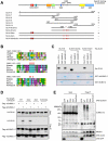
Fig. 1. Binding of SLX4 to SUMO-2/3 chains
(A) Purified GST, GST-SUMO-1, GST-SUMO-2, and GST-SUMO-2-2KA (200 μg of each) were incubated with extracts derived from HEK293T cells expressing HA-SLX4. The HA-SLX4 captured by GST pulldowns was detected by anti-HA antibody. (B-C) Purified GST, GST-SUMO-2, and GST-4xSUMO-2 (5 μg of each) were incubated with extracts derived from HA-SLX4-expressing cells (B) or untransfected cells (C). The HA-SLX4 (B) and endogenous SLX4 and MUS81 (C) captured by GST pulldowns were detected by Western blot. (D) HA-SLX4 and HA-RNF4 were coexpressed with Flag-1xSUMO-2 or Flag-4xSUMO-2 in HEK293T cells. Flag-tagged SUMO-2 was immunoprecipitated using anti-Flag antibody, and the HA-SLX4 and HA-RNF4 associated with SUMO-2 were detected by Western blot. (E) SFB-tagged GFP and SLX4 were immunoprecipitated using anti-Flag antibody under a non-denaturing condition. The SUMO-2/3 conjugates in the immunoprecipitates were analyzed using anti-SUMO-2/3 antibody. (F) SFB-SLX4 was immunoprecipitated using anti-Flag antibody under a denaturing or a non-denaturing condition.
Fig. 2
Fig. 2. SLX4 recognizes SUMO-2/3 chains via the SIMs
(A) A schematic representation of the domain structure of SLX4 and the SLX4 fragments tested in this study. A summary of the ability of the SLX4 fragments to specifically recognize SUMO-2 chains is shown on the right. (B) Amino acid sequences of the three SIMs of human SLX4 are aligned with the corresponding sequences of SLX4 homologues from higher vertebrates. The hydrophobic residues that are mutated in the SLX4 mutants are marked by *. (C) The three SIMs of SLX4-8 were disrupted by an internal deletion (SLX4-8-3simΔ) or point mutations (SLX4-8-3sim). His-SLX4-8 and its mutant derivatives were purified and tested for binding to purified GST-SUMO-2 and GST-4xSUMO-2. (D) HA-SLX4WT and the indicated SLX4 mutants were coexpressed with Flag-1xSUMO-2 or Flag-4xSUMO-2 in HEK293T cells. Flag-tagged SUMO-2 was immunoprecipitated, and the HA-SLX4 proteins in the immunoprecipitates were analyzed using anti-HA antibody. *: two proteins non-specifically recognized by anti-Flag antibody. (E) SFB-tagged GFP, SLX4WT, and the SLX4 mutants were immunoprecipitated from cell extracts using anti-Flag antibody. The SUMO-2/3 conjugates and MUS81 in the immunoprecipitates were analyzed by Western blot.
Fig. 3
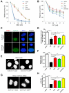
Fig. 3. The SIMs and UBZs of SLX4 are functionally distinct
(A) SLX4-null cells complemented with vector, SLX4WT, SLX43sim, SLX42ubz, and SLX43sim2ubz were tested for MMC sensitivity. (B) SLX4-null cells complemented with vector, SLX4WT, SLX43sim, SLX42ubz, and SLX43sim2ubz were tested for CPT sensitivity. (C-D) SLX4-null cells complemented with vector, SLX4WT, SLX43sim, SLX42ubz, and SLX43sim2ubz were analyzed by immunostaining of 53BP1 and Cyclin A. Representative images of cells are shown in (C). The fractions of G1 cells (Cyclin A-negative) with multiple 53BP1 foci (>2) were quantified in the indicated cell populations (D). Error bars: S.E.M.; n=40 (>200 G1 cells were analyzed in each population). (E- F) DNA of SLX4-null cells complemented with vector or SLX4WT was stained with DAPI (E). The levels of micronuclei in the indicated cell populations were quantified and normalized to the numbers of nuclei analyzed (F). Error bars: S.E.M.; n=40 (>1,300 nuclei were analyzed in each population). (G-H) The bulky anaphase bridge of an SLX4-null cell complemented with vector and the normally segregated chromosomes of an SLX4-null cell complemented with SLX4WT (G). The fractions of mitotic cells with bulky anaphase bridges were quantified in the indicated cell populations (H). Error bars: S.D.; n=2 (>50 mitotic cells were analyzed in each population).
Fig. 4
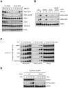
Fig. 4. The SUMO binding of SLX4 is important for processing CPT-induced replication intermediates
(A) U2OS cells were transfected with control or SLX4 siRNA and treated with 1 μM CPT. At the indicated time points, the phosphorylation of RPA32 and H2AX was analyzed with phospho-specific antibodies. Knockdown of SLX4 is shown in Fig. S4A. (B) U2OS cells were transfected with the indicated siRNAs. The CPT-induced phosphorylation of RPA32 was analyzed as in (A). Knockdown of MUS81 is shown in Fig. S4C. (C) SLX4-null cells complemented with vector, SLX4WT, SLX42ubz, SLX43sim, and SLX43sim2ubz were treated with 0.1 μM CPT. The CPT-induced phosphorylation of RPA32 was analyzed at the indicated time points. (D) SLX4-null cells complemented with SLX4WT were transfected with control or MUS81 siRNA. Cells were treated with 0.1 μM CPT, and the phosphorylation of RPA32 was analyzed.
Fig. 5
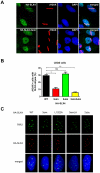
Fig. 5. SUMO binding of SLX4 promotes its localization to laser-induced DNA damage and ALT telomeres
(A) U2OS cells expressing HA-tagged SLX4WT and SLX43sim were microirradiated with UV laser and analyzed by immunostaining of HA and γH2AX 1 hr later. The expression of SLX4WT and SLX43sim is shown in Fig. S5A. (B) U2OS cells expressing HA-tagged SLX4WT, SLX43sim, SLX42ubz, and SLX43sim2ubz were microirradiated with UV laser and analyzed as in (A). The fractions of γH2AX-positive cells with HA stripes were quantified. Error bars: S.E.M.; n=2. The expression of SLX4WT and the SLX4 mutants and the cell-cycle profiles of various cell populations are shown in Fig. S5A, S5D. (C) The colocalization of HA-tagged SLX4WT, SLX43sim, SLX4L1022A, SLX43simLA, and SLX42ubz with endogenous TRF2 was analyzed in U2OS cells. Representative images of each cell line are shown. The expression of SLX4WT and the SLX4 mutants is shown in Fig. S5L.
Fig. 6
Fig. 6. Localization of SLX4 to laser-induced DNA damage requires DNA-end resection and MDC1
A-B) U2OS cells expressing HA-SLX4WT were treated with control or CtIP siRNA (A), or with 25 μM Mirin (B). The localization of HA-SLX4WT to DNA damage stripes was analyzed 1 hr after microirradiation and quantified as in Fig. 5B. Error bars: S.E.M.; n=2. Knockdown of CtIP and its effects on the cell cycle are shown in Fig. S6A-B. (C-D) The localizations of HA-SLX4WT and RPA32 were analyzed 1 hr after microirradiation (C). The fractions of HA-SLX4WT stripe-positive cells with or without RPA32 stripes were quantified (D). Error bars: S.E.M.; n=2. (E) U2OS cells expressing HA-SLX4WT were treated with control or MDC1 siRNA. The localization of HA-SLX4WT was analyzed as in Fig. 5B. Error bars: S.E.M.; n=2. Knockdown of MDC1 and its effects on the cell cycle are shown in Fig. S6B-C. (F) U2OS cells expressing HA-SLX4WT were treated with control siRNA or both CtIP and MDC1 siRNAs, and analyzed as in Fig. 5B. Error bars: S.E.M.; n=2. Knockdown of CtIP and MDC1 is shown in Fig. S6E.
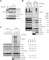
Fig. 7
Fig. 7. SLX4 interacts with TRF2, RPA and MRN in a UBC9- and SIM-regulated manner
(A) SFB-tagged GFP, SLX4WT, SLX43sim, SLX4L1022A, and SLX43simLA were coexpressed with myc-TRF2 in U2OS cells. SFB-tagged proteins were captured using anti-Flag antibody, and the associated myc-TRF2 and MUS81 were detected by Western blot. The relative abundance of myc-TRF2 pulled down by SFB-SLX4 variants was shown. (B) HeLa cells were transfected with control or UBC9 siRNA and plasmids expressing SFB-GFP or SFB-SLX4WT as indicated. SFB-tagged proteins were immunoprecipitated with anti-Flag antibody. The indicated proteins in inputs and immunoprecipitates were analyzed by Western blot. The relative abundance of proteins pulled down by SFB-SLX4WT in control and UBC9 knockdown cells is shown. (C) HEK293T cells expressing SFB-tagged GFP, SLX4WT, SLX42ubz, SLX43sim, or SLX43sim2ubz were treated with 1 μM CPT for 1 hr and subjected to immunoprecipitation using anti-Flag antibody. The indicated proteins in the immunoprecipitates were analyzed by Western blot. The relative abundance of proteins pulled down by SFB-SLX4 variants is shown.

Comment in

References

    1. Andersen SL, Bergstralh DT, Kohl KP, LaRocque JR, Moore CB, Sekelsky J. Drosophila MUS312 and the vertebrate ortholog BTBD12 interact with DNA structure-specific endonucleases in DNA repair and recombination. Mol Cell. 2009;35:128–135. - PMC - PubMed
    1. Armstrong AA, Mohideen F, Lima CD. Recognition of SUMO-modified PCNA requires tandem receptor motifs in Srs2. Nature. 2012;483:59–63. - PMC - PubMed
    1. Baba D, Maita N, Jee JG, Uchimura Y, Saitoh H, Sugasawa K, Hanaoka F, Tochio H, Hiroaki H, Shirakawa M. Crystal structure of thymine DNA glycosylase conjugated to SUMO-1. Nature. 2005;435:979–982. - PubMed
    1. Bekker-Jensen S, Lukas C, Kitagawa R, Melander F, Kastan MB, Bartek J, Lukas J. Spatial organization of the mammalian genome surveillance machinery in response to DNA strand breaks. J Cell Biol. 2006;173:195–206. - PMC - PubMed
    1. Bunting SF, Nussenzweig A. End-joining, translocations and cancer. Nat Rev Cancer. 2013;13:443–454. - PMC - PubMed

Publication types

MeSH terms