. 2015 Feb 20;290(8):4896-4907.
doi: 10.1074/jbc.M114.588566. Epub 2014 Dec 22.

A mutation within the extended X loop abolished substrate-induced ATPase activity of the human liver ATP-binding cassette (ABC) transporter MDR3

Affiliations

A mutation within the extended X loop abolished substrate-induced ATPase activity of the human liver ATP-binding cassette (ABC) transporter MDR3

Marianne Kluth et al. J Biol Chem. .

Abstract

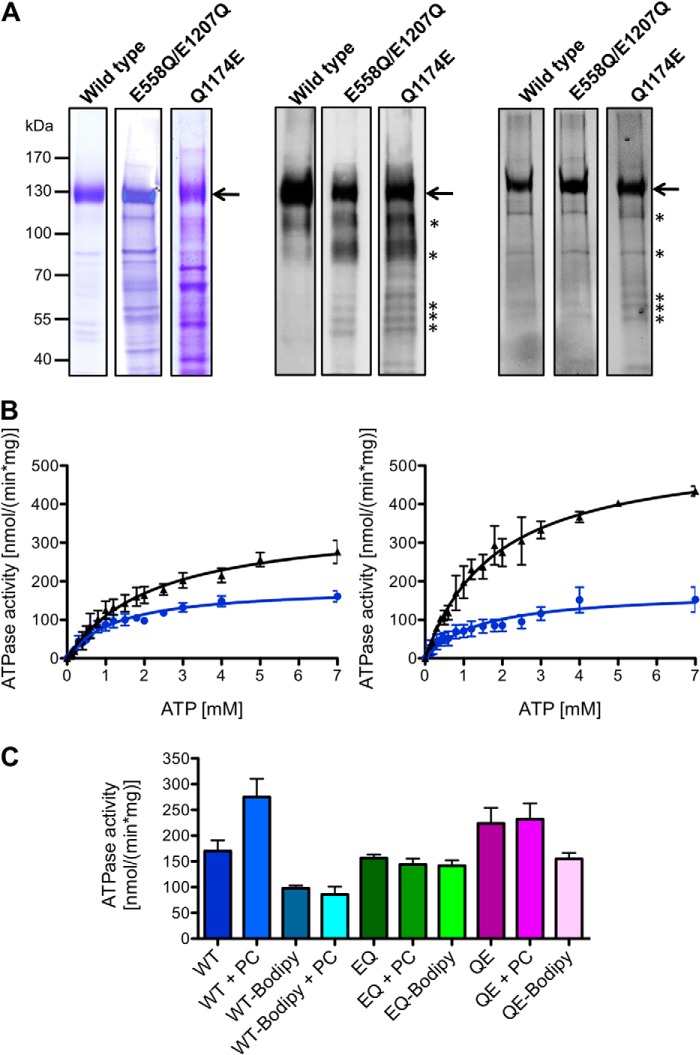
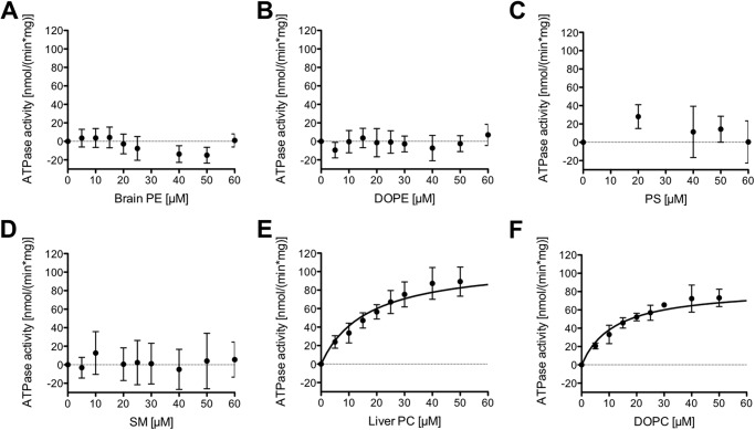
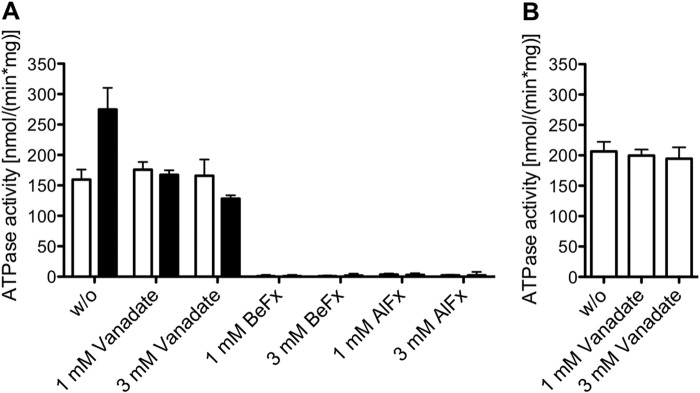
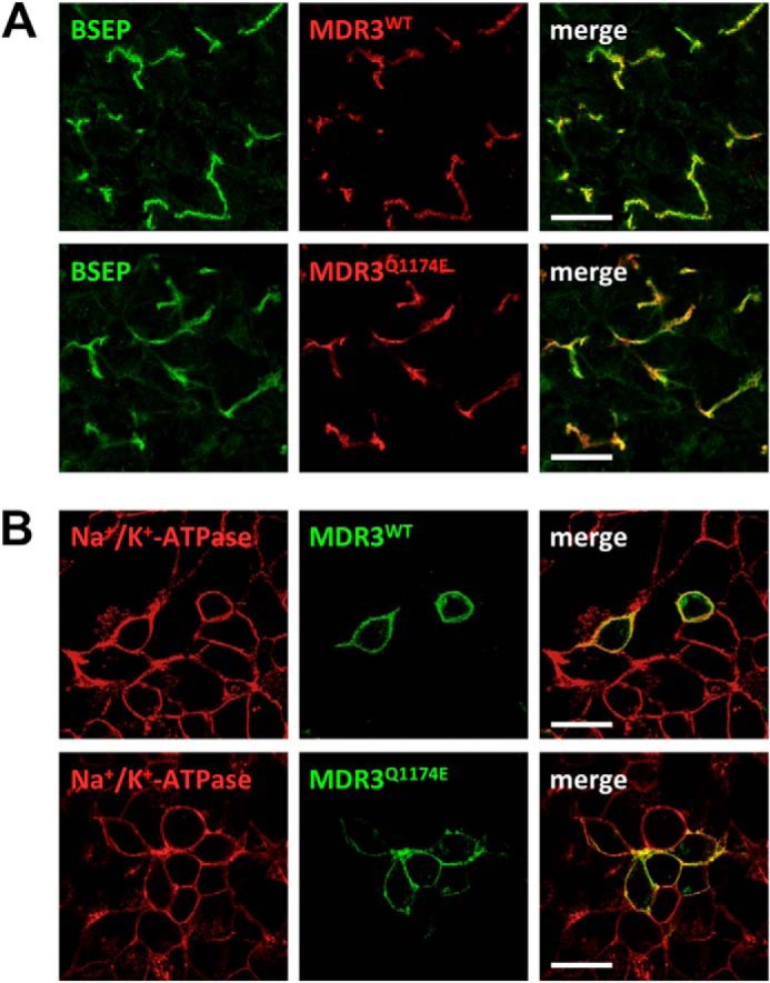
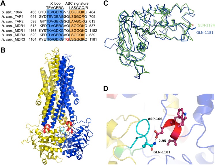
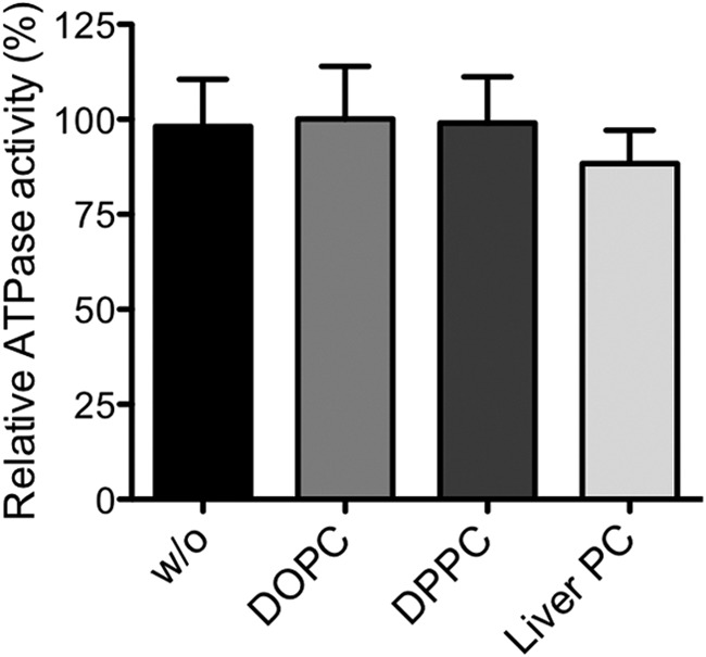
The human multidrug resistance protein 3 (MDR3/ABCB4) belongs to the ubiquitous family of ATP-binding cassette (ABC) transporters and is located in the canalicular membrane of hepatocytes. There it flops the phospholipids of the phosphatidylcholine (PC) family from the inner to the outer leaflet. Here, we report the characterization of wild type MDR3 and the Q1174E mutant, which was identified previously in a patient with progressive familial intrahepatic cholestasis type 3 (PFIC-3). We expressed different variants of MDR3 in the yeast Pichia pastoris, purified the proteins via tandem affinity chromatography, and determined MDR3-specific ATPase activity in the presence or absence of phospholipids. The ATPase activity of wild type MDR3 was stimulated 2-fold by liver PC or 1,2-dioleoyl-sn-glycero-3-phosphatidylethanolamine lipids. Furthermore, the cross-linking of MDR3 with a thiol-reactive fluorophore blocked ATP hydrolysis and exhibited no PC stimulation. Similarly, phosphatidylethanolamine, phosphatidylserine, and sphingomyelin lipids did not induce an increase of wild type MDR3 ATPase activity. The phosphate analogues beryllium fluoride and aluminum fluoride led to complete inhibition of ATPase activity, whereas orthovanadate inhibited exclusively the PC-stimulated ATPase activity of MDR3. The Q1174E mutation is located in the nucleotide-binding domain in direct proximity of the leucine of the ABC signature motif and extended the X loop, which is found in ABC exporters. Our data on the Q1174E mutant demonstrated basal ATPase activity, but PC lipids were incapable of stimulating ATPase activity highlighting the role of the extended X loop in the cross-talk of the nucleotide-binding domain and the transmembrane domain.

Keywords: ABC Transporter; ATPase; Lipid Transport; Liver Injury; MDR1; MDR3; Multidrug Transporter; PFIC-3; Transmission Interface; X Loop.


Figures

FIGURE 1.
FIGURE 1.
A, human wild type MDR3, the E558Q/E1207Q double mutant, and the Q1174E mutant purified from P. pastoris. The MDR3 variants containing a C-terminal His6 tag and a calmodulin-binding peptide were expressed in the yeast P. pastoris and purified as described under “Experimental Procedures.” 10 μg of purified MDR3 was resolved on a 7% SDS-PAGE and either stained with Coomassie Brilliant Blue (left panel) or detected by immunoblotting (middle panel) using the monoclonal anti-P-gp C219 antibody. MDR3 was cross-linked by the thiol-reactive maleimide-BODIPY fluorophore (MDR3-BODIPY) and analyzed by fluorescence imaging using excitation wavelength at 488 nm and emission wavelength at 520 nm (right panel). Molecular mass markers are shown on the left. MDR3 is indicated with an arrow, and degradation products of MDR3 are shown with asterisks. B, ATPase activity of purified wild type MDR3 (black triangle) and MDR3-BODIPY (blue circles) in the absence (left panel) or presence (right panel) of DOPC lipids. C, ATPase activity of purified wild type MDR3 (blue), the ATPase-deficient MDR3 EQ/EQ mutant (green), and the Q1174E mutant (magenta) and the corresponding cross-linked BODIPY derivatives in the presence of 2 mm ATP and 300 μm DOPC (PC).
FIGURE 2.
FIGURE 2.
Concentration dependence of the ATPase activity of MDR3 in the presence of different kinds of lipids: A, brain PE lipids; B, DOPE lipids; C, PS lipids; D, SM lipids; E, liver PC lipids; and F, DOPC lipids, respectively. The ATPase activity was started by addition of 2 mm ATP and assayed for 40 min at 37 °C. The ATPase activity in the absence of lipids (159 ± 14 nmol min−1 mg−1) was subtracted from the ATPase activity in the presence of lipids. The data represent the average of at least six independent experiments (mean ± S.D.).
FIGURE 3.
FIGURE 3.
Inhibition of MDR3 ATPase activity by phosphate analogues. A, ATPase activity of purified wild type MDR3 was measured without (w/o, white column) and with 300 μm DOPC lipids (black column) in the presence of orthovanadate, BeFx, and AlFx. The reaction mixture contained 2 mm ATP and inhibitors at a final concentration of 1 or 3 mm. The data are means ± S.D. of at least four independent experiments. B, ATPase activity of Q1174E mutant in the presence of orthovanadate. The data represent the average of at least two independent experiments (mean ± S.D.).
FIGURE 4.
FIGURE 4.
MDR3 mutation Q1174E does not affect protein localization in vivo or in vitro. A, sample from a liver with normal MDR3 expression (upper panel) and from the patient carrying the heterozygous Q1174E mutation (lower panel). The green fluorescence corresponds to bile salt export pump (BSEP), which acts as a canalicular marker, and the red fluorescence represents MDR3. B, transient expression of human wild type (upper panel) and Q1174E (lower panel) MDR3-eYFP (green) after transfection into HEK293 cells. Cells were fixed after 48 h and stained for the Na+/K+-ATPase as a plasma membrane marker (red). Scale bars, 20 μm.
FIGURE 5.
FIGURE 5.
A, alignment of the amino acid sequence of MDR3 with selected human ABC transporters. The X loop motif is shaded in blue and the ABC signature motif in orange. Gln-1174 is colored in red. B, homology model of human MDR3 based on the structure of Sav1866 (Protein Data Bank code 2HYD) and the amino acid sequence of isoform B of MDR3 (51). One transporter half, consisting of transmembrane domain (TMD) and nucleotide-binding domain (NBD), is shown in blue and the other in yellow. The coupling helices are highlighted in red, and the X loop is colored in cyan. C, overlay of the MDR3 NBD2 of the isoform A (green) and isoform B (blue). Gln-1174 of the isoform A is a rotamer of Gln-1181 of the isoform B. D, close-up view of the interface between TMD and NBD2. Gln-1174 of the X loop is shown in stick representation (cyan).
FIGURE 6.
FIGURE 6.
ATPase activity of the purified Q1174E mutant was measured without (w/o) lipids and with DOPC, DPPC, or liver PC lipids as described under “Experimental Procedures.” None of these lipids stimulated the ATPase activity of the Q1174E mutant. One hundred percent activity represents 223 ± 25 nmol min−1 mg−1.

Similar articles

Cited by

References

    1. Oude Elferink R. P., Paulusma C. C. (2007) Function and pathophysiological importance of ABCB4 (MDR3 P-glycoprotein). Pflugers Arch. 453, 601–610 - PubMed
    1. Smit J. J., Schinkel A. H., Oude Elferink R. P., Groen A. K., Wagenaar E., van Deemter L., Mol C. A., Ottenhoff R., van der Lugt N. M., van Roon M. A. (1993) Homozygous disruption of the murine mdr2 P-glycoprotein gene leads to a complete absence of phospholipid from bile and to liver disease. Cell 75, 451–462 - PubMed
    1. Smith A. J., de Vree J. M., Ottenhoff R., Oude Elferink R. P., Schinkel A. H., Borst P. (1998) Hepatocyte-specific expression of the human MDR3 P-glycoprotein gene restores the biliary phosphatidylcholine excretion absent in Mdr2(−/−) mice. Hepatology 28, 530–536 - PubMed
    1. Smith A. J., Timmermans-Hereijgers J. L., Roelofsen B., Wirtz K. W., van Blitterswijk W. J., Smit J. J., Schinkel A. H., Borst P. (1994) The human MDR3 P-glycoprotein promotes translocation of phosphatidylcholine through the plasma membrane of fibroblasts from transgenic mice. FEBS Lett. 354, 263–266 - PubMed
    1. van Helvoort A., Smith A. J., Sprong H., Fritzsche I., Schinkel A. H., Borst P., van Meer G. (1996) MDR1 P-glycoprotein is a lipid translocase of broad specificity, while MDR3 P-glycoprotein specifically translocates phosphatidylcholine. Cell 87, 507–517 - PubMed

Publication types

MeSH terms

LinkOut - more resources