. 2015 Jan 13;4(1):7-15.
doi: 10.1016/j.stemcr.2014.11.003. Epub 2014 Dec 18.

Genetic and functional diversity of propagating cells in glioblastoma

Affiliations

Genetic and functional diversity of propagating cells in glioblastoma

Sara G M Piccirillo et al. Stem Cell Reports. .

Abstract

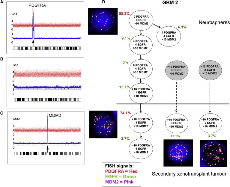
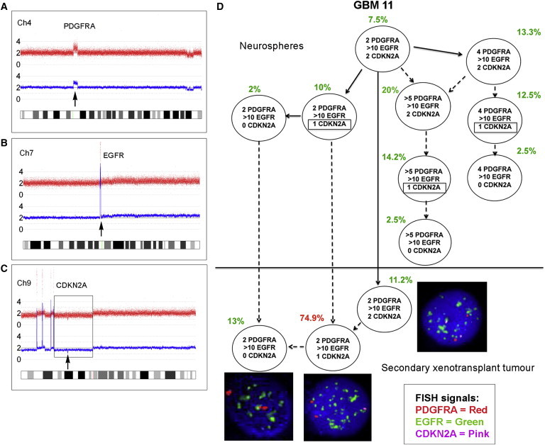
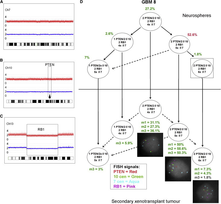
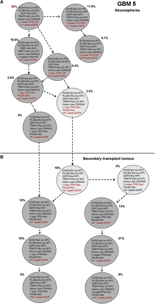
Glioblastoma (GBM) is a lethal malignancy whose clinical intransigence has been linked to extensive intraclonal genetic and phenotypic diversity and the common emergence of therapeutic resistance. This interpretation embodies the implicit assumption that cancer stem cells or tumor-propagating cells are themselves genetically and functionally diverse. To test this, we screened primary GBM tumors by SNP array to identify copy number alterations (a minimum of three) that could be visualized in single cells by multicolor fluorescence in situ hybridization. Interrogation of neurosphere-derived cells (from four patients) and cells derived from secondary transplants of these same cells in NOD-SCID mice allowed us to infer the clonal and phylogenetic architectures. Whole-exome sequencing and single-cell genetic analysis in one case revealed a more complex clonal structure. This proof-of-principle experiment revealed that subclones in each GBM had variable regenerative or stem cell activity, and highlighted genetic alterations associated with more competitive propagating activity in vivo.


Figures

Figure 1
Figure 1
Subclonal Genetic Structure of Neurosphere Cells and Tumor-Propagating Cells Derived from Primary Tumor GBM 2 (A–C) SNP 6 array profiles of DNA from GBM 2 primary tumor showing high-level PDGFRA amplification (A), whole chromosome 7 gain (B), and high-level MDM2 amplification (C). (D) Subclonal genetic structure in the neurospheres (top) and after secondary transplantation in a single mouse (m3) (bottom). Subclones represented by gray circles were not present in the neurospheres above the threshold detection level. FISH images are shown next to their respective genotype. Red type indicates the major clone. Solid arrows show probable derivation of subclones. Dashed arrows indicate possible alternative derivation of subclones. FISH images were captured at 100× magnification. See also Figure S1 and Tables S1, S2, S3, and S4.
Figure 2
Figure 2
Subclonal Genetic Structure of Neurosphere Cells and Tumor-Propagating Cells Derived from Primary Glioblastoma GBM 11 (A–C) SNP 6 array profiles of DNA from GBM 11 primary tumor showing PDGFRA amplification (A), high-level EGFR amplification (B), and homozygous CDKN2A deletion (C) comprised of a large deletion of one allele (box) and focal deletion of the second allele (arrow). (D) Subclonal structure of neurosphere cells (top) and tumor cells after secondary transplant in a single mouse (m3) (bottom). Potentially three different CDKN2A deletions occur in different subclones in the neurospheres (indicated by boxes). Solid arrows show probable derivation of subclones. Dashed arrows indicate possible alternative derivation of subclones. FISH images were captured at 100× magnification. See also Figure S1 and Tables S1, S2, S3, and S4.
Figure 3
Figure 3
Identical Subclonal Genetic Structure of Neurosphere Cells and Tumor-Propagating Cells Derived from Primary Tumor GBM 8 (A–C) SNP 6 array profiles showing chromosome 7 (A), focal PTEN loss (B), and a large deletion of chromosome 13, including RB1 (C). There was also loss of heterozygosity (LOH) for the whole of 17p (including the TP53 gene) without any copy number change (uniparental disomy [UPD]). (D) FISH analysis of GBM 8 neurospheres using a range of centromere probes revealed that these were nearly triploid, with two PTEN and RB1 signals corresponding to a loss of one copy of each locus. The neurospheres showed a branched subclonal structure with four subclones above the FISH detection threshold (2%) at the time of injection into primary mice (top). All of these read out in the tumors of at least one secondary transplanted mouse (bottom). One further subclone detected in all mice was present in the neurospheres at a level below the cutoff for FISH (1.8%) (box). m1, m2, m3: three replicate mice, each injected with 1 × 106 neurosphere cells. Solid arrows show the probable derivation of subclones. Dashed arrows indicate the possible alternative derivation of subclones. FISH images were captured at 100× magnification. See also Figure S1 and Tables S1, S2, S3, and S4.
Figure 4
Figure 4
Single-Cell Analysis of Selected Mutations and CNAs Identified by Exome Sequencing in GBM 5 (A and B) Subclonal genetic architecture in neurospheres (A) and tumor-propagating cells derived from GBM 5 after xenotransplantation (B, mouse 2). A total of 240 neurosphere cells and 100 cells from the secondary xenotransplant tumor were evaluated. Mutations and CNAs are given within the circles; additional mutations and CNAs in individual subclones are indicated in red. See also Figures S2–S4 and Tables S1, S2, S3, and S4.

References

    1. Anderson K., Lutz C., van Delft F.W., Bateman C.M., Guo Y., Colman S.M., Kempski H., Moorman A.V., Titley I., Swansbury J. Genetic variegation of clonal architecture and propagating cells in leukaemia. Nature. 2011;469:356–361. - PubMed
    1. Andersson U., Schwartzbaum J., Wiklund F., Sjöström S., Liu Y., Tsavachidis S., Ahlbom A., Auvinen A., Collatz-Laier H., Feychting M. A comprehensive study of the association between the EGFR and ERBB2 genes and glioma risk. Acta Oncol. 2010;49:767–775. - PubMed
    1. Bao S., Wu Q., Sathornsumetee S., Hao Y., Li Z., Hjelmeland A.B., Shi Q., McLendon R.E., Bigner D.D., Rich J.N. Stem cell-like glioma cells promote tumor angiogenesis through vascular endothelial growth factor. Cancer Res. 2006;66:7843–7848. - PubMed
    1. Bonavia R., Inda M.M., Cavenee W.K., Furnari F.B. Heterogeneity maintenance in glioblastoma: a social network. Cancer Res. 2011;71:4055–4060. - PMC - PubMed
    1. Chen R., Nishimura M.C., Bumbaca S.M., Kharbanda S., Forrest W.F., Kasman I.M., Greve J.M., Soriano R.H., Gilmour L.L., Rivers C.S. A hierarchy of self-renewing tumor-initiating cell types in glioblastoma. Cancer Cell. 2010;17:362–375. - PubMed

Publication types

MeSH terms