. 2016 Jan;75(1):19-31.
doi: 10.1002/mrm.25577. Epub 2014 Dec 22.

Concentric rings K-space trajectory for hyperpolarized (13)C MR spectroscopic imaging

Affiliations

Concentric rings K-space trajectory for hyperpolarized (13)C MR spectroscopic imaging

Wenwen Jiang et al. Magn Reson Med. 2016 Jan.

Abstract

Purpose: To develop a robust and rapid imaging technique for hyperpolarized (13)C MR Spectroscopic Imaging and investigate its performance.

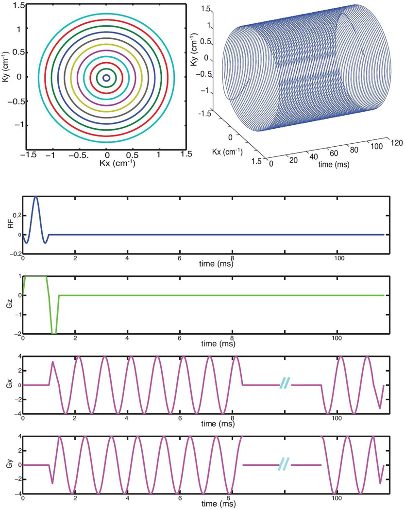
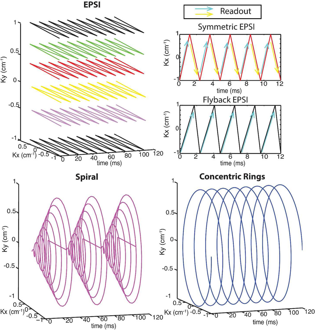
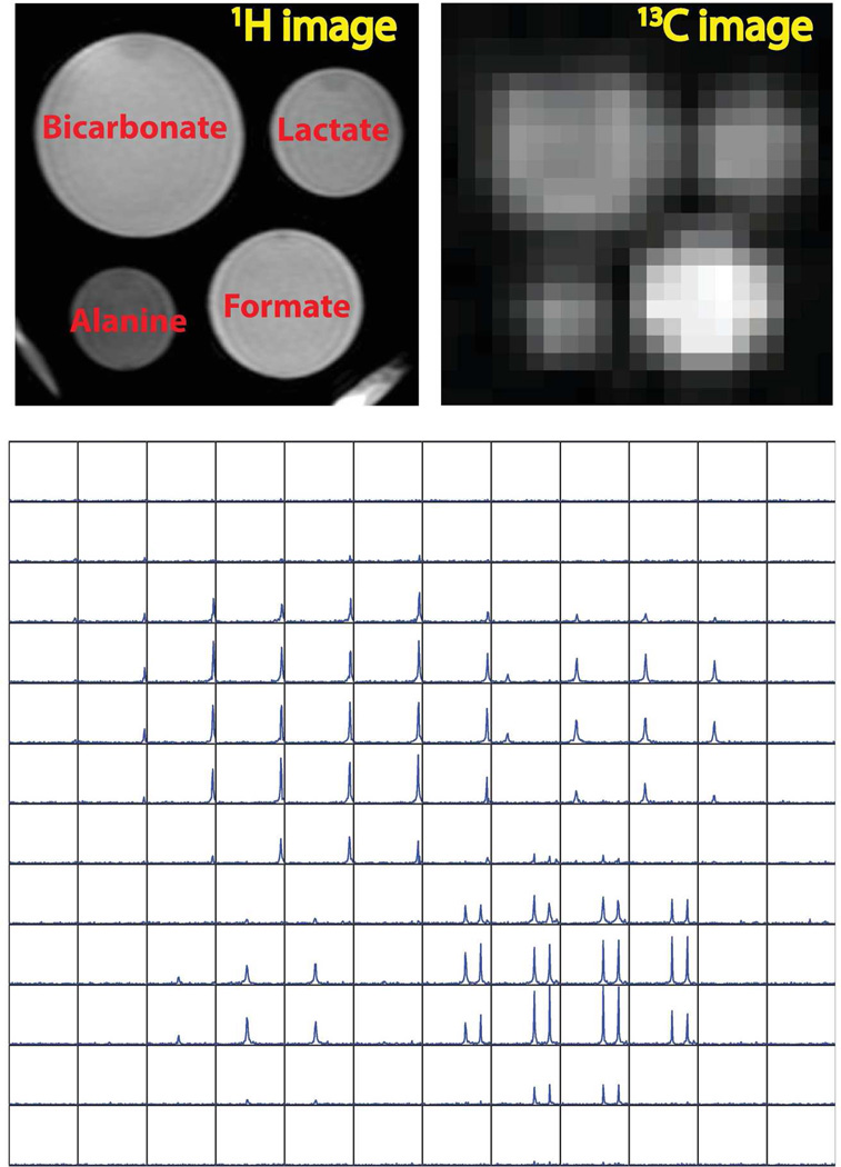
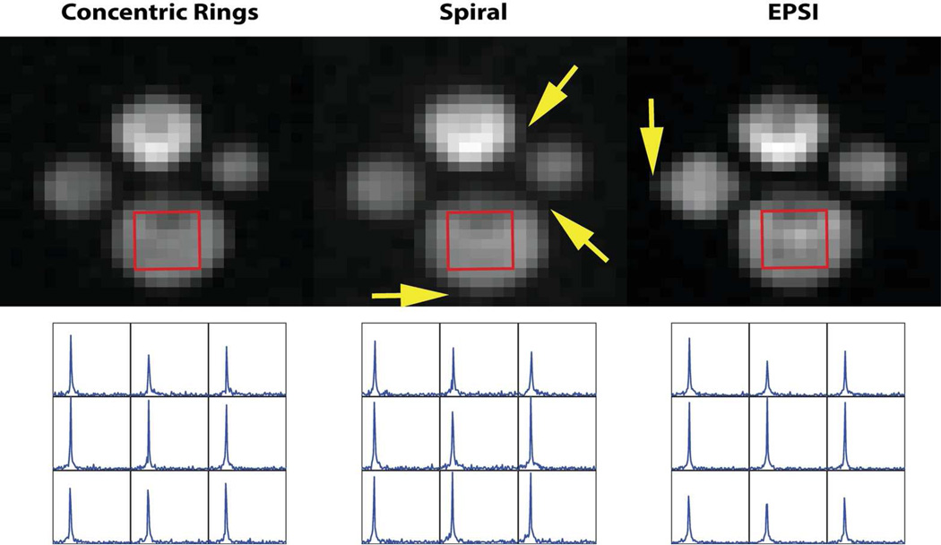
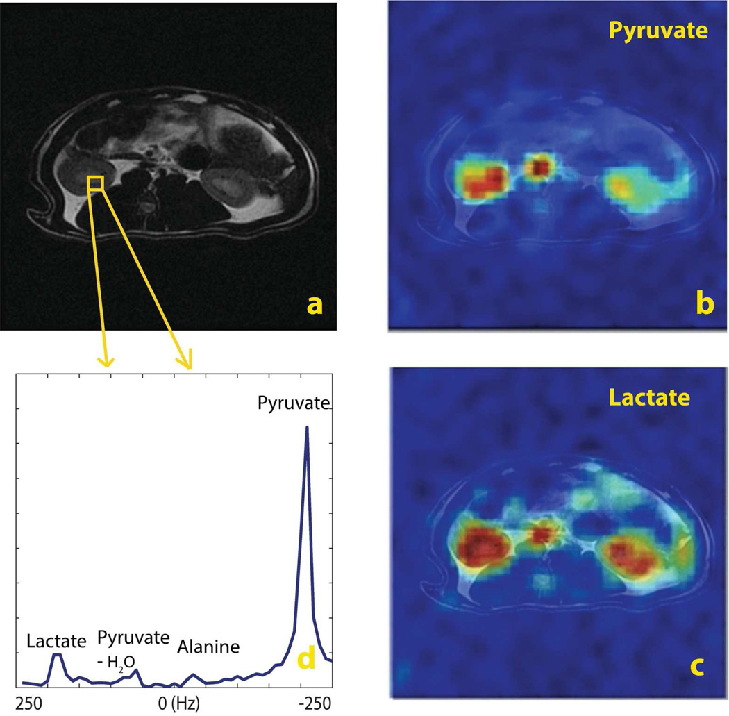
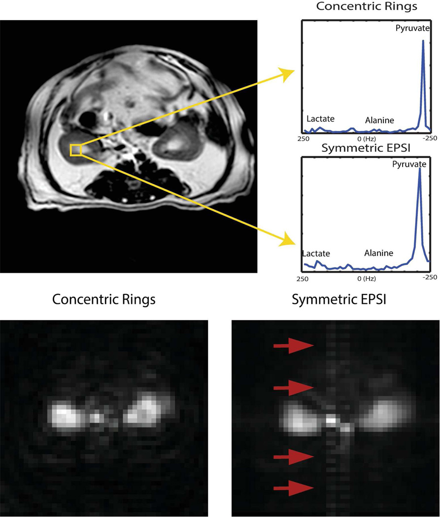
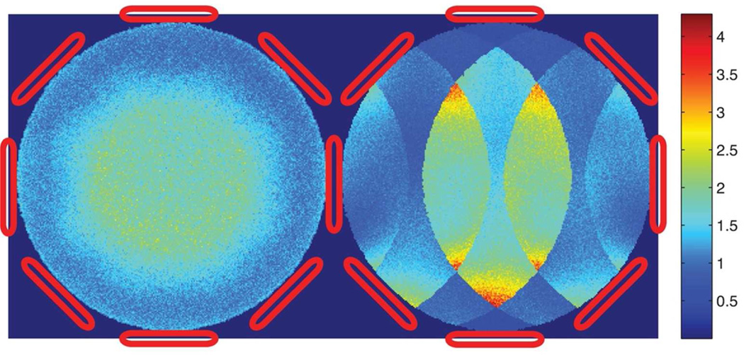
Methods: A concentric rings readout trajectory with constant angular velocity is proposed for hyperpolarized (13)C spectroscopic imaging and its properties are analyzed. Quantitative analyses of design tradeoffs are presented for several imaging scenarios. The first application of concentric rings on (13)C phantoms and in vivo animal hyperpolarized (13)C MR Spectroscopic Imaging studies were performed to demonstrate the feasibility of the proposed method. Finally, a parallel imaging accelerated concentric rings study is presented.

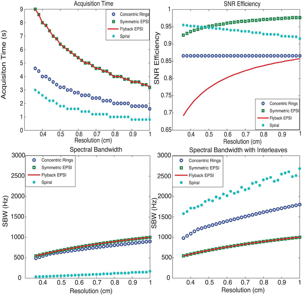
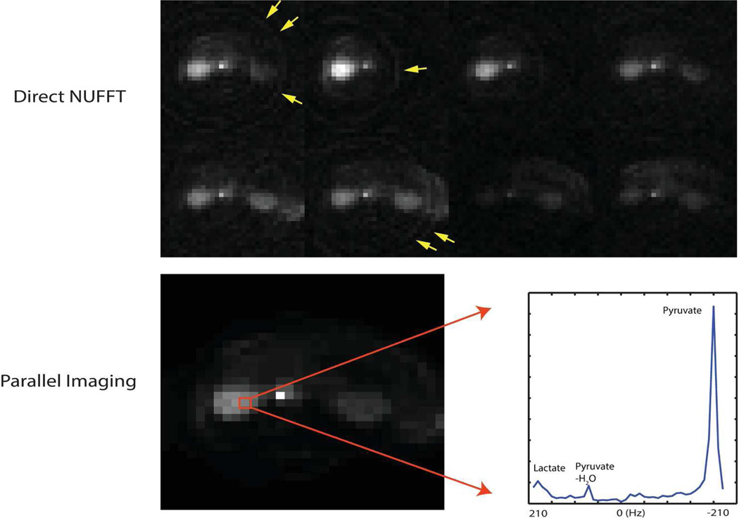
Results: The concentric rings MR Spectroscopic Imaging trajectory has the advantages of acquisition timesaving compared to echo-planar spectroscopic imaging. It provides sufficient spectral bandwidth with relatively high efficiency compared to echo-planar spectroscopic imaging and spiral techniques. Phantom and in vivo animal studies showed good image quality with half the scan time and reduced pulsatile flow artifacts compared to echo-planar spectroscopic imaging. Parallel imaging accelerated concentric rings showed advantages over Cartesian sampling in g-factor simulations and demonstrated aliasing-free image quality in a hyperpolarized (13)C in vivo study.

Conclusion: The concentric rings trajectory is a robust and rapid imaging technique that fits very well with the speed, bandwidth, and resolution requirements of hyperpolarized (13)C MR Spectroscopic Imaging.

Keywords: concentric rings; hyperpolarized 13C; non-Cartesian trajectory; parallel imaging; spectroscopic imaging.


Figures

Figure 1
Figure 1
Concentric rings trajectory and sequence design: top-left shows the spatial concentric rings k-space trajectory; top-right shows the spatial-spectral k-space trajectory; bottom shows the concentric rings GRE sequence for axial images.
Figure 2
Figure 2
K-space trajectories of EPSI, spiral and concentric rings spectroscopic imaging: the arrows illustrates the readout directions for both symmetric EPSI and flyback EPSI; for symmetric EPSI, we use different-colored arrows to differentiate the odd/even echoes for reconstruction.
Figure 3
Figure 3
Comparison of concentric rings, EPSI and spiral spectroscopic imaging: top-left shows the acquisition time; top-right shows the SNR efficiency; bottom-left and bottom-right show the SBW and SBW with spectral interleaves. CRT requires half of the total acquisition time compared with EPSI trajectories, offers about 87% SNR efficiency, and provides much wider spectral bandwidth than flyback EPSI and symmetric EPSI. Although nominally spirals are the most efficient trajectories, offering the best acquisition time and spectral bandwidth benefit while sacrificing the least SNR, they are limited by susceptibility to gradient infidelities.
Figure 4
Figure 4
13C phantom study using CRT: (top-left) 1H image localizer; (top-right) 13C 2D image via projection on spectral domain; (bottom) spatial-spectral display for spectroscopic imaging. The displayed 13C 2D image has the nominal spatial resolution of 1.83×1.83 mm2 after zero padding. The reconstructed spatial-spectral (2D spatial+1D spectral) data matrix (22×22×100) was cropped to 12×12×100, and the SBW for each voxel was 1000 Hz. This was a non-hyperpolarized study, with the TR = 5 s and total acquisition time of 1min 50 s.
Figure 5
Figure 5
Comparison of 13C phantom spectroscopic imaging using CRT, Spiral and EPSI: the top row (from left to right) images were obtained from CRT, Spiral and EPSI respectively. They were created from spectroscopic images via projection along the spectral domain. The displayed 13C 2D image has a nominal spatial resolution of 1.83×1.83 mm2 after zero padding and was cropped into a 5×5 cm2 ROI. The arrows show the slight blurring of spiral and EPSI. The bottom row (from left to right) plots are of 13C bicarbonate spectra obtained from CRT, Spiral and EPSI, respectively, from the same 9 voxels.
Figure 6
Figure 6
In vivo results using concentric rings in a normal rat (axial): a.1H T2-weighted localizer; b. [1-13C] pyruvate image; c. [1-13C] lactate image; d. the 13C spectrum of a selected voxel with 500 Hz SBW. MRSI was acquired with a spatial resolution of 3.67×3.67 mm2. Pyruvate and lactate images were twice zero padded to have a resolution of 1.83×1.83 mm2. For display purposes, the intensity of lactate image was scaled up by 7. The total scan time was 2.2 s.
Figure 7
Figure 7
In vivo results in an axial kidney slice using concentric rings and symmetric EPSI: (top-left)1H T2-weighted localizer; (top-right) spectrum from concentric rings trajectory and spectrum from EPSI trajectory respectively, both sequences captured the conversion of pyruvate with comparable SNR; Bottom figures show pulsatile flow effects on concentric rings and EPSI: (bottom-left) 2D image from the concentric rings and (bottom-right) 2D image from EPSI, via projection along spectral domain.
Figure 8
Figure 8
Simulated g-factor map of 4× undersampled concentric rings trajectory (on the left) and Cartesian trajectory counterpart, such as EPSI, (on the right) for a simulated 8-channel array. The isotropic non-Cartesian undersampling pattern of the concentric rings trajectory results in less coherent noise amplification than the Cartesian counterpart.
Figure 9
Figure 9
Parallel imaging in vivo results in an axial kidney slice using CRT with an 8-channel phased-array rat coil: the spectroscopic imaging CRT was spatially undersampled by 1.45, with a spatial resolution of 2.5×2.5 mm2, 8×8 cm2 FOV and 420 Hz SBW. The top row shows individual coil images of the undersampled CRT using a direct NUFFT reconstruction, where the coil images were generated by projection along the spectral domain. The 8 coils were distributed isotropically around the animal in the axial plane. The bottom row shows the parallel imaging reconstruction result. The 2D image was obtained from projection along the spectral domain and the spectrum from a selected kidney voxel was displayed to show the resolution of various metabolites, including lactate resulting from metabolic conversion. The total scan time was 3.52 s.

References

    1. Kurhanewicz J, Vigneron DB, Brindle K, Chekmenev EY, Comment A, Cunningham CH, DeBerardinis RJ, Green GG, Leach MO, Rajan SS, et al. Analysis of cancer metabolism by imaging hyperpolarized nuclei: prospects for translation to clinical research. Neoplasia (New York, NY) 2011;13:81. - PMC - PubMed
    1. ArdenkjærLarsen JH, Fridlund B, Gram A, Hansson G, Hansson L, Lerche MH, Servin R, Thaning M, Golman K. Increase in signal-to-noise ratio of > 10,000 times in liquid-state nmr. Proceedings of the National Academy of Sciences. 2003;100:10158–10163. - PMC - PubMed
    1. Nelson SJ, Kurhanewicz J, Vigneron DB, Larson PEZ, Harzstark AL, Ferrone M, van Criekinge M, Chang JW, Bok R, Park I, Reed G, Carvajal L, Small EJ, Munster P, Weinberg VK, ArdenkjaerLarsen JH, Chen AP, Hurd RE, Odegardstuen LI, Robb FJ, Tropp J, Murray JA. Metabolic imaging of patients with prostate cancer using hyperpolarized [1-13c]pyruvate. Science Translational Medicine. 2013;5:198ra108. - PMC - PubMed
    1. Cunningham CH, Vigneron DB, Chen AP, Xu D, Nelson SJ, Hurd RE, Kelley DA, Pauly JM. Design of flyback echo-planar readout gradients for magnetic resonance spectroscopic imaging. Magnetic resonance in medicine. 2005;54:1286–1289. - PubMed
    1. Yen YF, Kohler S, Chen A, Tropp J, Bok R, Wolber J, Albers M, Gram K, Zierhut M, Park I, Zhang V, Hu S, Nelson S, Vigneron D, Kurhanewicz J, Dirven H, Hurd R. Imaging considerations for in vivo 13c metabolic mapping using hyperpolarized 13c-pyruvate. Magnetic Resonance in Medicine. 2009;62:1–10. - PMC - PubMed

Publication types