Review
. 2014 Dec 22;6(12):5093-134.
doi: 10.3390/v6125093.

Phocine distemper virus: current knowledge and future directions

Affiliations
Review

Phocine distemper virus: current knowledge and future directions

Pádraig J Duignan et al. Viruses. .

Abstract

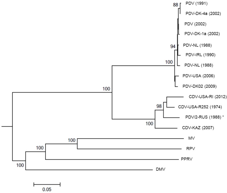
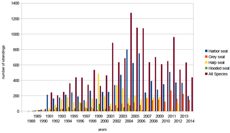
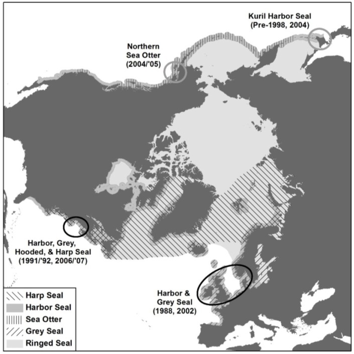
Phocine distemper virus (PDV) was first recognized in 1988 following a massive epidemic in harbor and grey seals in north-western Europe. Since then, the epidemiology of infection in North Atlantic and Arctic pinnipeds has been investigated. In the western North Atlantic endemic infection in harp and grey seals predates the European epidemic, with relatively small, localized mortality events occurring primarily in harbor seals. By contrast, PDV seems not to have become established in European harbor seals following the 1988 epidemic and a second event of similar magnitude and extent occurred in 2002. PDV is a distinct species within the Morbillivirus genus with minor sequence variation between outbreaks over time. There is now mounting evidence of PDV-like viruses in the North Pacific/Western Arctic with serological and molecular evidence of infection in pinnipeds and sea otters. However, despite the absence of associated mortality in the region, there is concern that the virus may infect the large Pacific harbor seal and northern elephant seal populations or the endangered Hawaiian monk seals. Here, we review the current state of knowledge on PDV with particular focus on developments in diagnostics, pathogenesis, immune response, vaccine development, phylogenetics and modeling over the past 20 years.


Figures

Figure 1
Figure 1
Evolutionary relationships of phocine distemper virus (PDV) isolates to the other morbilliviruses. The neighbor-joining method [46] was used to generate a phylogenetic tree based on the hemagglutinin (H) glycoprotein sequences of seventeen selected PDV, CDV, MV, RPV, PPRV and DMV strains (branch length 1.59). Positions containing gaps were eliminated. Bootstrap analysis [47] was used to indicate the percentage number of trees from 1000 replicates in which the virus H glycoprotein sequences clustered equivalently. Branch lengths are identical to the evolutionary distances, determined using the p-distance method [48], used to infer the phylogenetic tree. Sequence alignments and phylogenetic analysis was performed using MEGA5 [49]. PDV accession numbers (Z36979.1; AF479276.1; AF479277.1; AF479274.1; D10371.1; KC802221; AJ224707.1; HQ007902.1 and FJ648456.1), PDV/2-RUS (1988) * (X84998.1) is generally accepted to be a CDV isolate although the name has not been changed. CDV accession numbers (HM046486.1, USA-RI and USA-R252, in submission). Other morbilliviruses, accession numbers, MV (HM439386.1), RPV (NC006296.2), PPRV (FJ750563.1) and DMV (FJ648457.1), were included to show the evolutionary distances across the genus. Feline morbillivirus was not included in this analysis as it is questionable if this virus is a true morbillivirus.
Figure 2
Figure 2
(A) Grey seal (Halichoerus grypus) cerebrum showing expanded Virchow Robin space containing lymphocytes. Activated endothelial cell (arrow). H&E stain (B) Grey seal cerebrum with marked non-suppurative meningoencephalitis. H&E stain (C) Grey seal cerebral white matter with focal malacia centrally and peripheral focal hemorrhage. H&E stain (D) Grey seal cerebrum with acidophilic intranuclear inclusion bodies (INIB, arrow) and intracytoplasmic inclusion bodies (ICIB, arrow heads). H&E stain (E) Grey seal hippocampus with acute focal neuronal necrosis. H&E stain (F) Harp seal (Pagophilus groenlandicus) cerebrum with numerous INIBs (arrows). H&E stain; (G) Harp seal cerebrum showing laminar distribution of morbillivirus antigen. Immunohistochemistry (IHC) using primary antibody against the nucleocapsid (N) of canine distemper virus (VMRD, Inc., Pullman, WA, USA); (H) Harp seal cerebrum. Neurons with strong staining of morbillivirus antigen in cytoplasm and nuclei (arrows). IHC stain as described. The grey seal tissues are from a pup that stranded in February 2006 in Maine, USA, with neurological clinical signs [56]. The harp seal tissue is from a juvenile that stranded on Prince Edward Island, Canada, in May 1991 with similar clinical signs [53].
Figure 3
Figure 3
Pinniped strandings by species for the US Atlantic coast for the period 1988 to 2014. The peaks in strandings in 1991/1992; 1998/1999, and from 2004 to the end of 2007 were associated with confirmed PDV infection in harbor [54], harp and hooded seals [55], and grey seals [56].
Figure 4
Figure 4
North polar azimuthal equidistant projection showing the location of 1988 and 2002 PDV epidemics in the northeastern Atlantic (lower right black oval); two mortality events in the northwestern Atlantic in which PDV infection was confirmed (left black oval); putative PDV-associated Kuril seal mortalities, Hokkaido, Japan (upper right grey circle); and presumptive PDV mortality in northern sea otters in southern Alaska (upper left grey circle). Distribution ranges or harbor, grey, harp, ringed seals and sea otters are shown.

References

    1. Grachev M.A., Kumarev V.P., Mamaev L.V., Zorin V.L., Baranova L.V., Denikina N.N., Belikov S.I., Petrov E.A., Kolesnik V.S., Kolesnik R.S., et al. Distemper virus in Baikal seals. Nature. 1989;338:209. doi: 10.1038/338209a0. - DOI - PubMed
    1. Osterhaus A.D.M.E., Groen J., Uytdehaag F.G.C.M., Visser I.K.G., Van De Bildt M.W.G., Bergman A., Klingeborn B. Distemper virus in Baikal seals. Nature. 1989;338:209–210. doi: 10.1038/338209b0. - DOI - PubMed
    1. Heide-Jorgensen M.-P., Harkonen T., Dietz R., Thompson P.M. Retrospective of the 1988 European Seal Epizootic. Dis. Aquat. Org. 1992;13:37–62. doi: 10.3354/dao013037. - DOI
    1. Cosby S.L., Mcquaid S., Duffy N., Lyons C., Rima B.K., Allan G.M., Mccullough S.J., Kennedy S., Smyth J.A., Mcneilly F., et al. Characterization of a seal morbillivirus. Nature. 1988;336:115–116. doi: 10.1038/336115b0. - DOI - PubMed
    1. Mahy B.W.J., Barrett T., Evans S., Anderson E.C., Bostock C.J. Characterization of a seal morbillivirus. Nature. 1988;336:115. doi: 10.1038/336115a0. - DOI - PubMed

Publication types

MeSH terms

LinkOut - more resources