. 2015 Jun;29(6):1390-401.
doi: 10.1038/leu.2014.347. Epub 2014 Dec 23.

A novel patient-derived tumorgraft model with TRAF1-ALK anaplastic large-cell lymphoma translocation

Collaborators, Affiliations

A novel patient-derived tumorgraft model with TRAF1-ALK anaplastic large-cell lymphoma translocation

F Abate et al. Leukemia. 2015 Jun.

Abstract

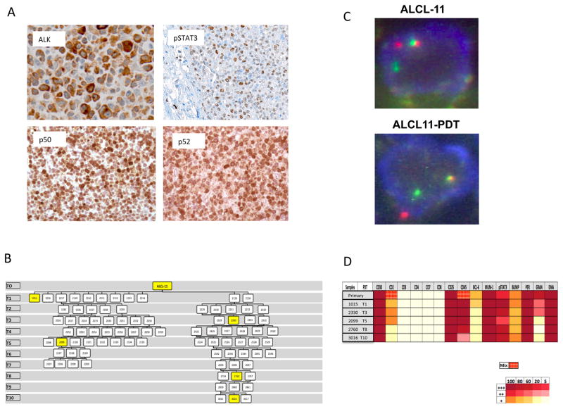
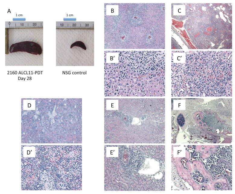
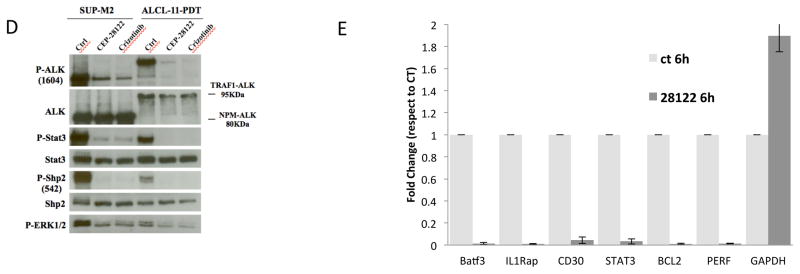
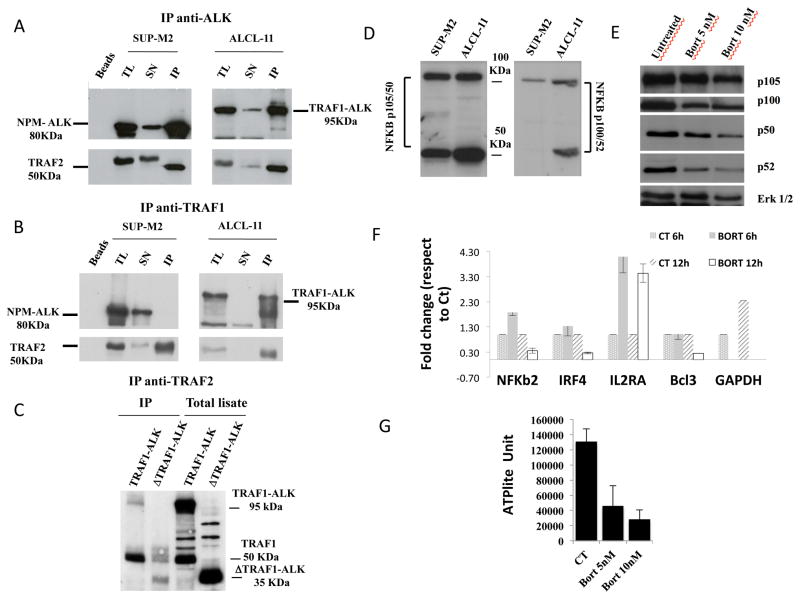
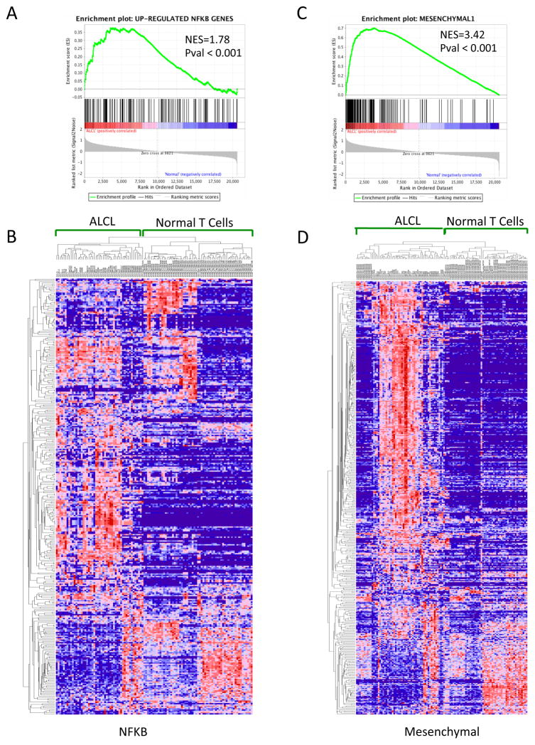
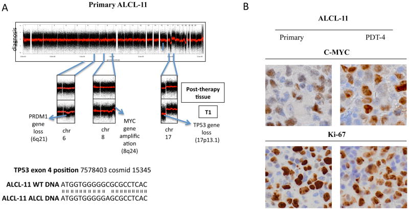
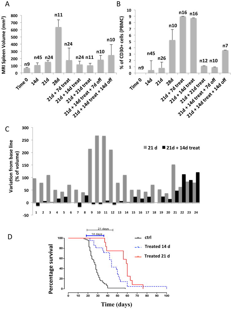
Although anaplastic large-cell lymphomas (ALCL) carrying anaplastic lymphoma kinase (ALK) have a relatively good prognosis, aggressive forms exist. We have identified a novel translocation, causing the fusion of the TRAF1 and ALK genes, in one patient who presented with a leukemic ALK+ ALCL (ALCL-11). To uncover the mechanisms leading to high-grade ALCL, we developed a human patient-derived tumorgraft (hPDT) line. Molecular characterization of primary and PDT cells demonstrated the activation of ALK and nuclear factor kB (NFkB) pathways. Genomic studies of ALCL-11 showed the TP53 loss and the in vivo subclonal expansion of lymphoma cells, lacking PRDM1/Blimp1 and carrying c-MYC gene amplification. The treatment with proteasome inhibitors of TRAF1-ALK cells led to the downregulation of p50/p52 and lymphoma growth inhibition. Moreover, a NFkB gene set classifier stratified ALCL in distinct subsets with different clinical outcome. Although a selective ALK inhibitor (CEP28122) resulted in a significant clinical response of hPDT mice, nevertheless the disease could not be eradicated. These data indicate that the activation of NFkB signaling contributes to the neoplastic phenotype of TRAF1-ALK ALCL. ALCL hPDTs are invaluable tools to validate the role of druggable molecules, predict therapeutic responses and implement patient specific therapies.


Conflict of interest statement

CONFLICT OF INTEREST

Authors must declare to not have any competing financial interests in relation to the work described.

Figures

Fig 1
Fig 1. The ALCL-11 Patient Derived Tumorgraft line mimics its corresponding primary tissues sample
(A) Immunohistochemistry stains performed on primary diagnostic sample (ALCL-11) show a strong ALK cytoplasmic staining with an antibody (D5F3, Cell Signaling Technology) specific for an intracytoplasmic peptide of the human ALK gene (x 400). Tissue sections stained with specific antibody against human phospoSTAT3 (pSTAT3, x100), p50 and p52 demonstrate a variable number lymphoma cells with nuclear positivity (x400). These analyses were performed on the diagnostic/primary tissue sample of the ALCL-11 patient. (B) PDT engraftment and serial tumor propagation in NSG mice. Two distinct engraftments are depicted with their serial passages. Intersecting lines define the relationship among different tumors. Yellow boxes show mice the lymphoma tissue samples used for IHC stains. (C) FISH analysis was performed on the primary tissue and in a representative PDT (ALCL-11 PDT1-2152) sample using a commercial kit and break apart set recognizing the 2p23 regions (Abbott Molecular/Vysis LSI ALK Break Apart FISH Probe Kit). (D) Immunohistochemical stains were performed with specific antibodies recognizing T-cell associated and/or restricted antigens on the primary and serial PDT tissue samples (see Table S1). Percentage of positive cells and intensity of staining are indicated in the above diagram. Broken patterns indicate the presence of heterogeneous staining patterns.
Figure 2
Figure 2. ALCL11 harbors the novel TRAF1-ALK fusion protein and leads to the constitutive activation of ALK signaling pathway
(A) TRAF1–ALK gene fusion was defined by RNAseq (with 29 split reads overlapping junction breakpoint across TRAF1 and ALK). The genomic coordinates are shown. TRAF1 exon 5 and ALK exon 20 reading frames were conserved (central panel). The TRAF1-ALK chimera includes the TRAF1 and Coiled-coil domains, fused to the ALK intra-cytoplasmic region (lower panel). (B) TRAF1-ALK fusion transcripts are detected in primary, leukemic and ALCL11-PDT samples by RT-PCR. ALK + (SUP-M2) and ALK- (MAC1) lines were used as controls. Negative controls correspond to samples lacking cDNA templates. (C) TRAF1 BA assay (see supplementary T2S) was designed to validate molecular results by FISH. Case NE 433870L showed a normal hybridization pattern of TRAF1 BA in all analyzed interphase cells. To test if the TRAF1-ALK fusion might occur by an insertion of the 3′ALK within the TRAF1 locus in case 2, we applied probes covering the 5′end of TRAF1 (SO-labeled) and the 3′end of ALK (SG-labeled) (Upper right panel). Examination with MYC, TP53 and BLIMP1 of NE 433870L diagnostic tissue showed a normal hybridization pattern for all three probes (data not shown). ALCL-11 revealed split TRAF1 BA signals in approximately 50% of interphase cells (Lower panel); (D) TRAF1-ALK signaling is inhibited by anti-ALK inhibitors. PDT cells (PDT-3-2330) and SUP-M2 as control were treated in vitro (6hr, Crizotinib or CEP28122, 200nM). Total cell lysates were immune-blotted with the indicated antibodies. (E) Panel shows the normalized levels of mRNA expression of STAT3 and representative STAT3-responsive genes in untreated or (anti-ALK) CEP28122 treated ALCL-11 leukemic cells (200nM, 6hr treatment). Expression levels were determined using a qRT-PCR approach as previously described {Piva, 2010 #8623;Agnelli, 2012 #599}. Data are depicted as 2^ddCt.5. Expression levels GAPDH of untreated (reference value) and CEP28122 treated cells are reported.
Figure 3
Figure 3. TRAF1-ALK leads to the constitutive activation of NFkB transcription members
(A–B) Lysates from a representative NPM-ALK cell line (SUP-M2 and Karpas 299) and leukemic ALCL-11 cells were immune-precipitated with an anti-ALK (Panel A) and anti-TRAF1 (B) antibodies. Immuno-complexes were resolved and then blotted with the indicated antibodies. Immuno-precipitates in presence of mouse serum are depicted as well (Beads). TL: Total Lysates, SN: Supernatant and IP Immune Precipitation. Protein molecular weights are shown. (C) Lysates from HEK293T cells, transfected with the full length TRAF1-ALK and ΔTRAF1-ALK, were immuno-precipitated with an anti-TRAF2 antibodies. Immuno-complexes were resolved and blotted with anti-TRAF1 antibodies. The expression of the corresponding proteins prior the immune-precipitation is shown (Total lysate) Predicted protein molecular weights are indicated. (D) ALCL-11 cells show constitutive activation of the NFkB pathway. Primary ALCL-11 cells were immune-blotted with antibodies recognizing p105/p50 and p100/p52 NFkB. NPM-ALK+ ALCL cell lines were used as control. (E) Luciferase Expression of transfected HEK-293T cells. Cells were transfected with the indicated cassettes expressing CD30, NFkB-ROS, CD30+NPM-ALK (active form), CD30+K210R NPM_ALK (inactive form) and CD30+ EML4-ALK. (F) Knock-down of p50 or p52 impairs the NFkB mediated luciferase expression via CD30 signaling. HEK-293T cells were transfected with the indicated cassettes in presence of a CD30 expression vector. (G) Protein expression of untreated and treated (Bortezomib 5nM/10nM) ALCL-11-PDTs was determined at 36 hr by Western Blotting with specific antibodies. (H) Panel shows the normalized levels of mRNA expression of NFkB-regulated genes in untreated (control) or Bortezomib treated ALCL-11 leukemic cells (10nM, at 6 and 12 hr of culture). Expression levels were determined using a qRT-PCR approach as previously described {Piva, 2010 #8623;Agnelli, 2012 #599}. Data are depicted as 2^ddCt. Expression levels GAPDH of 6 hr treated (reference value) and 12 hr treated cells are reported. (J) ALCL-11 cells are sensitive to Bortezomib. Primary cells (1×10⁵/ml) were treated with increasing dose of the drug, overtime. Data have been normalized to control DMSO treated cells. DMSO viability decreased over time with a 30–40% spontaneous cell death at 36hrs.
Figure 4
Figure 4
Figure 5
Figure 5. GSEA identifies distinct subsets of ALCL among PTCL and normal samples
(A and B) Unsupervised hierarchical clustering (NFKB and Mesenchymal1 gene set) classifies ALCL in two distinct groups, respectively NKFBclus1/NKFBclus1 and MesClus1/MesClus2, (Pearson correlation distance measure, pairwise average-linkage clustering) with a significant association between the two groups (pvalue < 0.0001, Fisher exact test) (C and D) Overall survival (OS) analysis of the two molecularly defined clusters for both NFKB and Mesenchymal gene set (M1=Mch1, M2=Mch2, NK1=NFKB1, NK2=NFKB2, see figure S7) in an independent cohort of 46 ALCL. Tables show a significant correlation between the NFKB/Mesenchymal groups and the ALK status in ALCL.
Figure 6
Figure 6. CGH analysis of primary and PDT ALCL-11 show the emergency of subclonal population lacking TP53 and MYC amplification
(A) SNP-array plots of ALCL11-PDT identified a los at 17p13.1 (TP53) and 6q21 (PRDM1) and a gene amplification at 8q24 (MYC). X-axis: chromosome localization and physical mapping; Y-axis: signal indicating copy number status at each locus. DNA sequencing of normal and pathological DNA derived form Patient ALCL-11. (B) IHC stains of primary and PDTs (ALCL-11-PDT-T4) tissue samples by an anti-MYC (Y69, Epitomics, Burlingame, CA) and Ki-67 (MIB-1, Dako Italia S.p.A, Italy) Mab. Original magnification x400.
Figure 7
Figure 7. Single therapy with anti-ALK inhibitor leads to a partial response of ALCL1-11-PDT mice
(A) ALCL-11 PDT2-6 (injected with 1×10⁶ lymphoma cells, i.v) were analyzed using a dedicated MRI unit to determine the volume of the spleen as a surrogate of lymphoma/leukemia burden. Mice were treated with CEP28122, 100mg/kg/BID starting day 21th from the i.v. inoculum and scanned a different intervals (7, 14 or 21 days). A cohort of treated mice (14 days) were then released and followed overtime (7 or 14 days off). Numbers of mice are indicated. (B) ALCL-11 PDT2-6 (injected with 1×10⁶ lymphoma cells, i.v) were bled and blood circulating human CD45+CD30+ cells were determined by flow cytometry. Mice were treated with CEP28122, 100mg/kg/BID as indicate above. Numbers of mice are indicated. (C) Effect of CEP28122 treatment in ALCL-11 PDT. Waterfall plot of mice injected with 1×10⁶ ALCL-11 cells (i.v. day 21, grey bars) and CEP28122 response (day 21+14, back bars) after two weeks of treatment, compared with tumor volume at baseline (day 21), in 24 cases. (D) Overall survival of ALCL-11-PDT treated with anti-ALK (CEP28122). Mice were randomized and treated as indicated with 100mg/kg/BID CEP28122 as indicated.

References

    1. Falini B, Martelli MP. Anaplastic large cell lymphoma: changes in the World Health Organization classification and perspectives for targeted therapy. Haematologica. 2009;94(7):897–900. - PMC - PubMed
    1. Swerdlow SHCE, Harris NL, Jaffe ES, Pileri SA, Stein H, Thiele J, et al. WHO classification of tumors of haemotolopoietic and lymphoid tissues. 4. Stylus Publishing, LLC; Sterling, VA: 2008.
    1. Savage KJ, Harris NL, Vose JM, Ullrich F, Jaffe ES, Connors JM, et al. ALK- anaplastic large-cell lymphoma is clinically and immunophenotypically different from both ALK+ ALCL and peripheral T-cell lymphoma, not otherwise specified: report from the International Peripheral T-Cell Lymphoma Project. Blood. 2008;111(12):5496–5504. - PubMed
    1. Schmitz N, Trumper L, Ziepert M, Nickelsen M, Ho AD, Metzner B, et al. Treatment and prognosis of mature T-cell and NK-cell lymphoma: an analysis of patients with T-cell lymphoma treated in studies of the German High-Grade Non-Hodgkin Lymphoma Study Group. Blood. 2010;116(18):3418–3425. - PubMed
    1. Monaco S, Tsao L, Murty VV, Nandula SV, Donovan V, Oesterheld J, et al. Pediatric ALK+ anaplastic large cell lymphoma with t(3;8)(q26.2;q24) translocation and c-myc rearrangement terminating in a leukemic phase. Am J Hematol. 2007;82(1):59–64. - PubMed

Publication types

MeSH terms