

A systematic analysis of neonatal mouse heart regeneration after apical resection
- PMID: 25533939
- PMCID: PMC4302033
- DOI: 10.1016/j.yjmcc.2014.12.011
A systematic analysis of neonatal mouse heart regeneration after apical resection
Abstract
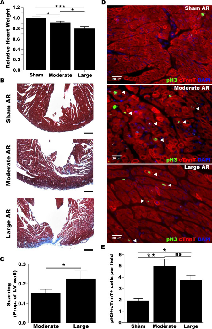
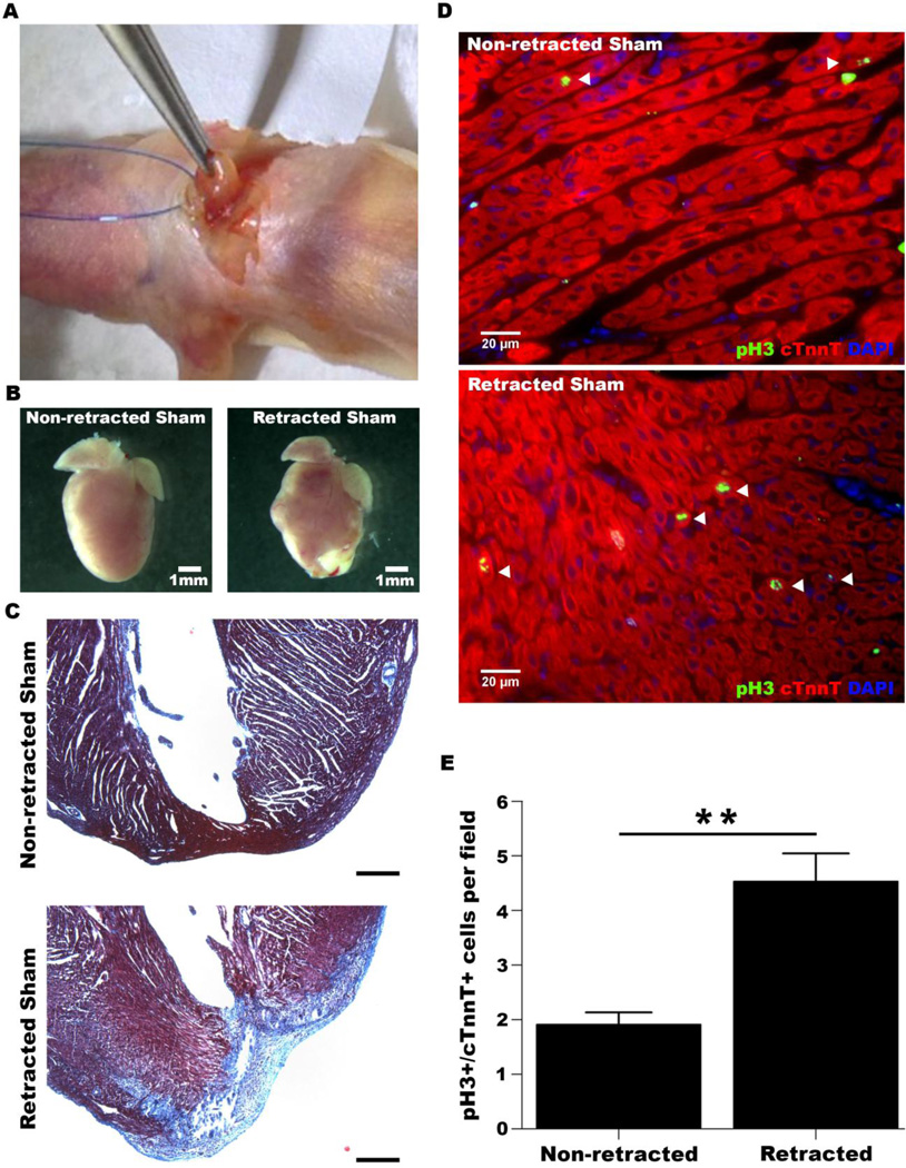
The finding that neonatal mice are able to regenerate myocardium after apical resection has recently been questioned. We determined if heart regeneration is influenced by the size of cardiac resection and whether surgical retraction of the ventricular apex results in an increase in cardiomyocyte cell cycle activity. We performed moderate or large apical ventricular resections on neonatal mice and quantified scar infiltration into the left ventricular wall at 21 days post-surgery. Moderately resected hearts had 15±2% of the wall infiltrated by a collagen scar; significantly greater scar infiltration (23±4%) was observed in hearts with large resections. Resected hearts had higher levels of cardiomyocyte cell cycle activity relative to sham hearts. Surgically retracting the ventricle often resulted in fibrosis and induced cardiomyocyte cell cycle activity that were comparable to that of resected hearts. We conclude that apical resection in neonatal mice induces cardiomyocyte cell cycle activity and neomyogenesis, although scarring can occur. Surgical technique and definition of approach to assessing the extent of regeneration are both critical when using the neonatal mouse apical resection model.
Keywords: Apical resection; Cardiac regeneration; Fibrosis; Neomyogenesis.
Copyright © 2014 Elsevier Ltd. All rights reserved.
Figures
Comment in
-
Cardiac regeneration - Alchemy, science, and a wee bit of magic?J Mol Cell Cardiol. 2015 Apr;81:10-1. doi: 10.1016/j.yjmcc.2015.01.016. Epub 2015 Jan 31. J Mol Cell Cardiol. 2015. PMID: 25647276 No abstract available.
-
Comments to the article "A systematic analysis of neonatal mouse heart regeneration after apical resection".J Mol Cell Cardiol. 2015 May;82:59. doi: 10.1016/j.yjmcc.2015.02.022. Epub 2015 Mar 5. J Mol Cell Cardiol. 2015. PMID: 25746439 No abstract available.
-
Response to "Comment to the article 'A systematic analysis of neonatal mouse heart regeneration after apical resection'".J Mol Cell Cardiol. 2015 May;82:184-5. doi: 10.1016/j.yjmcc.2015.03.003. Epub 2015 Mar 11. J Mol Cell Cardiol. 2015. PMID: 25771145 No abstract available.
References
Publication types
MeSH terms
Grants and funding
LinkOut - more resources
Full Text Sources
Other Literature Sources
Medical
Miscellaneous
