. 2015 Jan 29;517(7536):621-5.
doi: 10.1038/nature14112. Epub 2014 Dec 24.

Lineage-negative progenitors mobilize to regenerate lung epithelium after major injury

Affiliations

Lineage-negative progenitors mobilize to regenerate lung epithelium after major injury

Andrew E Vaughan et al. Nature. .

Abstract

Broadly, tissue regeneration is achieved in two ways: by proliferation of common differentiated cells and/or by deployment of specialized stem/progenitor cells. Which of these pathways applies is both organ- and injury-specific. Current models in the lung posit that epithelial repair can be attributed to cells expressing mature lineage markers. By contrast, here we define the regenerative role of previously uncharacterized, rare lineage-negative epithelial stem/progenitor (LNEP) cells present within normal distal lung. Quiescent LNEPs activate a ΔNp63 (a p63 splice variant) and cytokeratin 5 remodelling program after influenza or bleomycin injury in mice. Activated cells proliferate and migrate widely to occupy heavily injured areas depleted of mature lineages, at which point they differentiate towards mature epithelium. Lineage tracing revealed scant contribution of pre-existing mature epithelial cells in such repair, whereas orthotopic transplantation of LNEPs, isolated by a definitive surface profile identified through single-cell sequencing, directly demonstrated the proliferative capacity and multipotency of this population. LNEPs require Notch signalling to activate the ΔNp63 and cytokeratin 5 program, and subsequent Notch blockade promotes an alveolar cell fate. Persistent Notch signalling after injury led to parenchymal 'micro-honeycombing' (alveolar cysts), indicative of failed regeneration. Lungs from patients with fibrosis show analogous honeycomb cysts with evidence of hyperactive Notch signalling. Our findings indicate that distinct stem/progenitor cell pools repopulate injured tissue depending on the extent of the injury, and the outcomes of regeneration or fibrosis may depend in part on the dynamics of LNEP Notch signalling.


Figures

Extended Data Figure 1
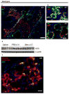
Extended Data Figure 1. Characterization of influenza-induced Krt5+ cells
a–c, Airway (c) and alveolar (a–b) Krt5+ cells strongly express β4 after influenza injury. d, FACS plot of epithelial (EpCam+) cells from tamoxifen-treated Krt5-CreERT2/tdTomato mice at day 15 post-influenza, demonstrating β4 expression in nearly all traced (tdTomato+) cells. e–f, Most Krt5+ cells co-express ΔNp63 (e) and Krt14 (f). g–h, Expanded Krt5+ cells are invariably associated with abundant CD45+ inflammatory cells (g) and few if any remaining normal E-cadherin+ epithelial cells other than the Krt5+ cells themselves (h). i, Krt5+ cells are unlabeled in SPC-CreERT2/mTmG mice. Inset in (i) demonstrates appropriate labeling of type II cells in an uninjured region of the same lung. j–k, Krt5+ cells are not fluorescent after trachea transplantation from tdTomato donor. Basal cells in transplanted section of trachea retained fluorescence (j, inset in k). Scale bars = 100 μm in a and 20 μm in all others.
Extended Data Figure 2
Extended Data Figure 2. Influenza-induced Krt5+ cells arise in both airways and alveoli and migrate across, around, and through airway and parenchymal tissue
a–b, Krt5+ cells are detected in alveoli as early as day 5 and are found in larger clusters over time. c–d, Krt5+ cells similarly arise in airways in greater abundance with time. e, Distinct alveolar and airway expansion is apparent 11 days after infection. f, Freeze-frames of live imaging from a Krt5-CreERT2/tdTomato mouse 11 days post-influenza, wherein tdTomato+ cells migrate from their original location (white box) outward. See Supplementary Video 1a. g, Freeze-frames from a small airway in the same mouse; arrow denotes a single cell crossing the basement membrane. See Supplementary Video 2. Scale bars = 20 μm in a–b and g, 100 μm in all other panels.
Extended Data Figure 3
Extended Data Figure 3. Characterization of bleomycin-induced Krt5+ cells
a–c, β4+ Krt5+ cells also arise after bleomycin injury and express ΔNp63 (b–c). d, Western blotting demonstrating more pronounced and reproducible Krt5 induction after influenza injury than bleomycin at day 11 or 17, respectively. Each lane was loaded with whole lung lysate from a single mouse; average % lung area corresponding to a band in influenza-injured mice is 3.6 ± 0.5% (n=13 mice quantified, see Fig. 3G as an example). e, Lineage tracing of bleomycin-injured Krt5-CreERT2 mice reveal traced (tdTomato+) type II cells expressing SPC and cells morphologically resembling type I cells. 31% of Krt5-Cre traced cells express SPC by day 50 post-bleomycin (n=3 mice, 264 Krt5-CreERT2-labeled cells counted). Scale bars = 100 μm in a and 20 μm in all others. Full scan of western blot in d is available as Supplementary Figure 1.
Extended Data Figure 4
Extended Data Figure 4. Krt5+ cells do not arise from CC10-expressing progenitors but rather upregulate CC10 during expansion
a, Krt5+ cells express detectable levels of CC10 (top) compared to isotype control (bottom) in alveolar clusters (a). b, Representative image of CC10-CreERT2 lineage trace wherein waiting only 7 days after tamoxifen administration prior to influenza injury results in significant labeling of Krt5+ cells (quantified in Figure 1D). c, Strong CC10 expression in Krt5-CreERT2 traced (tdTomato+) cells by day 22 post-influenza. For comparison see single channel images (c′ and c″) of the same region. Scale bars = 20 μm.
Extended Data Figure 5
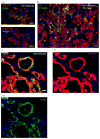
Extended Data Figure 5. Heterogeneity of the LNEP-containing CC10− β4+ population
a, Rare Krt5-CreERT2 traced (tdTomato+) cells were observed in uninjured distal lung airways that lacked Krt5 staining compared to trachea basal cells (inset) in the same section. All distal tdTomato+ cells express ΔNp63 but most ΔNp63+ cells are untraced (see Fig. 2C). b, Cytospins of sorted CC10− β4+ cells reveal the presence of abundant multiciliated cells (green, acetylated tubulin+) and a small fraction of ΔNp63+ cells (red). c, qRT-PCR analysis of mature lineage genes and genes of interest in all populations. n= 3 biological replicates, Mean ± S.D. d, PCA plot of cells sequenced in Fig. 2B, demonstrating that p63+ cells in the CC10− β4+ population (outlined, *) cluster with multi-ciliated cells. e, CD200 is not expressed by FoxJ1-CreERT2-labeled multi-ciliated cells, highlighting its use in excluding such cells. f, Cytospin of Foxj1-CreERT2-labeled β4+ cells demonstrating faithful selection for multi-ciliated cells (198 cells quantified). g, Gating on CD14 expression within the Epcam+ β4+ CD200+ population excludes CC10-expressing club cells. Scale bars = 20 μm.
Extended Data Figure 6
Extended Data Figure 6. Orthotopic transplantation of LNEPs reveals their multipotency and differentiation appropriate to the local microenvironment
a, Several distinct areas of LNEP engraftment (red) reflect differentiation in response to location. Left dashed box demonstrates SPC expression in engrafted cells with nearby endogenous SPC-expressing cells (white); far right dashed box demonstrates Krt5 expression in engrafted cells and nearby endogenous Krt5-expressing cells (green). b–c, Cells in regions of SPC+ differentiation (b) lack Hes1 expression (b′), while those in areas of Krt5+ differentiation (c) strongly express Hes1 (c′). d, Distinct areas of LNEP engraftment demonstrate an inverse relationship between SPC expression (d) and Hes1 expression (d′) in probable single clones. e, Examination of transplanted cells 5 days after engraftment demonstrate abundant Edu incorporation (see Methods) indicative of proliferation. At this time point cells can be identified co-expressing β4 and SPC (e′, circled). f–g, Krt5+ cells and CC10+ cells were often found clustered in single regions of engraftment. h, Many engrafted cells in Fig. 2E are also SPC positive. i, β4- type II cells engraft in small clusters and only express SPC. j–k, CC10+ cells engraft but do not express SPC, CC10, or Krt5. l, Multi-ciliated cells engraft but only persist as isolated single cells, losing acetylated tubulin expression. Scale bars = 100 μm in a and 20 μm in all other panels.
Extended Data Figure 7
Extended Data Figure 7. Transplantation of β4+ CD14+ CD200+ and Krt5-CreERT2-traced cells recapitulates multipotency of the heterogenous CC10− β4+ population
a, Single channels images from Fig. 2H demonstrate Krt5 expression in transplanted β4+ CD14+ CD200+ cells. b–c, Transplanted β4+ CD14+ CD200+ can also differentiate towards type II cells (b) and club cells (c). d–e, Transplantation of rare Krt5-CreERT2-traced cells from uninjured mice resulting in donor-derived Krt5+ cell expansion indistinguishable from endogenous expansion. d–e are representative images from 4 attempted transplants, two of which exhibited engraftment in 2 or 4 individual lobes. Scale bars = 20 μm in all panels.
Extended Data Figure 8
Extended Data Figure 8. Notch activity in normal and injured lung
a, Uninjured Notch reporter mice (Cp-eGFP) show dim GFP in small airways and no detectable GFP in alveoli. b, Krt5+ cells arising in distal airways express GFP in Notch reporter mice 7 days after influenza infection. c–d, Some Krt5+ cells persist within Krt5-CreERT2 labeled (tdTomato+) cysts (d) long-term after influenza injury, while many traced cells express CC10 (c). e, Cysts rarely contain SPC+ type II cells (a′, arrows). f–g, Hes1 expression is maintained in Krt5-CreERT2 traced (GFP+) cysts cells 98 days post-influenza (f) but is absent in normal alveolar parenchyma from the same mice g). h, Representative images of Krt5+ cell expansion in vehicle (h) or DAPT (h′) treated mice at day 11 post-influenza, quantified in Fig. 3G. Scale bars = 20 μm in ag and 100 μm in h.
Extended Data Figure 9
Extended Data Figure 9. IPF and scleroderma lungs both contain Hes1+ honeycomb cysts, but scleroderma lungs also possess SPC/Krt5 co-expressing cells. Normal human lungs contain putative LNEPs and lack Hes1 in alveoli
ad, Honeycomb cysts in several IPF lungs; many Krt5+ cells as well as surrounding cystic epithelium demonstrate strong nuclear Hes1 signal. e, Region of scleroderma honeycombing similar to IPF lung. f, Scleroderma subpleural alveolar region with type II cell hyperplasia demonstrating cells co-expressing SPC and Krt5. g–h, Cystic epithelium in scleroderma lungs expresses Hes1 as in IPF. i, Krt5- ΔNp63+ cells (white outlines) distinct from Krt5+ ΔNp63+ basal cells (red outlines) are present in distal airways. j–k, Hes1 staining is apparent in small airways of normal lung (j) but very low in alveolar parenchyma (k). All images are from patient samples in addition to those shown in Fig. 4. Scale bars = 100 μm in e–f and 20 μm in all otherss.
Extended Data Figure 10
Extended Data Figure 10. Hierarchical cellular responses to injury severity and Notch-regulated LNEP dynamics
a, Distinct epithelial cell types contribute to regeneration depending on the severity of parenchymal injury. Examples of each are referenced. b, Notch signaling regulates the activation, expansion, and differentiation of LNEPs. Notch is required for activation and maintenance of LNEPs. Alveolar differentiation requires subsequent loss of Notch activity, while persistent Notch results in either airway differentiation or abnormal cystic honeycombing.
Figure 1
Figure 1. Injury-induced Krt5+ cells are derived from a lineage-negative precursor
a. Schematic depicting lineage analysis methodology. b–c. Krt5+ cells are untraced (GFP negative) after influenza injury in CC10-CreERT2/mTmG mice. d–e. Quantification of CC10 and SPC lineage tracing, expressed as percentage of cells counted bearing the respective lineage tag (see Methods). Short chase time after tamoxifen administration to CC10-CreERT2 mice results in significant trace in Krt5+ cells (e) (Supplemental Discussion). Means ± S.D., n=7 CC10-CreERT2 and n=3 SPC-CreERT2 mice quantified. f–g, A small fraction of Krt5+ cells bear Krt5-Cre trace (tdTomato+), quantified in (g) (n=3 Krt5-CreERT2 mice) h, Krt5+ cells are not fluorescent after lung transplantation from a wild-type donor into a tdTomato recipient. Non-transplanted lung tissue retained fluorescence (inset). Image representative of n=1 lung transplant. Scale bars = 20 μm. Source data available online.
Figure 2
Figure 2. Isolation and transplantation of a lineage-negative distal epithelial population
a, FACS segregation of epithelial (EpCam+) cells by β4 expression and a CC10-CreERT2 lineage tag (GFP), demonstrating a β4+ population distinct from club cells. b, Hierarchical clustering/heat map of RNA-seq transcriptomes from single CC10− β4+ cells (○) and distal Krt5-CreERT2 traced cells (Δ) (columns). Listed genes (rows) were selected from >1200 differentially expressed genes identified by ANOVA. c, Immunofluorescent staining for ΔNp63 in uninjured lungs from Krt5-CreERT2/tdTomato mice. Single cells from cytospins of the CC10− β4+ population demonstrate primary cilium (green) in a subset of non-multiciliated cells (c′). d, Schematic depicting orthotopic cell transplantation methodology. e, Transplantation of LNEPs combined from eGFP or tdTomato-expressing donors into a single recipient. Most engrafted regions are exclusively GFP+ (green) or tdTomato+ (red) suggesting clonal expansion. f–h, FACS isolation and transplantation of β4+ CD200+ CD14+ LNEPs (f). Transplanted cells differentiate into both SPC+ (g) and Krt5+ (h) cells, representative of n=3 transplants. i, FACS isolation and transplantation of Krt5-CreERT2-labeled LNEPS also differentiate into SPC+ (j) and Krt5+ (k) cells, representative of n=2 transplants. Scale bars = 20 μm except c′=10 μm and e = 100 μm.
Figure 3
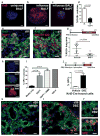
Figure 3. Activation and Krt5 expression by lineage-negative progenitors is Notch-dependent
a–c, LNEP colonies upregulate Krt5 only upon stimulation with BALF from influenza-injured mice (b) (n=6 experiments), a process blocked by γ-secretase inhibition (c) (n=4 experiments), quantified in (d). e–f, Hes1 (e) and Notch1 intracellular domain (f) are present in the nucleus of Krt5+ cells at day 11 indicating Notch activity. g, γ-secretase inhibition during influenza injury reduces Krt5+ cell activation/expansion as measured by fraction of lung section area; each dot = one section, two sections per mouse, n=5 (vehicle) or 4 (DAPT) mice per group. h–i, γ-secretase inhibition induces SPC expression in LNEPs in vitro, quantified in (i) (n=3 experiments). h, bottom panel, Krt5-CreERT2 lineage label could be observed in SPC+ cells after DAPT treatment in vitro. j–l, Representative images of Notch inhibition in vivo via intranasal administration of DBZ results in a significant increase in Krt5-CreERT2-traced SPC+ cells (l) versus labeled cells in vehicle-treated mice (k) post-influenza, quantified in (j) (n= 2 mice/group, >900 cells quantified per mouse in 2 sections from 2 separate lobes). IB = “influenza BALF”. Scale bars = 20 μm. Means ± S.D. Source data for (g) and (j) available online.
Figure 4
Figure 4. Persistent Notch activity promotes cystic honeycombing in both mouse and human
a, Krt5-CreERT2-traced (tdTomato+) cells develop into cystic structures at late time points post-influenza. b, Cyst cells demonstrate nuclear expression of Hes1 indicative of persistent Notch signaling. c, Lung from IPF patient bearing honeycomb cysts with mutually exclusive Krt5+ and SPC+ cells. d, IPF honeycomb cysts with nuclear Hes1 in Krt5+ cells and surrounding epithelium, similar to mouse (b). e, SPC+ type II cells in hyperplastic foci infrequently express Hes1, quantified in (f) (n=8 patients, means ± S.D). g, Scleroderma lung demonstrating sub-pleural Krt5+ and SPC+ cell expansion with many Krt5+/SPC+ double positive cells (right). Scale bars = 20 μm except c where scale bar = 100 μm.

Comment in

References

    1. Clevers H. The Intestinal Crypt, A Prototype Stem Cell Compartment. Cell. 2013;154:274–284. - PubMed
    1. Gurtner GC, Werner S, Barrandon Y, Longaker MT. Wound repair and regeneration. Nature. 2008;453:314–321. - PubMed
    1. Tanimizu N, Mitaka T. Re-evaluation of liver stem/progenitor cells. Organogenesis. 2014;10:0–1. - PMC - PubMed
    1. King RS, Newmark PA. The cell biology of regeneration. The Journal of Cell Biology. 2012;196:553–562. - PMC - PubMed
    1. Desai TJ, Brownfield DG, Krasnow MA. Alveolar progenitor and stem cells in lung development, renewal and cancer. Nature. 2014;507:190–194. - PMC - PubMed

Publication types

MeSH terms

Associated data