Comparative Study
. 2015 Mar:108:124-37.
doi: 10.1016/j.neuroimage.2014.12.039. Epub 2014 Dec 20.

Virtual dissection and comparative connectivity of the superior longitudinal fasciculus in chimpanzees and humans

Affiliations
Comparative Study

Virtual dissection and comparative connectivity of the superior longitudinal fasciculus in chimpanzees and humans

Erin E Hecht et al. Neuroimage. 2015 Mar.

Abstract

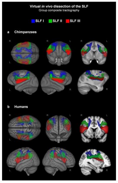
Many of the behavioral capacities that distinguish humans from other primates rely on fronto-parietal circuits. The superior longitudinal fasciculus (SLF) is the primary white matter tract connecting lateral frontal with lateral parietal regions; it is distinct from the arcuate fasciculus, which interconnects the frontal and temporal lobes. Here we report a direct, quantitative comparison of SLF connectivity using virtual in vivo dissection of the SLF in chimpanzees and humans. SLF I, the superior-most branch of the SLF, showed similar patterns of connectivity between humans and chimpanzees, and was proportionally volumetrically larger in chimpanzees. SLF II, the middle branch, and SLF III, the inferior-most branch, showed species differences in frontal connectivity. In humans, SLF II showed greater connectivity with dorsolateral prefrontal cortex, whereas in chimps SLF II showed greater connectivity with the inferior frontal gyrus. SLF III was right-lateralized and proportionally volumetrically larger in humans, and human SLF III showed relatively reduced connectivity with dorsal premotor cortex and greater extension into the anterior inferior frontal gyrus, especially in the right hemisphere. These results have implications for the evolution of fronto-parietal functions including spatial attention to observed actions, social learning, and tool use, and are in line with previous research suggesting a unique role for the right anterior inferior frontal gyrus in the evolution of human fronto-parietal network architecture.

Keywords: Cerebral asymmetry; Diffusion tensor imaging; Evolution; Laterality; Tractography; White matter.


Figures

Figure 1
Figure 1. Portions of SLF I, II, and III visible in the DTI color map before tractography
(a) Coronal slice in representative chimpanzee and human subjects showing all 3 tracts. (b) Parasagittal slices showing each tract. Note the medial-lateral crossing fibers in the inferior frontal sections of SLF II and especially SLF III in humans (white arrow).
Figure 2
Figure 2. SLF tracts in individual subjects
Parasagittal slices in representative chimpanzee and human subjects showing SLF I (top, blue), SLF II (middle, green), and SLF III (bottom, red).
Figure 3
Figure 3. Group composite images of SLF tracts
Results in individual subjects were thresholded at .1% of the waytotal, binarized, registered to template space, and summed, so that in these composite images, intensity corresponds to the number of subjects with above-threshold connectivity at that voxel. Group composite tracts were thresholded to show only above-threshold connectivity common to at least 50% of subjects. (a) Chimpanzees. (b) Humans. The right-most images in each row are 2D slices; the rest are 3D renderings of white matter tracts onto the brain surface.
Figure 4
Figure 4. Quantification of SLF tracts
(a) Regions of interest used to quantify frontal cortical connectivity. (b) Blue, green, and red bands represent proportion of total SLF frontal connectivity from SLF I, II, and III, respectively. Pie charts show connectivity of each frontal region relative to the entire SLF (percent of the entire SLF’s total frontal gray matter terminations). (c) Radar plots show connectivity of each frontal region relative to a particular branch of the SLF (percent of that particular tract’s total frontal gray matter terminations). IFG, inferior frontal gyrus. DLPFC, dorsolateral prefrontal cortex. PMd, dorsal premotor cortex. PMv, ventral premotor cortex. Anatomical boundaries for each ROI are listed in Table 1. Panel a is modified with permission from Hecht et al., J Neurosci 2013 33(35):14117-34.
Figure 5
Figure 5. Quantification of each branch of the SLF in chimpanzees and humans
Proportional volume measurements for each tract are expressed relative to the entire SLF summed across both hemispheres.
Figure 6
Figure 6. Lateralization of the frontal terminations of SLF III
(a) In chimpanzees, the gray matter terminations of SLF III occur mainly in the ventral precentral gyrus in both hemispheres. (b) In humans, the anterior termination of the left SLF III is occurs largely in the pars opercularis of the inferior frontal gyrus, while the right SLF III terminates more anteriorly, in the pars triangularis and pars orbitalis. (c) In chimpanzees, PMv connections outweigh IFG connections in both hemispheres. (d) In humans, IFG connections are significantly greater than PMv connections in both hemispheres.
Figure 7
Figure 7. Diagram of the frontal connectivity of the superior longitudinal fasciculus in chimpanzees and humans
(a) Chimpanzees. (b) Humans. The width of the main body of each tract is proportional to the volume of that tract’s white matter relative to the total white matter of the SLF. The widths of the cortical terminations of each tract are proportional to the volume of gray matter connectivity of that tract within that region relative to the total gray matter connectivity of the SLF. All measurements represent average measurements across both hemispheres, except for the inferior frontal terminations of SLF III, which are depicted separately for the left and right hemisphere. The pattern of SLF I connectivity was similar across species. In SLF II, humans showed more DLPFC connectivity and less IFG connectivity. In SLF III, humans showed more IFG connectivity and less PMd connectivity. Humans also showed a lateralization effect in the inferior frontal terminations of SLF III which was not apparent in chimpanzees, namely, an extension of right SLF III into the more anterior aspects of the inferior frontal gyrus.

References

    1. Andersson JLR, Jenkinson M, Smith S. Non-linear optimisation: FMRIB technical report TR07JA1. 2007 from www.fmrib.ox.ac.uk/analysis/techrep.
    1. Aron AR, Robbins TW, Poldrack RA. Inhibition and the right inferior frontal cortex. Trends Cogn Sci. 2004;8(4):170–177. - PubMed
    1. Aron AR, Robbins TW, Poldrack RA. Inhibition and the right inferior frontal cortex: one decade on. Trends Cogn Sci. 2014 - PubMed
    1. Assmus A, Marshall JC, Ritzl A, Noth J, Zilles K, Fink GR. Left inferior parietal cortex integrates time and space during collision judgments. Neuroimage. 2003;20(Suppl 1):S82–88. - PubMed
    1. Badre D, D’Esposito M. Is the rostro-caudal axis of the frontal lobe hierarchical? Nat Rev Neurosci. 2009;10(9):659–669. - PMC - PubMed

Publication types

LinkOut - more resources