

Comparison of in vivo 3D cone-beam computed tomography tooth volume measurement protocols
- PMID: 25534123
- PMCID: PMC4274349
- DOI: 10.1186/s40510-014-0069-2
Comparison of in vivo 3D cone-beam computed tomography tooth volume measurement protocols
Abstract
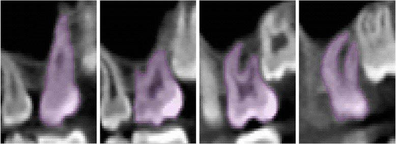
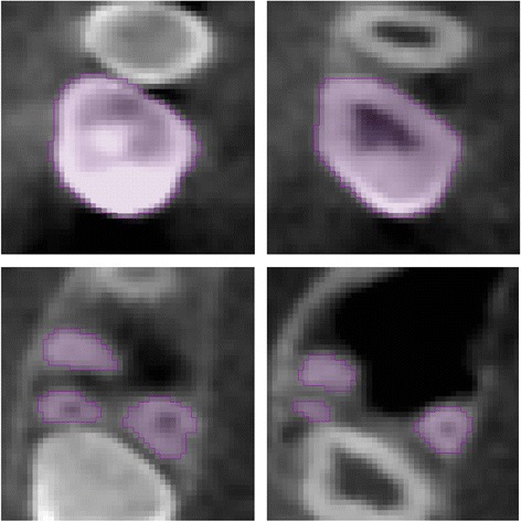
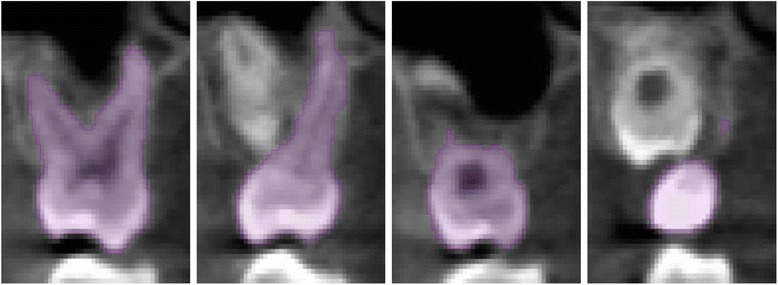
Background: The objective of this study is to analyze a set of previously developed and proposed image segmentation protocols for precision in both intra- and inter-rater reliability for in vivo tooth volume measurements using cone-beam computed tomography (CBCT) images.
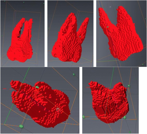
Methods: Six 3D volume segmentation procedures were proposed and tested for intra- and inter-rater reliability to quantify maxillary first molar volumes. Ten randomly selected maxillary first molars were measured in vivo in random order three times with 10 days separation between measurements. Intra- and inter-rater agreement for all segmentation procedures was attained using intra-class correlation coefficient (ICC).
Results: The highest precision was for automated thresholding with manual refinements.
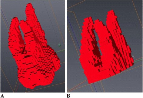
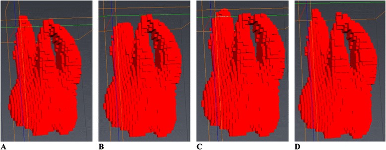
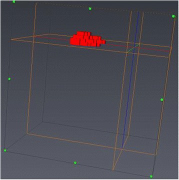
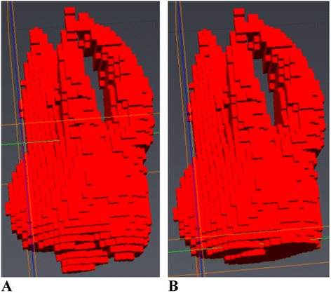
Conclusions: A tooth volume measurement protocol for CBCT images employing automated segmentation with manual human refinement on a 2D slice-by-slice basis in all three planes of space possessed excellent intra- and inter-rater reliability. Three-dimensional volume measurements of the entire tooth structure are more precise than 3D volume measurements of only the dental roots apical to the cemento-enamel junction (CEJ).
Figures
References
-
- de Freitas MR, Beltrao RT, Janson G, Henriques JF, Chiqueto K. Evaluation of root resorption after open bite treatment with and without extractions. Am J Orthod Dentofacial Orthop. 2007;132(2):143.e15–143.e22. - PubMed
Publication types
MeSH terms
LinkOut - more resources
Full Text Sources
Other Literature Sources
