. 2015 Apr;6(2):217-29.
doi: 10.1007/s13244-014-0367-8. Epub 2014 Dec 23.

A practical approach to imaging the axilla

Affiliations

A practical approach to imaging the axilla

V Dialani et al. Insights Imaging. 2015 Apr.

Abstract

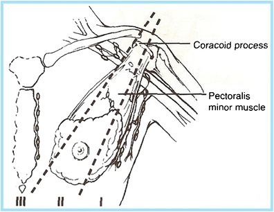
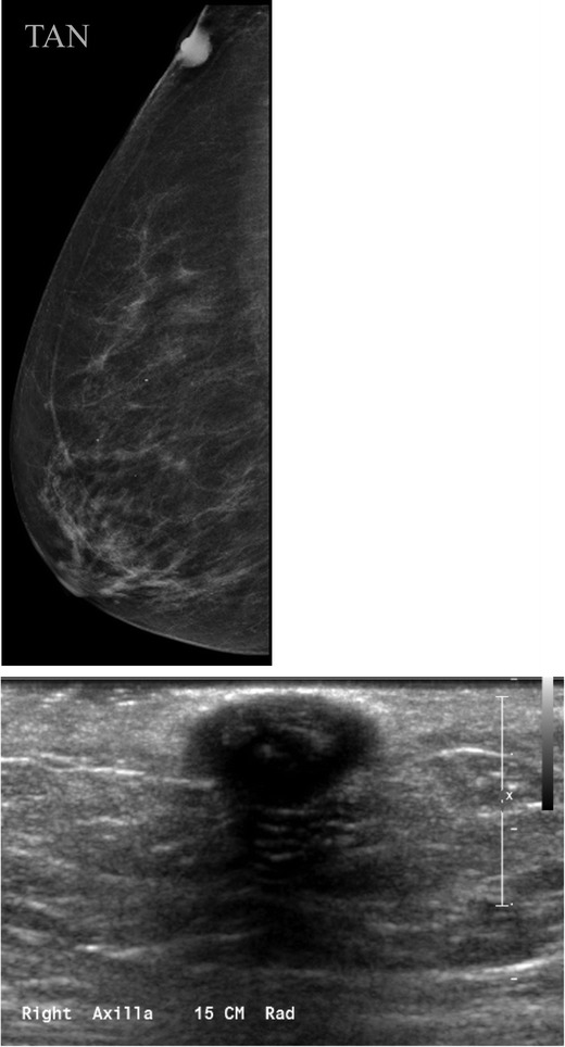
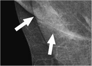
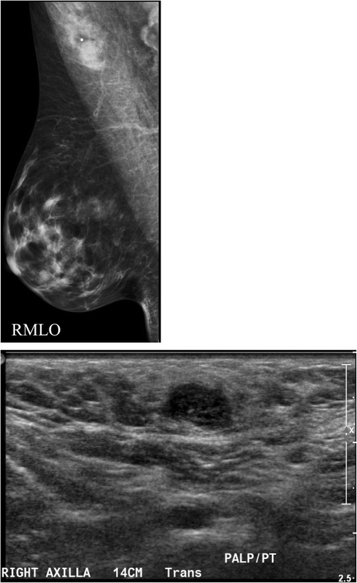
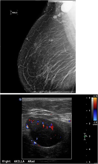
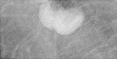
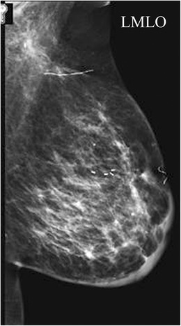
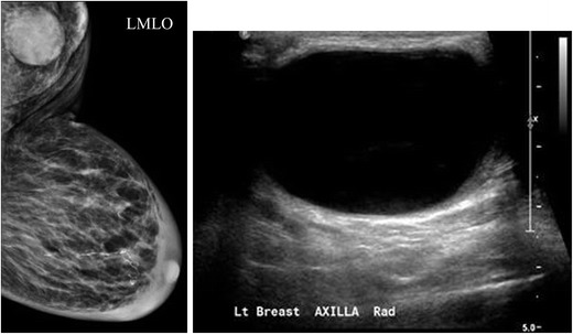
Imaging of the axilla typically occurs when patients present with axillary symptoms or newly diagnosed breast cancer. An awareness of the axillary anatomy is essential in order to generate an accurate differential diagnosis and guide patient management. The purpose of this article is to review the indications for axillary imaging, discuss the logistics of the scanning technique and percutaneous interventions, and present the imaging findings and management of a variety of breast diseases involving the axilla. Teaching points • Knowledge of normal axillary anatomy aids in determining the aetiology of an axillary mass. • The differential diagnosis of an axillary mass is broad and can be subdivided by the location of the lesion. • Imaging evaluation of the axilla usually entails diagnostic mammography and targeted ultrasound. • FNA or core needle biopsies are safe and accurate methods for diagnosis and guiding management.


Figures

Fig. 1
Fig. 1
The axilla is composed of the axillary artery and vein, brachial plexus, lymph nodes, fat, accessory breast tissue, skin and subcutaneous glands. The lymph node-bearing area is divided into three regions: level I: lymph nodes lateral and inferior to the pectoralis minor muscle; level II: lymph nodes under the pectoralis minor muscle; level III: lymph nodes deep and medial to the medial border of the pectoralis minor muscle
Fig. 2
Fig. 2
Sebaceous cyst or epidermal inclusion cyst. a Tangential view reveals that this lesion is located within in the skin. b Ultrasound confirms a hypoechoic mass within the skin
Fig. 3
Fig. 3
Antiperspirant artefact: magnified view shows radiopaque densities overlying the axilla
Fig. 4
Fig. 4
Accessory breast tissue with fibroadenoma: a BB placed on the skin in the area of concern to the patient. MLO views show hetereogeneously dense 4 cm asymmetry of similar density and morphology to breast tissue. b Targeted sonographic evaluation of the right axilla in the area of concern demonstrates fibroglandular breast tissue and a hypoechoic 8-mm solid mass. Biopsy of the nodule using a no-throw vacuum-assisted biopsy device revealed fibroadenoma
Fig. 5
Fig. 5
Normal lymph node: On ultrasound, lymph nodes typically are smooth, gently lobulated ovals with a hypoechoic cortex measuring less than 3 mm in thickness with a central echogenic hilum
Fig. 6
Fig. 6
Abnormal lymph nodes: characteristics concerning for malignancy: a absence of the fatty hilum and b increased focal cortical thickness greater than 3 mm with colour Doppler US that shows hyperaemic blood flow in the hilum and central cortex or abnormal (non-hilar cortical) blood flow
Fig. 7
Fig. 7
A 47-year-old female with a palpable left breast lump. a Left MLO view with magnified image of the left axilla demonstrates a 1.4 × 1-cm lymph node with eccentric cortical thickening and cortical calcifications. b Transverse image of left axillary lymph node confirms echogenic foci within the thickened cortex suspicious for malignancy. FNA was performed with aspirates sent for routine cytology and flow cytometry. Final diagnosis: metastatic carcinoma
Fig. 8
Fig. 8
A 54-year-old female with remote history of right breast cancer and a left knee sarcoma now for screening. a MLO view demonstrates a focally thickened non-enlarged left axillary lymph node. b Ultrasound confirms a 1.4-cm lymph node with focal cortical thickening of 6 mm and (c) internal vascular flow. FNA was performed for diagnosis. Aspirates were sent for routine cytology and flow cytometry. Final diagnosis: reactive lymph node
Fig. 9
Fig. 9
A 55-year-old African American female for screening. a MLO views of the breasts demonstrate a dense enlarged right axillary lymph node and several abnormal-appearing intramammary lymph nodes. Right axillary ultrasound confirms a large fatty replaced lymph node with non-hilar internal vascular flow. This is highly suspicious for malignancy in a patient with breast cancer; however this patient had a negative mammogram and so an ultrasound-guided core biopsy was performed for diagnosis. Final diagnosis: sarcoidosis
Fig. 10
Fig. 10
A 56-year-old female treated with gold therapy for rheumatoid arthritis. Right MLO (magnified image) of two right axillary lymph nodes reveals fine punctate radiopaque densities secondary to previous gold therapy for rheumatoid arthritis
Fig. 11
Fig. 11
A 66-year-old female, 7 months out from lumpectomy, sentinel lymph node biopsy and radiation therapy, presents for first post-treatment mammogram. Diffuse left breast skin and trabecular thickening accompanied by post-surgical distortion in the breast and axilla are visible. Final diagnosis: Expected post-treatment changes following breast cancer diagnosis
Fig. 12
Fig. 12
A 80-year-old female with a history of sarcoidosis presenting with a left axillary mass 1 year following axillary surgery of a benign node. a MLO view and (b) ultrasound of the left axilla demonstrates a 4.1 × 2.7 × 3.5-cm well-circumscribed predominantly anechoic structure with internal debris consistent with a complicated cyst. Aspiration was performed for symptomatic relief. Final diagnosis: lymphocele of the left breast demonstrates extensive skin and trabecular thickening and a well-circumscribed left axillary mass
Fig. 13
Fig. 13
A 50-year-old female with focal 1-cm asymmetry in the left axilla region. a MLO views demonstrate left axillary irregular asymmetry, which persisted on spot compression. b Ultrasound of the left axilla demonstrated a 12-mm irregular hypoechoic hypovascular mass in the left axilla. Ultrasound-guided core needle biopsy was performed. Final diagnosis: granular cell tumour
Fig. 14
Fig. 14
A 64-year-old asymptomatic female presented for a screening mammogram. MLO view of the left breast shows an 8-mm well-defined ovoid mass in the left upper outer quadrant (arrow). Final diagnosis: schwannoma
Fig. 15
Fig. 15
A 43-year-old female presents with a stable left axillary mass for 3–4 years, which the patient reports to fluctuate with her menstrual cycle. MLO and CC views reveal no focal mammographic abnormality (not shown here). b Ultrasound demonstrated a complex oval mass with cystic and solid components lying within the muscle. Colour Doppler analysis demonstrates some peripheral blood flow. c Axial STIR and d axial post-gadolinium sequences demonstrate an ovoid lesion within the latissmus dorsi muscle at the level of the axilla, which was hypointense to muscle on T1-weighted images (not shown here) and shows increased signal intensity on the fluid-sensitive STIR sequence shown here. Post-gadolinium imaging shows some internal enhancement. CT-guided needle biopsy was performed. Final diagnosis: intramuscular myxoma of the latissimus dorsi

References

    1. Ecanow JS, et al. Axillary staging of breast cancer: what the radiologist should know. Radiographics. 2013;33(6):1589–1612. doi: 10.1148/rg.336125060. - DOI - PubMed
    1. Mainiero MB, et al. Axillary ultrasound and fine-needle aspiration in the preoperative evaluation of the breast cancer patient: an algorithm based on tumor size and lymph node appearance. AJR Am J Roentgenol. 2010;195(5):1261–1267. doi: 10.2214/AJR.10.4414. - DOI - PubMed
    1. Krishnamurthy S, et al. Role of ultrasound-guided fine-needle aspiration of indeterminate and suspicious axillary lymph nodes in the initial staging of breast carcinoma. Cancer. 2002;95(5):982–988. doi: 10.1002/cncr.10786. - DOI - PubMed
    1. Damera A, et al. Diagnosis of axillary nodal metastases by ultrasound-guided core biopsy in primary operable breast cancer. Br J Cancer. 2003;89(7):1310–1313. doi: 10.1038/sj.bjc.6601290. - DOI - PMC - PubMed
    1. Abdsaleh S, Azavedo E, Lindgren PG. Ultrasound-guided large needle core biopsy of the axilla. Acta Radiol. 2004;45(2):193–196. doi: 10.1080/02841850410003563. - DOI - PubMed

LinkOut - more resources