

N-linked sugar-regulated protein folding and quality control in the ER
- PMID: 25534658
- PMCID: PMC4474783
- DOI: 10.1016/j.semcdb.2014.12.001
N-linked sugar-regulated protein folding and quality control in the ER
Abstract
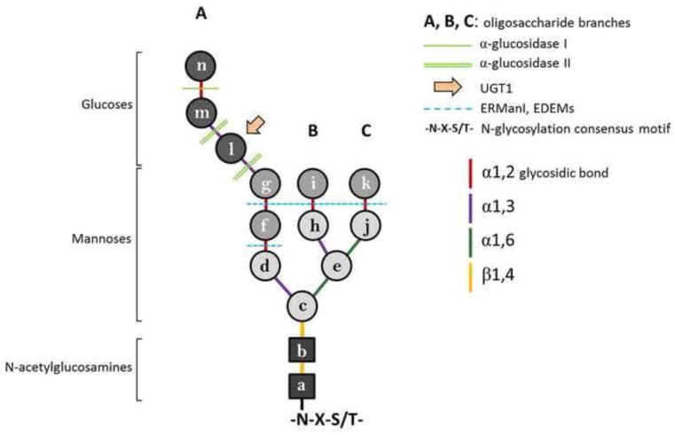
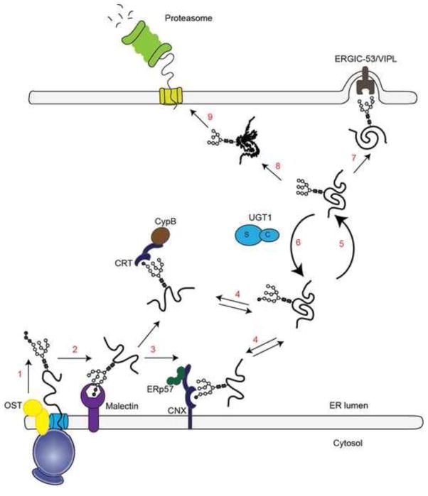
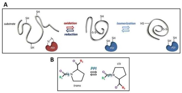
Asparagine-linked glycans (N-glycans) are displayed on the majority of proteins synthesized in the endoplasmic reticulum (ER). Removal of the outermost glucose residue recruits the lectin chaperone malectin possibly involved in a first triage of defective polypeptides. Removal of a second glucose promotes engagement of folding and quality control machineries built around the ER lectin chaperones calnexin (CNX) and calreticulin (CRT) and including oxidoreductases and peptidyl-prolyl isomerases. Deprivation of the last glucose residue dictates the release of N-glycosylated polypeptides from the lectin chaperones. Correctly folded proteins are authorized to leave the ER. Non-native polypeptides are recognized by the ER quality control key player UDP-glucose glycoprotein glucosyltransferase 1 (UGT1), re-glucosylated and re-addressed to the CNX/CRT chaperone binding cycle to provide additional opportunity for the protein to fold in the ER. Failure to attain the native structure determines the selection of the misfolded polypeptides for proteasome-mediated degradation.
Keywords: Calnexin; Calreticulin; Endoplasmic reticulum; N-glycosylation; Protein folding and quality control; UDP-glucose glycoprotein glucosyltransferase 1.
Copyright © 2014 Elsevier Ltd. All rights reserved.
Figures
References
-
- Palade G. Intracellular aspects of the process of protein synthesis. Science. 1975;189:867. - PubMed
-
- Ghaemmaghami S, Huh WK, Bower K, Howson RW, Belle A, Dephoure N, et al. Global analysis of protein expression in yeast. Nature. 2003;425:737–41. - PubMed
-
- Anfinsen CB. Principles that Govern the Folding of Protein chains. Science. 1973;181:223–30. - PubMed
-
- Hartl FU. Molecular chaperones in cellular protein folding. Nature. 1996;381:571–9. - PubMed
Publication types
MeSH terms
Substances
Grants and funding
LinkOut - more resources
Full Text Sources
Other Literature Sources
Research Materials
Miscellaneous
