. 2015 Mar;59(3):1525-33.
doi: 10.1128/AAC.04261-14. Epub 2014 Dec 22.

Protection against Klebsiella pneumoniae using lithium chloride in an intragastric infection model

Affiliations

Protection against Klebsiella pneumoniae using lithium chloride in an intragastric infection model

Nina Tsao et al. Antimicrob Agents Chemother. 2015 Mar.

Abstract

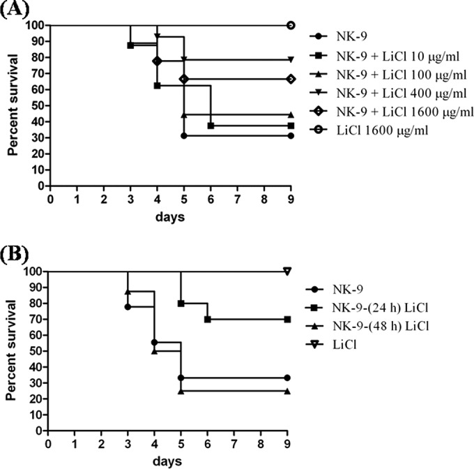
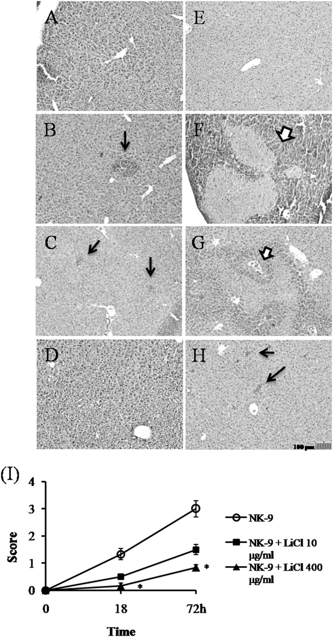
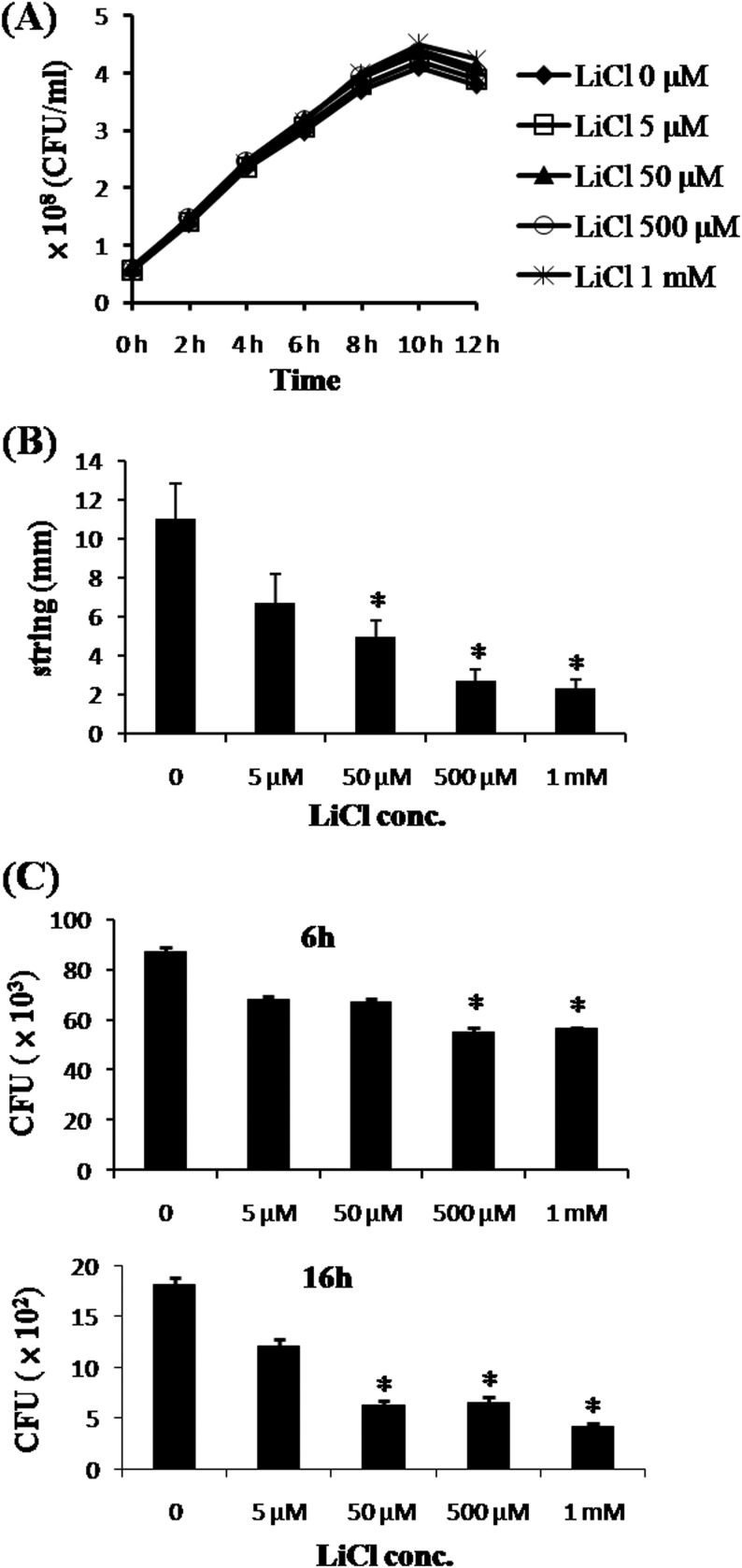
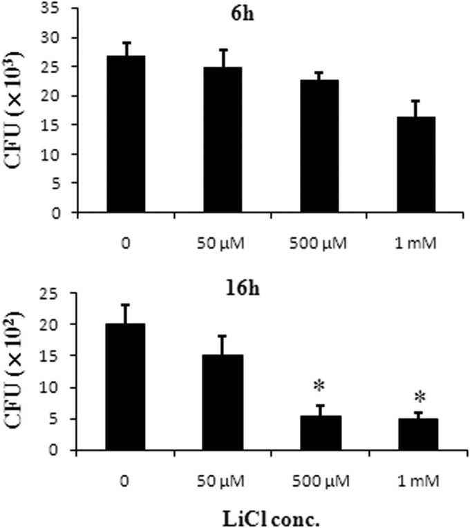
Intragastric Klebsiella pneumoniae infections of mice can cause liver abscesses, necrosis of liver tissues, and bacteremia. Lithium chloride, a widely prescribed drug for bipolar mood disorder, has been reported to possess anti-inflammatory properties. Using an intragastric infection model, the effects of LiCl on K. pneumoniae infections were examined. Providing mice with drinking water containing LiCl immediately after infection protected them from K. pneumoniae-induced death and liver injuries, such as necrosis of liver tissues, as well as increasing blood levels of aspartate aminotransferase and alanine aminotransferase, in a dose-dependent manner. LiCl administered as late as 24 h postinfection still provided protection. Monitoring of the LiCl concentrations in the sera of K. pneumoniae-infected mice showed that approximately 0.33 mM LiCl was the most effective dose for protecting mice against infections, which is lower than the clinically toxic dose of LiCl. Surveys of bacterial counts and cytokine expression levels in LiCl-treated mice revealed that both were effectively inhibited in blood and liver tissues. Using in vitro assays, we found that LiCl (5 μM to 1 mM) did not directly interfere with the growth of K. pneumoniae but made K. pneumoniae cells lose the mucoid phenotype and become more susceptible to macrophage killing. Furthermore, low doses of LiCl also partially enhanced the bactericidal activity of macrophages. Taken together, these data suggest that LiCl is an alternative therapeutic agent for K. pneumoniae-induced liver infections.


Figures

FIG 1
FIG 1
Inhibition of K. pneumoniae-induced death by LiCl. (A) Oral administration of LiCl inhibiting K. pneumoniae NK-9-induced death in B6 mice in a dose-dependent manner. Groups of eight to 16 B6 mice were inoculated, via the intragastric route, with 1 × 10⁹ K. pneumoniae NK-9 cells per mouse. Various doses of LiCl were administered with the drinking water immediately postinfection. LiCl alone (1,600 μg/ml) had no effect on mice. Survival curves were compared for significance using the log rank test for the NK-9 plus LiCl (400 μg/ml) group versus the NK-9 group (P = 0.0087). (B) Therapeutic effects of LiCl against K. pneumoniae NK-9 infections in B6 mice. Groups of eight to 10 B6 mice were inoculated intragastrically with 1 × 10⁹ K. pneumoniae NK-9 cells per mouse. LiCl (400 μg/ml) was administered with the drinking water at 24 h or 48 h postinfection. Survival curves were compared for significance using the log rank test for the NK-9 plus LiCl (24 h postinfection) group versus the NK-9 group (P = 0.048).
FIG 2
FIG 2
Inhibition of K. pneumoniae-induced liver damage by LiCl. Groups of six B6 mice were inoculated intragastrically with 1 × 10⁹ K. pneumoniae NK-9 cells per mouse. Various concentrations of LiCl were administered as described for Fig. 1A. The mice were sacrificed at 18 or 72 h postinfection, and liver sections were prepared as described in Materials and Methods. (A to H) Representative tissue sections. (A) Drinking water only, without K. pneumoniae, with sacrifice at 72 h. (B) Drinking water plus K. pneumoniae, with sacrifice at 18 h. (C) LiCl (10 μg/ml) plus K. pneumoniae, with sacrifice at 18 h. (D) LiCl (400 μg/ml) plus K. pneumoniae, with sacrifice at 18 h. (E) LiCl (400 μg/ml) without K. pneumoniae, with sacrifice at 72 h. (F) Drinking water plus K. pneumoniae, with sacrifice at 72 h. (G) LiCl (10 μg/ml) plus K. pneumoniae, with sacrifice at 72 h. (H) LiCl (400 μg/ml) plus K. pneumoniae, with sacrifice at 72 h. Thick arrows, necrotic regions; thin arrows, liver abscesses. Magnification, ×100. (I) Degree of liver inflammation determined by histological examination, as described in Materials and Methods. *, P < 0.05, compared with K. pneumoniae-treated mice.
FIG 3
FIG 3
In vitro effects of LiCl on K. pneumoniae NK-9 cells. (A) Effects of LiCl on the in vitro growth of K. pneumoniae. K. pneumoniae NK-9 cells (5.8 × 10⁷ CFU/ml) were incubated with various concentrations of LiCl for different times, and the amounts of bacteria at each time point were determined by counting colonies on LB agar plates, as described in Materials and Methods. (B) Effects of LiCl on the mucoid phenotype of K. pneumoniae. K. pneumoniae NK-9 cells were cultured on agar plates containing various concentrations of LiCl, and the mucoid strings of the colonies were determined as described in Materials and Methods. (C) Clearance of LiCl-pretreated K. pneumoniae by macrophages. The same amounts of K. pneumoniae NK-9 cells were pretreated with various concentrations of LiCl for 3 h. Subsequently, LiCl-pretreated K. pneumoniae cells were infected for 2 h with RAW264.7 cells (4 × 10⁵ cells/well) at a ratio of bacteria to RAW264.7 cells of 250:1. After removal of the extracellular bacteria with gentamicin, the live intracellular bacteria were quantitated at 6 or 16 h postinfection, as described in Materials and Methods. *, P < 0.05, compared with the K. pneumoniae-infected group not treated with LiCl.
FIG 4
FIG 4
Effects of LiCl on the bactericidal activity of macrophages. RAW264.7 cells (4 × 10⁵ cells/well) were cocultured for 2 h with K. pneumoniae NK-9 cells at a ratio of bacteria to RAW264.7 cells of 200:1. Subsequently, the free bacteria outside the cells were washed, DMEM containing gentamicin and various concentrations of LiCl was added to the cells, and the cells were cultured for another 4 h or 14 h. At the indicated times, the cells were lysed and the live intracellular bacteria were counted as described in Materials and Methods. *, P < 0.05, compared with the group not treated with LiCl.

References

    1. Podschun R, Ullmann U. 1998. Klebsiella spp. as nosocomial pathogens: epidemiology, taxonomy, typing methods, and pathogenicity factors. Clin Microbiol Rev 11:589–603. - PMC - PubMed
    1. Wang JH, Liu YC, Lee SS, Yen MY, Chen YS, Wang JH, Wann SR, Lin HH. 1998. Primary liver abscess due to Klebsiella pneumoniae in Taiwan. Clin Infect Dis 26:1434–1438. doi:10.1086/516369. - DOI - PubMed
    1. Hu BS, Lau YJ, Shi ZY, Lin YH. 1999. Necrotizing fasciitis associated with Klebsiella pneumoniae liver abscess. Clin Infect Dis 29:1360–1361. doi:10.1086/313471. - DOI - PubMed
    1. Lai YC, Yang SL, Peng HL, Chang HY. 2000. Identification of genes present specifically in a virulent strain of Klebsiella pneumoniae. Infect Immun 68:7149–7151. doi:10.1128/IAI.68.12.7149-7151.2000. - DOI - PMC - PubMed
    1. Lau YJ, Hu BS, Wu WL, Lin YH, Chang HY, Shi ZY. 2000. Identification of a major cluster of Klebsiella pneumoniae isolates from patients with liver abscess in Taiwan. J Clin Microbiol 38:412–414. - PMC - PubMed

Publication types

MeSH terms

LinkOut - more resources