. 2015 Jul;17(7):953-64.
doi: 10.1093/neuonc/nou330. Epub 2014 Dec 21.

Preclinical evaluation of dasatinib alone and in combination with cabozantinib for the treatment of diffuse intrinsic pontine glioma

Affiliations

Preclinical evaluation of dasatinib alone and in combination with cabozantinib for the treatment of diffuse intrinsic pontine glioma

Nathalène Truffaux et al. Neuro Oncol. 2015 Jul.

Abstract

Background: Platelet-derived growth factor receptor A is altered by amplification and/or mutation in diffuse intrinsic pontine glioma (DIPG). We explored in vitro on new DIPG models the efficacy of dasatinib, a multi-tyrosine kinase inhibitor targeting this receptor.

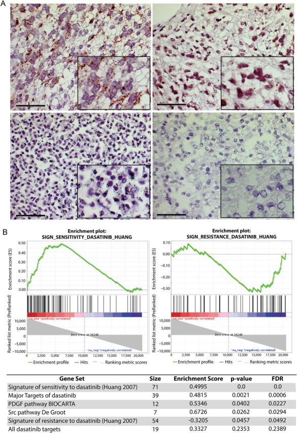
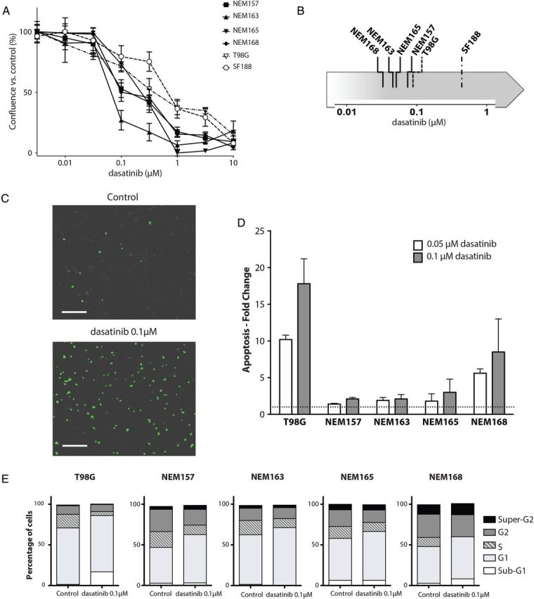
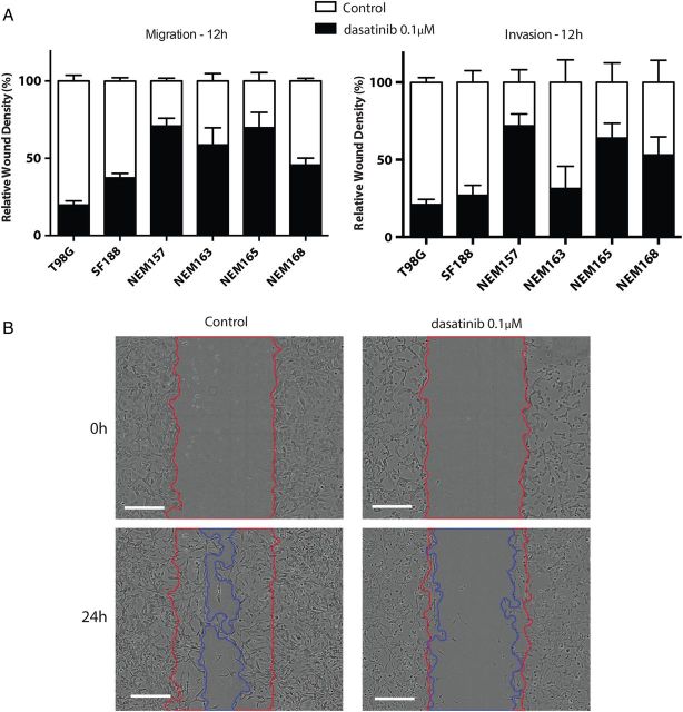
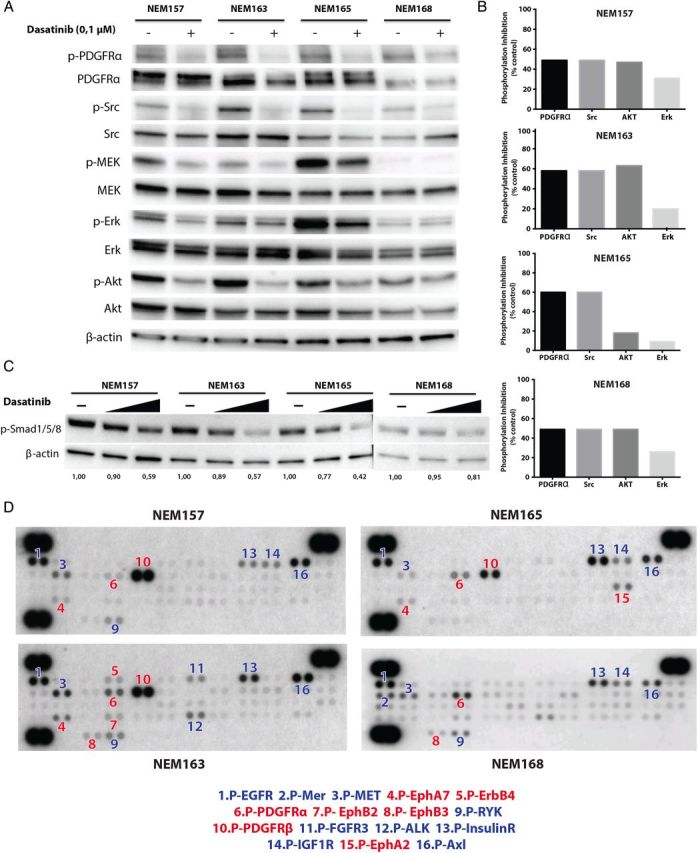
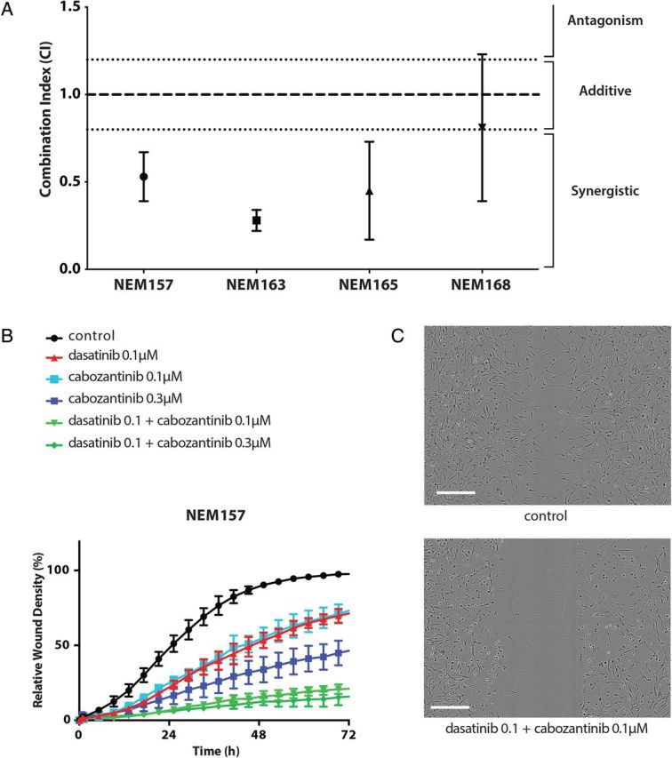
Methods: Gene expression profiles were generated from 41 DIPGs biopsied at diagnosis and compared with the signature associated with sensitivity/resistance to dasatinib. A panel of 12 new DIPG cell lines were established from biopsy at diagnosis, serially passaged, and characterized by gene expression analyses. Effects of dasatinib (1-10 μM) on proliferation, invasion, and cytotoxicity were determined on 4 of these cell lines using live-cell imaging and flow cytometry assays. Downstream signaling and receptor tyrosine kinases (RTKs) were assessed by western blot and phospho-RTK array. The effect of the combination with the c-Met inhibitor cabozantinib was studied on cellular growth and invasion analyzed by the Chou-Talaly method.

Results: DIPG primary tumors and cell lines exhibited the gene expression signature of sensitivity to dasatinib. Dasatinib reduced proliferation (half-maximal inhibitory concentration = 10-100 nM) and invasion (30%-60% reduction) at 100 nM in 4/4 cultures and induced apoptosis in 1 of 4 DIPG cell lines. Activity of downstream effectors of dasatinib targets including activin receptor 1 was strongly reduced. Since multiple RTKs were activated simultaneously in DIPG cell lines, including c-Met, which can be also amplified in DIPG, the benefit of the combination of dasatinib with cabozantinib was explored for its synergistic effects on proliferation and migration/invasion in these cell lines.

Conclusion: Dasatinib exhibits antitumor effects in vitro that could be increased by the combination with another RTK inhibitor targeting c-Met.

Keywords: ACVR1; PDGFRA; Src; brainstem; preclinical model.


Figures

Fig. 1.
Fig. 1.
Frequent activation of PDGFRA and prediction of sensitivity to dasatinib in DIPG patients. (A) Phospho-PDGFRA proximity ligation assay. Upper panels represent tumors from DIPG patients (#4 and #11, described in the Supplementary Table S1). Lower panels represent positive and negative controls. Positive immunochemical staining appears as cytoplasmic brown dots. Scale bar = 50 µM. (B) GSEA plot comparing DIPG gene expression profile with the signature described for sensitivity and resistance to dasatinib (upper panels) and summary of other significant gene sets (FDR ≤ 0.25) related to dasatinib targets (lower panel).
Fig. 2.
Fig. 2.
Effect of dasatinib on DIPG cell line in vitro. (A) DIPG cell lines (NEM), T98G, and SF188 were treated with the indicated doses of dasatinib or vehicle for 3 days and growth was measured using the confluence algorithm. Results are the mean percent inhibition compared with control cells ± SEM of 3 experiments carried out in triplicate. (B) Representative classification of dasatinib sensitivity by IC50 determination for each cell line. (C) Contrast phase fluorescent images of NEM168 12 h posttreatment. Nuclear staining indicates loss of membrane integrity. Scale bar = 300 µM. (D) Cells were plated in complete medium with or without the indicated dose of dasatinib or vehicle for 72 h, stained with annexin V and propidium iodide, and analyzed by flow cytometer, as described in Materials and Methods. Apoptosis detected in the control cells was set to 1 and is represented by the dashed line. Results are the mean ± SD of duplicate representatives of 3 experiments carried out. (E) Distribution of cells in sub-G1, G1, S, G2, and super-G2 phases was determined after propidium iodide staining and by using the Dean-Jet-Fox algorithm in FlowJo software.
Fig. 3.
Fig. 3.
Effect of dasatinib on cell migration and cell invasion. Cells were seeded in 96-well plates and incubated until subconfluent. A wound was scratched across each well (WoundMaker, Essen BioScience), and BD Matrigel was then added (or not for migration assay) with treatment. Wound closure was automatically imaged each 6 h and calculated as a percentage of wound confluence that the cell gained. (A) Quantitative wound-repair analysis at 12 h after dasatinib treatment (0.1 μM) for migration and invasion assay, respectively. (B) Contrast-phase images of NEM168 invasion at 24 h. Red and blue lines correspond to the frontline of the scratch wound at 0 h and 24 h, respectively. Scale bar = 300 µM.
Fig. 4.
Fig. 4.
Targets downstream of dasatinib and potential escape mechanisms. (A) Effect of dasatinib on target activation and downstream signaling was assessed by western blot. Cells were treated with the indicated doses of dasatinib or vehicle for 1 h, in complete medium. Cells were lysed in Tris NaCl EDTA NP40 buffer, and equal protein (30 μg) was analyzed by western blotting on 4%–15% sodium dodecyl sulfate–polyacrylamide electrophoresis gels with the indicated antibodies. (B) Phosphorylation inhibition induced by dasatinib was calculated by densitometric analysis for each DIPG cell line. (C) Effect of dasatinib (1 μM and 10 μM) on Smad1/5/8 signaling. (D) Multiple RTKs were activated simultaneously in glioma cell lines. Whole-cell extracts from the DIPG cell lines were incubated on RTK antibody arrays, and phosphorylation status was determined by subsequent incubation with anti-phosphotyrosine horseradish peroxidase. Each RTK is spotted in duplicate: the pairs of dots in each corner are positive controls. Each pair of positive RTK dots is denoted by a numeral, with the identity of the corresponding RTKs listed below the arrays, in red and blue for dasatinib target or not, respectively. EGFR, epidermal growth factor receptor; FGFR3, fibroblast growth factor receptor 3; ALK, anaplastic lymphoma receptor tyrosine kinase; IGF1R, insulin-like growth factor 1 receptor.
Fig. 5.
Fig. 5.
Synergistic effect of dasatinib and cabozantinib combination in vitro. (A) Synergistic effect at median effect of dasatinib and submicromolar dose of c-Met inhibitor (cabozantinib) (0.75 μM) on cell growth in vitro. Cells were treated with increasing concentrations of drugs either alone or concurrently at their equipotent molar ratio and combination indices (CIs) calculated by the method of Chou and Talaly (CI calculates synergism <0.8; additivity between >0.8 and <1.2; antagonism >1.2. All values are given as mean ± SD of at least 3 independent experiments). (B) Time course of relative wound density of the high invasive NEM157 cell line. (C) Contrast-phase images of NEM157 invasion at 24 h in control and combined condition (upper and lower panel, respectively). Scale bar = 300 µM.

References

    1. Hargrave D, Bartels U, Bouffet E. Diffuse brainstem glioma in children: critical review of clinical trials. Lancet Oncol. 2006;7(3):241–248. - PubMed
    1. Warren KE. Diffuse intrinsic pontine glioma: poised for progress. Front Oncol. 2012;2:205. - PMC - PubMed
    1. Sturm D, Witt H, Hovestadt V, et al. Hotspot mutations in H3F3A and IDH1 define distinct epigenetic and biological subgroups of glioblastoma. Cancer Cell. 2012;22(4):425–437. - PubMed
    1. Paugh BS, Broniscer A, Qu C, et al. Genome-wide analyses identify recurrent amplifications of receptor tyrosine kinases and cell-cycle regulatory genes in diffuse intrinsic pontine glioma. J Clin Oncol. 2011;29(30):3999–4006. - PMC - PubMed
    1. MacDonald TJ. Pediatric glioma: role of platelet derived growth factor receptor. In: Hayat MA, ed., Pediatric Cancer, Vol. 2:Teratoid/Rhabdoid, Brain Tumors, and Glioma. New York: Springer; 2012:259–267.

Publication types

MeSH terms