. 2015 Mar;83(3):942-9.
doi: 10.1128/IAI.02928-14. Epub 2014 Dec 22.

Enterohemorrhagic Escherichia coli colonization of human colonic epithelium in vitro and ex vivo

Affiliations

Enterohemorrhagic Escherichia coli colonization of human colonic epithelium in vitro and ex vivo

Steven B Lewis et al. Infect Immun. 2015 Mar.

Abstract

Enterohemorrhagic Escherichia coli (EHEC) is an important foodborne pathogen causing gastroenteritis and more severe complications, such as hemorrhagic colitis and hemolytic uremic syndrome. Pathology is most pronounced in the colon, but to date there is no direct clinical evidence showing EHEC binding to the colonic epithelium in patients. In this study, we investigated EHEC adherence to the human colon by using in vitro organ culture (IVOC) of colonic biopsy samples and polarized T84 colon carcinoma cells. We show for the first time that EHEC colonizes human colonic biopsy samples by forming typical attaching and effacing (A/E) lesions which are dependent on EHEC type III secretion (T3S) and binding of the outer membrane protein intimin to the translocated intimin receptor (Tir). A/E lesion formation was dependent on oxygen levels and suppressed under oxygen-rich culture conditions routinely used for IVOC. In contrast, EHEC adherence to polarized T84 cells occurred independently of T3S and intimin and did not involve Tir translocation into the host cell membrane. Colonization of neither biopsy samples nor T84 cells was significantly affected by expression of Shiga toxins. Our study suggests that EHEC colonizes and forms stable A/E lesions on the human colon, which are likely to contribute to intestinal pathology during infection. Furthermore, care needs to be taken when using cell culture models, as they might not reflect the in vivo situation.


Figures

FIG 1
FIG 1
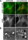
Different adherence phenotypes of EHEC on polarized and nonpolarized T84 cells. Confluent T84 cells on coverslips were infected with strain TUV93-0 for 5 h. Shown are representative images from two independent experiments performed in duplicate. (A) Immunofluorescence staining for actin (green) and E. coli (red). (Top) Merged images; (bottom) actin staining as a separate channel. Bars = 10 μm. (B) Scanning electron micrographs showing EHEC-associated microvillous effacement (white arrows) on polarized cells and actin pedestal formation (black arrows) on nonpolarized cells. Bars = 2 μm.
FIG 2
FIG 2
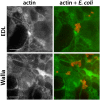
EHEC bacteria do not recruit actin in polarized T84 cells. T84 cells differentiated on Transwell membranes were infected with strain EDL933 or Walla-1 for 5 to 9 h. Immunofluorescence staining for actin (green) and E. coli (red). (Right) Merged images; (left) actin staining as a separate channel. Representative images after 9 h of infection from two independent experiments performed in duplicate. Bars = 5 μm.
FIG 3
FIG 3
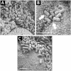
EHEC bacteria colonize human ileal and colonic biopsy samples. Endoscopic biopsy samples from the terminal ileum or transverse colon were infected with EDL933 for 8 h. (A) Scanning electron micrograph showing EHEC bacteria adhering to the terminal ileum and surrounded by elongated microvilli. (B) On the colon, a zone of microvillous effacement (arrows) was evident around adhering bacteria, and adjacent microvilli displayed a normal length. (C) An adherence phenotype similar to that shown in panel B was evident on pediatric colonic biopsy samples. Images are representative of those from three (A and B) and two (C) independent experiments performed in duplicate. Bars = 2 μm.
FIG 4
FIG 4
Colonization of colonic epithelium by EHEC is not affected by Stx production. (A) Scanning electron microscopy of biopsy samples from the transverse colon infected with Stx-negative strain TUV93-0 or 85-170 for 8 h. Images are representative of those from two independent experiments performed in duplicate. Bar = 2 μm. (B) Colonic biopsy samples were infected with wild-type (WT) EDL933 or an isogenic Stx deletion mutant (Δstx) for 8 h. Samples were viewed by scanning electron microscopy, and epithelial colonization was quantified by recording the presence or absence of adherent bacteria in approximately 250 fields of view. Colonization is expressed as the percentage of the fields of view containing adherent bacteria. Data are shown as means ± SEMs from two independent experiments performed in triplicate. (C) Polarized T84 cells were infected with wild-type EDL933 or EDL933 Δstx for 6 h. The numbers of adherent bacteria were quantified by plating serial dilutions of cell lysates and determining the numbers of CFU. Colonization is expressed as the percentage of adherent bacteria relative to the inoculum. Data are shown as means ± SEMs from five independent experiments performed in duplicate.
FIG 5
FIG 5
EHEC colonization of colonic biopsy samples is dependent on intimin and T3S. Scanning electron micrographs of biopsy samples from the transverse colon infected with wild-type (WT) EDL933 or isogenic EspA, EscN, or intimin (eae) mutants for 8 h. Images are representative of those from four independent experiments performed in duplicate. Bars = 10 μm.
FIG 6
FIG 6
EHEC bacteria form typical A/E lesions on human colonic biopsy samples. Colonic biopsy samples were infected with EDL933 for 8 h. Immunofluorescence staining was performed for EspA (A) or Tir (B) in green and E. coli in red. (C) Transmission electron micrograph showing intimate EHEC adherence to host cell membrane and loss of microvilli. Images are representative of those from two independent experiments performed in duplicate. Bars = 2 μm (A, B) or 0.5 μm (C).
FIG 7
FIG 7
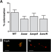
Adherence of EHEC to polarized T84 cells is independent of intimin and T3S and does not involve Tir translocation. (A) Polarized T84 cells were infected with wild-type (WT) EDL933 or isogenic EspA, EscN, or intimin (eae) mutants for 6 h. The numbers of adherent bacteria were quantified by plating serial dilutions of cell lysates and determining the number of CFU. Colonization is expressed as the percentage of adherent bacteria relative to the inoculum. Data are shown as means ± SEMs from four independent experiments performed in duplicate. (B) Immunofluorescence staining of polarized T84 cells infected with EDL933 for 6 h. Green, EspA and Tir; red, E. coli. Images are representative of those from two independent experiments performed in duplicate. Bars = 5 μm.
FIG 8
FIG 8
EHEC A/E lesion formation on colonic biopsy samples is suppressed by oxygen-rich conditions. IVOC of colonic biopsy samples with EDL933 was performed for 8 h under high (oxygen) or atmospheric (air) oxygen levels. Samples were viewed by scanning electron microscopy, and epithelial colonization was quantified by recording the presence or absence of adherent bacteria in approximately 250 fields of view. Colonization is expressed as the percentage of fields of view containing adherent bacteria. Data are shown as means ± SEMs from two independent experiments performed in triplicate. *, P < 0.05.

References

    1. Nataro JP, Kaper JB. 1998. Diarrheagenic Escherichia coli. Clin Microbiol Rev 11:142–201. - PMC - PubMed
    1. Tarr PI, Gordon CA, Chandler WL. 2005. Shiga-toxin-producing Escherichia coli and haemolytic uraemic syndrome. Lancet 365:1073–1086. doi:10.1016/S0140-6736(05)71144-2. - DOI - PubMed
    1. Croxen MA, Law RJ, Scholz R, Keeney KM, Wlodarska M, Finlay BB. 2013. Recent advances in understanding enteric pathogenic Escherichia coli. Clin Microbiol Rev 26:822–880. doi:10.1128/CMR.00022-13. - DOI - PMC - PubMed
    1. Karmali MA, Petric M, Lim C, Fleming PC, Arbus GS, Lior H. 1985. The association between idiopathic hemolytic uremic syndrome and infection by verotoxin-producing Escherichia coli. J Infect Dis 151:775–782. doi:10.1093/infdis/151.5.775. - DOI - PubMed
    1. Bergan J, Dyve Lingelem AB, Simm R, Skotland T, Sandvig K. 2012. Shiga toxins. Toxicon 60:1085–1107. doi:10.1016/j.toxicon.2012.07.016. - DOI - PubMed

Publication types

MeSH terms

LinkOut - more resources