. 2015 Mar;83(3):934-41.
doi: 10.1128/IAI.02768-14. Epub 2014 Dec 22.

Dynamics and establishment of Clostridium difficile infection in the murine gastrointestinal tract

Affiliations

Dynamics and establishment of Clostridium difficile infection in the murine gastrointestinal tract

Mark J Koenigsknecht et al. Infect Immun. 2015 Mar.

Abstract

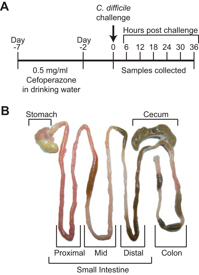
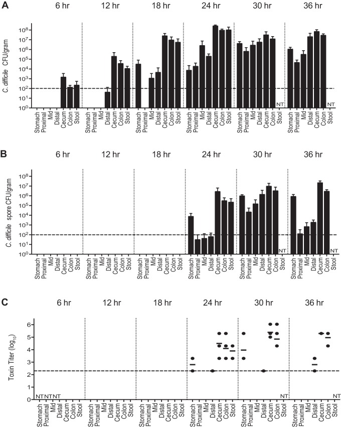
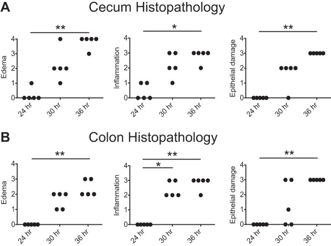
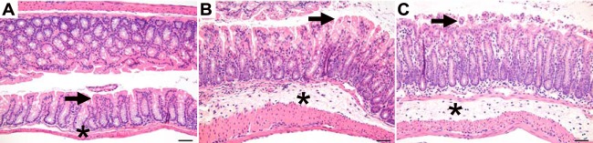
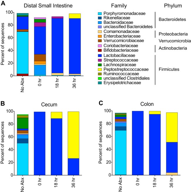
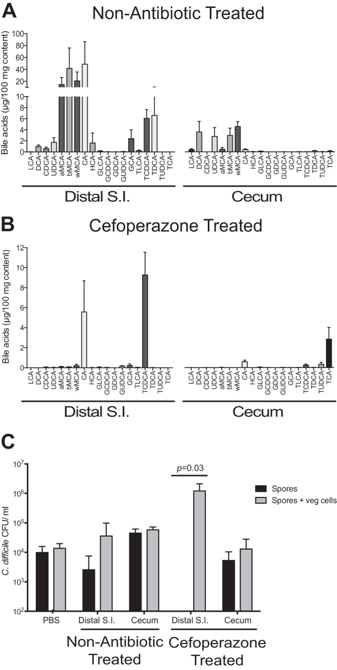
Clostridium difficile infection (CDI) following antibiotic therapy is a major public health threat. While antibiotic disruption of the indigenous microbiota underlies the majority of cases of CDI, the early dynamics of infection in the disturbed intestinal ecosystem are poorly characterized. This study defines the dynamics of infection with C. difficile strain VPI 10463 throughout the gastrointestinal (GI) tract using a murine model of infection. After inducing susceptibility to C. difficile colonization via antibiotic administration, we followed the dynamics of spore germination, colonization, sporulation, toxin activity, and disease progression throughout the GI tract. C. difficile spores were able to germinate within 6 h postchallenge, resulting in the establishment of vegetative bacteria in the distal GI tract. Spores and cytotoxin activity were detected by 24 h postchallenge, and histopathologic colitis developed by 30 h. Within 36 h, all infected mice succumbed to infection. We correlated the establishment of infection with changes in the microbiota and bile acid profile of the small and large intestines. Antibiotic administration resulted in significant changes to the microbiota in the small and large intestines, as well as a significant shift in the abundance of primary and secondary bile acids. Ex vivo analysis suggested the small intestine as the site of spore germination. This study provides an integrated understanding of the timing and location of the events surrounding C. difficile colonization and identifies potential targets for the development of new therapeutic strategies.


Figures

FIG 1
FIG 1
Experimental design for the present study. (A) Timeline of the course of antibiotic treatment and infection. (B) Representative gastrointestinal tract defining which samples were collected and used for study. The small intestine was divided into sections containing proximal, middle, and distal sections as indicated by brackets.
FIG 2
FIG 2
Colonization, spore formation, and cytotoxin activity throughout the GI tract during CDI. (A) Total C. difficile isolated per g of content (n = 5 animals for each group). (B) Number of C. difficile spores isolated per g of content (n = 5 animals for each group). (C) Log10 reciprocal cytotoxin titer per g of content (n = 3 to 5 animals for each group). The dashed line going across the x axis represents the limit of detection. Values not shown are below the limit of detection. NT, not tested. The time denotes hours postchallenge with C. difficile.
FIG 3
FIG 3
Histopathology of the cecum and colon. Histological lesions were categorically scored from 0 (normal) to 4 (most severe) for edema, inflammation, and epithelial damage for the cecum (A) and colon (B). Significance was determined by nonparametric Kruskal-Wallis one-way ANOVA followed by Dunn’s posttest (*, P < 0.05; **, P < 0.005).
FIG 4
FIG 4
Histopathological changes in the colon throughout infection. Representative microscopic images of the colon at 24 h (A), 30 h (B), and 36 h (C) postchallenge are shown. The arrow identifies the epithelial surface, and the asterisk identifies the submucosa. The histological appearance is normal at 24 h (A) and shows submucosal edema, mucosal and submucosal inflammation, and epithelial vacuolation and erosion at 30 and 36 h (B and C). Bar, 50 μm.
FIG 5
FIG 5
Bacterial community membership before and after cefoperazone treatment, as well as during CDI. The bar plots show the mean percent abundances of the top bacterial families (≥1% relative abundance) of the distal small intestine (A), cecum (B), and colon (C). n = 3 to 5 animals for each group. NoAbx, non-antibiotic-treated mice; 0 h, cefoperazone-treated mice prior to C. difficile challenge; 18 h, cefoperazone-treated mice at 18 h postchallenge with C. difficile; 36 h, cefoperazone-treated mice at 36 h postchallenge with C. difficile. The Peptostreptococcaceae family of bacteria includes C. difficile.
FIG 6
FIG 6
Bile acid profile and ex vivo analysis before and after cefoperazone treatment. Bile acids were analyzed by liquid chromatography-mass spectrometry from distal small intestinal (S.I.) and cecal content of non-antibiotic-treated (A) and cefoperazone-treated (B) mice (n = 5 animals for each group). (C) Ex vivo germination and outgrowth of C. difficile spores were measured in distal small intestinal and cecal content from non-antibiotic-treated and cefoperazone-treated mice (n = 4 animals for each group). Black bars depict results for spores alone, and gray bars depict results for both spores and vegetative (veg) cells that were able to outgrow after 6 h of incubation. Significance between groups was determined by using the Mann-Whitney nonparametric t test. Error bars represent the means ± standard errors of the mean.

References

    1. Dubberke ER, Olsen MA. 2012. Burden of Clostridium difficile on the health care system. Clin Infect Dis 55(Suppl 2):S88–S92. doi:10.1093/cid/cis335. - DOI - PMC - PubMed
    1. Rupnik M, Wilcox MH, Gerding DN. 2009. Clostridium difficile infection: new developments in epidemiology and pathogenesis. Nat Rev Microbiol 7:526–536. doi:10.1038/nrmicro2164. - DOI - PubMed
    1. Paredes-Sabja D, Shen A, Sorg JA. 2014. Clostridium difficile spore biology: sporulation, germination, and spore structural proteins. Trends Microbiol 22:406–416. doi:10.1016/j.tim.2014.04.003. - DOI - PMC - PubMed
    1. Britton RA, Young VB. 2012. Interaction between the intestinal microbiota and host in Clostridium difficile colonization resistance. Trends Microbiol 20:313–319. doi:10.1016/j.tim.2012.04.001. - DOI - PMC - PubMed
    1. Buffie CG, Pamer EG. 2013. Microbiota-mediated colonization resistance against intestinal pathogens. Nat Rev Immunol 13:790–801. doi:10.1038/nri3535. - DOI - PMC - PubMed

Publication types

MeSH terms

LinkOut - more resources