Review
. 2015 Feb;94(2):241-51.
doi: 10.1177/0022034514562833. Epub 2014 Dec 22.

Role of dentin MMPs in caries progression and bond stability

Affiliations
Review

Role of dentin MMPs in caries progression and bond stability

A Mazzoni et al. J Dent Res. 2015 Feb.

Abstract

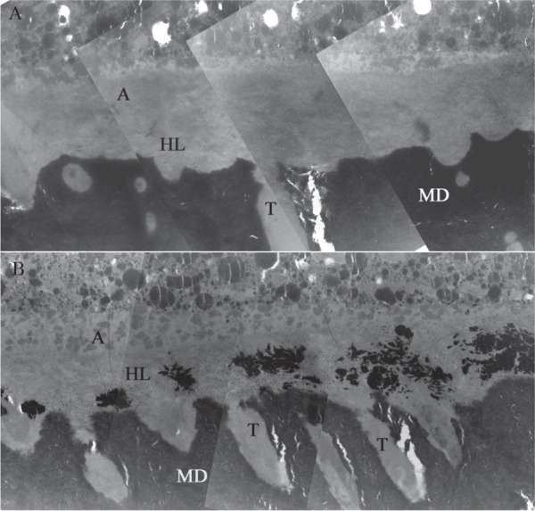
Dentin can be described as a biological composite with collagen matrix embedded with nanosized hydroxyapatite mineral crystallites. Matrix metalloproteinases (MMPs) and cysteine cathepsins are families of endopeptidases. Enzymes of both families are present in dentin and collectively capable of degrading virtually all extracellular matrix components. This review describes these enzymes and their presence in dentin, mainly focusing on their role in dentin caries pathogenesis and loss of collagen in the adhesive hybrid layer under composite restorations. MMPs and cysteine cathepsins present in saliva, mineralized dentin, and/or dentinal fluid may affect the dentin caries process at the early phases of demineralization. Changes in collagen and noncollagenous protein structure may participate in observed decreases in mechanical properties of caries-affected dentin and reduce the ability of caries-affected dentin to remineralize. These endogenous enzymes also remain entrapped within the hybrid layer during the resin infiltration process, and the acidic bonding agents themselves (irrespective of whether they are etch-and-rinse or self-etch) can activate these endogenous protease proforms. Since resin impregnation is frequently incomplete, denuded collagen matrices associated with free water (which serves as a collagen cleavage reagent for these endogenous hydrolase enzymes) can be enzymatically disrupted, finally contributing to the degradation of the hybrid layer. There are multiple in vitro and in vivo reports showing that the longevity of the adhesive interface is increased when nonspecific enzyme-inhibiting strategies are used. Different chemicals (i.e., chlorhexidine, galardin, and benzalkonium chloride) or collagen cross-linker agents have been successfully employed as therapeutic primers in the bonding procedure. In addition, the incorporation of enzyme inhibitors (i.e., quaternary ammonium methacrylates) into the resin blends has been recently promoted. This review will describe MMP functions in caries and hybrid layer degradation and explore the potential therapeutic role of MMP inhibitors for the development of improved intervention strategies for MMP-related oral diseases.

Keywords: cathepsins; collagen; degradation; dentin bonding agents; enzymes; tooth.


Conflict of interest statement

The authors declare no potential conflicts of interest with respect to the authorship and/or publication of this article.

Figures

Figure 1.
Figure 1.
Field emission in-lens scanning electron micrographs (FEI-SEMs) of unfixed, partially decalcified dentin, after a preembedding immunolabeling procedure with monoclonal antibodies for matrix metalloproteinase–2 (MMP-2) or MMP-9. The images were obtained by a combination of secondary electron and backscattered electron signals to simultaneously reveal immunogold labeling and related substrate morphology. Labeling can be identified as electron-dense white spots under the electron beam (pointers). (A, D) Low magnification view (20,000×) of the partially decalcified dentin surface showing open tubular orifices (T) surrounded by a thick collar of fibrillar organic matrix and intertubular porous dentin (ITD). MMP-2 and -9 labeling can be identified as mainly localized in peritubular dentin. (B, E) A higher magnification view (50,000×) of the partially decalcified surface: positive immunohistochemical staining identifying MMP-2 (B) and -9 (E) antibodies located along collagen fibrils. (C, F) High-magnification FEI-SEM micrographs (100,000×), revealing the relationship between MMP-2 and -9 and the collagen meshwork. The specimen shows a moderate labeling for MMP-9 (F) uniformly weaker than MMP-2 staining (C). Reprinted with permission from Mazzoni et al. (2009).
Figure 2.
Figure 2.
Cleavage mechanisms of matrix metalloproteinases (MMPs) and cathepsins. (A) The type I collagen molecule with approximate locations of the MMP cleavage sites (arrows above) and cysteine cathepsin K cleavage sites (arrows below). Arrowheads indicate telopeptidase cleavage activities, resulting in the loss of terminal telopeptides and release of the longer ICTP (by MMPs) and shorter CTX (by cathepsins) fragments. Collagenolytic MMPs always cleave the triple-helical part of the molecule into ¾ N-terminal and ¼ C-terminal fragments. Cysteine cathepsin K has multiple triple-helical cleavage sites, which makes it the most potent mammalian collagenolytic enzyme. (B) Orientation of type I collagen molecules in hard tissues. N- and C-terminal ends of successive molecules are separated by the gap zone, which is the site of intrafibrillar minerals (IF; Bertassoni et al. 2009). (C) The characteristic periodicity of collagen is caused by the overlapping of 4 (gap zone) or 5 (overlap zone) individual molecules. N- and C-terminal telopeptides reside in the overlap zone; therefore, their cleavage by enzymes with telopeptidase activity results in gradual loss of collagen periodicity.
Figure 3.
Figure 3.
Clinical and schematic view of carious dentin zones as they have been described in the literature (upper), and the effect of caries on dentin mechanical properties and wetness (lower). (Upper) Outer caries layer, usually called caries-infected dentin, has lost most of its mineral component, and the collagen matrix structure is decomposed. The tubular structure of the outermost part has disappeared, and the deeper part of this layer has lost its peritubular dentin. The inner caries layer, also called the caries-affected dentin, may have several zones depending on the distance from the lesion surface (upper left). Closer to the surface, the peritubular and intertubular dentin are partially demineralized. The width of the peritubular dentin, the mineral content of the intertubular dentin, and the amount of intratubular mineral crystals increase toward the pulp, and the dentin matrix is believed to be able to remineralize (upper right). While this part of the caries-affected dentin may appear transparent and is sometimes called sclerotic, the overall mineral content is markedly lower than in normal dentin, despite the presence of intratubular mineral crystals. The deepest part of the caries-affected dentin is a narrow subtransparent layer. The subtransparent layer may have little or no demineralization of the intertubular dentin, normal peritubular dentin, and a relatively high level of intratubular mineral crystals. The mineral content and the hardness of the subtransparent layer has been found to be higher than in normal dentin, and it has therefore been described as the “real sclerotic layer” (upper right). It should be noted that the absolute and relative width of the layers may vary significantly between the lesions and even within a lesion, and all the layers are not found in all dentinal caries lesions. (Lower) Shrinkage of normal dentin (A) is independent of water content, while caries-affected dentin (B) demonstrates a highly significant correlation between shrinkage and water content. Note differences in scales for both shrinkage and water content between normal and caries-affected dentin. The increase in water content and shrinkage of caries-affected dentin are dependent on the rate of demineralization, which can vary even within caries-affected dentin. The highly significant correlation between stiffness (D) and water content can also be seen in caries-affected but not in normal dentin (C). Again, note the differences in scales. The graphs demonstrate the problems potentially faced when bonding to caries-infected or -affected dentin: increased wetness, shrinkage following drying, and low mechanical strength. Caries-affected dentin has been shown to contain more detectable matrix metalloproteinases and cysteine cathepsins (Nascimento et al. 2011; Vidal et al. 2014), and poor infiltration of carious dentin (due to tubule occlusion of tubules) during adhesive procedures has also been demonstrated. This result in much unprotected collagen (more than in normal, intact dentin), whose proteases are fully activated collagen-degrading enzymes in and under the hybrid and adhesive layers. Figure 3A and B data adapted from Ito et al. (2005). Spearman bivariate correlation was used for the statistical analysis.
Figure 4.
Figure 4.
Field emission in-lens scanning electron micrographs (FEI-SEM) of sclerotic dentin after double immunolabeling with monoclonal antibodies for type I collagen and proteoglycans. The images were obtained by a combination of secondary electron and back-scattered electron signals. (A) Low magnification view of the surface of sclerotic dentin revealed partially patent tubular orifices that were surrounded by thick collars of peritubular fibrillar structures. Intertubular dentin was highly porous and was inhomogenously covered with the large (30-nm) gold particles used for labeling of antigenically intact collagen fibrils. Gold nanoparticles (15 nm) used for labeling chondroitin sulfate could not be discerned at this magnification. (B) A higher magnification view of the sclerotic intertubular dentin, showing the labeling patterns for type I collagen (solid arrows) and chondroitin sulphate (open arrows). Only a few nanoparticles specific for chondroitin sulfate could be visualized at this level of magnification. (C) Higher magnification view taken from the peritubular regions of sclerotic dentin after immunolabeling. Labeling for collagen fibrils (black arrows) was sparse in this region. A few clusters of 15-nm nanoparticles (open arrowheads) that were bound to the anti-chondroitin sulphate monoclonal antibodies could also be identified. (D) Another specimen showing sparse labeling for antigenically intact type I collagen. Labeling for proteoglycans could not be observed. (E) Very high magnification view of representative sclerotic intertubular dentin specimens after immunolabeling showing a specimen that exhibited moderately intense labeling for type I collagen and proteoglycans. Labeling of type I collagen was represented by the identification of larger (30-nm), discrete gold nanoparticles along the surface of the collagen fibrils (arrows). Labeling of proteoglycans was represented by smaller (15-nm) gold nanoparticles that appeared either as discrete particles or in clusters of 2 to 3 particles (pointers). Collagen banding was infrequently observed on the collagen fibrils in sclerotic dentin and, when present, appeared as very vague surface elevations (open arrowheads). The collagen fibrils appeared collapsed and swollen and exhibited extensive branching when compared with those observed in normal hard dentin (see G, H). (F) A collapsed and swollen collagen network from another specimen of sclerotic dentin that exhibited less intense immunolabeling of both type I collagen and proteoglycans. A banded collagen fibril could be seen in the foreground (open arrowheads; no open arrowheads in F). (G, H) FEI-SEM micrographs of the collagen fibrillar network in normal mineralized dentin after immunolabeling. Collagen fibrils appeared unmodified, with surface cross-banding features (arrow) and gold nanoparticles (pointers) along the fibrils. Gold nanoparticles specific for proteoglycans appeared as clusters of smaller electron-lucent particles around the collagen fibrils (open arrowheads). These clusters could be seen only at high magnification. Reprinted with permission from Suppa et al. (2006).
Figure 5.
Figure 5.
In situ zymogramic views of dentin, hybrid, and adhesive layer showing the endogenous enzymatic activity. (A) Tridimensional model of the acquired image in the green channel of the multiphoton confocal microscope superposed on images obtained with differential interference contrast showing intense fluorescence, produced by gelatin hydrolysis and expression of MMP activity, throughout the entire extension of the hybrid layer created with Scotchbond 1 XT (3M ESPE). (B) Higher magnification image model showing the gelatinolytic activity inside dentinal tubules visible as cylindrical tubes in deep dentin. The high tubule density is related to a very deep dentin portion. R = resin composite; HL = hybrid layer; D = dentin. Reprinted with permission from Mazzoni et al. (2012).
Figure 6.
Figure 6.
Interfacial nanoleakage expression of a hybrid layer created with or without the use of CHX as additional therapeutic primer. (A) Transmission electron microscopy (TEM) image obtained by combining numerous micrographs of a representative specimen treated with 0.2% chlorhexidine for 30 s, then bonded with Scotchbond 1 XT (3M ESPE) and stored for 2 y in artificial saliva at 37 °C. The adhesive (A) interface revealed only very few scattered particles of silver nanoleakage within the hybrid layers (HLs). MD = mineralized dentin; T = dentinal tubules; A = filled adhesive. Bar = 2 µm. (B) TEM image obtained combining numerous micrographs of a representative control specimen bonded with Scotchbond 1 XT and stored for 2 y in artificial saliva at 37 °C. This control adhesive interface reveals extensive interfacial silver nanoleakage due to individual silver grains and large clusters of silver deposits within the collagen fibrils of the HL. The presence of silver deposits reveals water-rich regions where collagen fibrils were hydrolyzed and replaced by water. Reprinted with permission from Breschi, Mazzoni, et al. (2010).

Similar articles

Cited by

References

    1. Arola D. 2008. Fracture and aging of dentine. In: Curtis RV, Watson TF, editors. Dental biomaterials: imaging, testing and modeling, Cambridge (UK): Woodhead Publishing Ltd; p. 314–342.
    1. Bedran-Russo AK, Pauli GF, Chen SN, McAlpine J, Castellan CS, Phansalkar RS, Aguiar TR, Vidal CM, Napotilano JG, Nam JW, et al. 2014. Dentin biomodification: strategies, renewable resources and clinical applications. Dent Mater. 30(1):62–76. - PMC - PubMed
    1. Bertassoni LE, Habelitz S, Kinney JH, Marshall SJ, Marshall GW., Jr. 2009. Biomechanical perspective on the remineralization of dentin. Caries Res. 43(1):70–77. - PMC - PubMed
    1. Boukpessi T, Menashi S, Camoin L, Tencate JM, Goldberg M, Chaussain-Miller C. 2008. The effect of stromelysin-1 (MMP-3) on non-collagenous extracellular matrix proteins of demineralized dentin and the adhesive properties of restorative resins. Biomaterials. 29(33):4367–4373. - PubMed
    1. Boushell LW, Nagaoka H, Nagaoka H, Yamauchi M. 2011. Increased matrix metalloproteinase-2 and bone sialoprotein response to human coronal caries. Caries Res. 45(5):453–459. - PMC - PubMed

Publication types

MeSH terms