

Amelogenins as potential buffers during secretory-stage amelogenesis
- PMID: 25535204
- PMCID: PMC4336156
- DOI: 10.1177/0022034514564186
Amelogenins as potential buffers during secretory-stage amelogenesis
Abstract
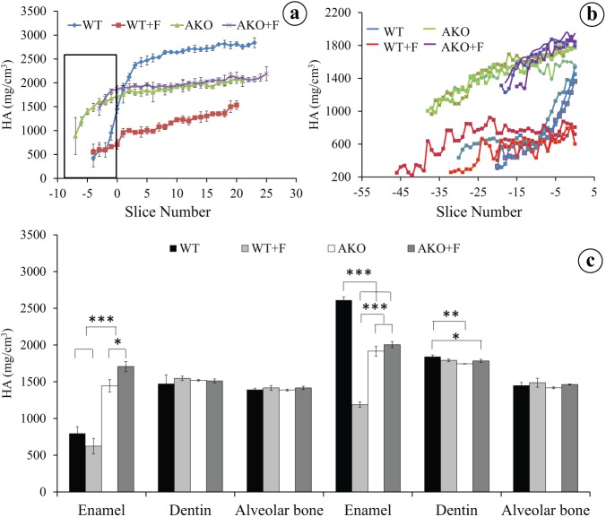
Amelogenins are the most abundant protein species in forming dental enamel, taken to regulate crystal shape and crystal growth. Unprotonated amelogenins can bind protons, suggesting that amelogenins could regulate the pH in enamel in situ. We hypothesized that without amelogenins the enamel would acidify unless ameloblasts were buffered by alternative ways. To investigate this, we measured the mineral and chloride content in incisor enamel of amelogenin-knockout (AmelX(-/-)) mice and determined the pH of enamel by staining with methyl-red. Ameloblasts were immunostained for anion exchanger-2 (Ae2), a transmembrane pH regulator sensitive for acid that secretes bicarbonate in exchange for chloride. The enamel of AmelX(-/-) mice was 10-fold thinner, mineralized in the secretory stage 1.8-fold more than wild-type enamel and containing less chloride (suggesting more bicarbonate secretion). Enamel of AmelX(-/-) mice stained with methyl-red contained no acidic bands in the maturation stage as seen in wild-type enamel. Secretory ameloblasts of AmelX(-/-) mice, but not wild-type mice, were immunopositive for Ae2, and stained more intensely in the maturation stage compared with wild-type mice. Exposure of AmelX(-/-) mice to fluoride enhanced the mineral content in the secretory stage, lowered chloride, and intensified Ae2 immunostaining in the enamel organ in comparison with non-fluorotic mutant teeth. The results suggest that unprotonated amelogenins may regulate the pH of forming enamel in situ. Without amelogenins, Ae2 could compensate for the pH drop associated with crystal formation.
Keywords: SLC4A2; alveolar bone; dentin; enamel; mineral density; pH control.
© International & American Associations for Dental Research 2014.
Conflict of interest statement
The authors declare no potential conflicts of interest with respect to the authorship and/or publication of this article.
Figures


Similar articles
-
Ameloblast Modulation and Transport of Cl⁻, Na⁺, and K⁺ during Amelogenesis.J Dent Res. 2015 Dec;94(12):1740-7. doi: 10.1177/0022034515606900. Epub 2015 Sep 24. J Dent Res. 2015. PMID: 26403673 Free PMC article.
-
Composition of mineralizing incisor enamel in cystic fibrosis transmembrane conductance regulator-deficient mice.Eur J Oral Sci. 2015 Feb;123(1):9-16. doi: 10.1111/eos.12163. Epub 2014 Dec 30. Eur J Oral Sci. 2015. PMID: 25557910 Free PMC article.
-
The anion exchanger Ae2 is required for enamel maturation in mouse teeth.Matrix Biol. 2008 Mar;27(2):119-27. doi: 10.1016/j.matbio.2007.09.006. Epub 2007 Oct 11. Matrix Biol. 2008. PMID: 18042363 Free PMC article.
-
Ion Transport by Ameloblasts during Amelogenesis.J Dent Res. 2017 Mar;96(3):243-253. doi: 10.1177/0022034516681768. Epub 2016 Dec 19. J Dent Res. 2017. PMID: 28221098 Review.
-
Bicarbonate Transport During Enamel Maturation.Calcif Tissue Int. 2017 Nov;101(5):457-464. doi: 10.1007/s00223-017-0311-2. Epub 2017 Aug 9. Calcif Tissue Int. 2017. PMID: 28795233 Free PMC article. Review.
Cited by
-
Ameloblast Modulation and Transport of Cl⁻, Na⁺, and K⁺ during Amelogenesis.J Dent Res. 2015 Dec;94(12):1740-7. doi: 10.1177/0022034515606900. Epub 2015 Sep 24. J Dent Res. 2015. PMID: 26403673 Free PMC article.
-
Cytocompatibility and bioactive potential of AH Plus Bioceramic Sealer: An in vitro study.Int Endod J. 2022 Oct;55(10):1066-1080. doi: 10.1111/iej.13805. Epub 2022 Aug 11. Int Endod J. 2022. PMID: 35950780 Free PMC article.
-
Dose-Dependent Rescue of KO Amelogenin Enamel by Transgenes in Vivo.Front Physiol. 2017 Nov 16;8:932. doi: 10.3389/fphys.2017.00932. eCollection 2017. Front Physiol. 2017. PMID: 29201008 Free PMC article.
-
The Importance of Connexin 43 in Enamel Development and Mineralization.Front Physiol. 2018 Jun 26;9:750. doi: 10.3389/fphys.2018.00750. eCollection 2018. Front Physiol. 2018. PMID: 30013481 Free PMC article.
-
Evaluation of calcium and magnesium contents in tooth enamel without any pathological changes: in vitro preliminary study.Odontology. 2018 Oct;106(4):369-376. doi: 10.1007/s10266-018-0353-6. Epub 2018 Mar 19. Odontology. 2018. PMID: 29556861 Free PMC article.
References
-
- Deutsch D, Haze-Filderman A, Blumenfeld A, Dafni L, Leiser Y, Shay B, Gruenbaum-Cohen Y, Rosenfeld E, Fermon E, Zimmermann B, et al. 2006. Amelogenin, a major structural protein in mineralizing enamel, is also expressed in soft tissues: brain and cells of the hematopoietic system. Eur J Oral Sci. 114(Suppl 1):183–189. - PubMed
Publication types
MeSH terms
Substances
Grants and funding
LinkOut - more resources
Full Text Sources
Other Literature Sources
Molecular Biology Databases
