. 2015 Feb;56(2):423-34.
doi: 10.1194/jlr.M055798. Epub 2014 Dec 22.

Secretion of fatty acid binding protein aP2 from adipocytes through a nonclassical pathway in response to adipocyte lipase activity

Affiliations

Secretion of fatty acid binding protein aP2 from adipocytes through a nonclassical pathway in response to adipocyte lipase activity

Meric Erikci Ertunc et al. J Lipid Res. 2015 Feb.

Abstract

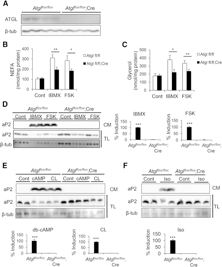
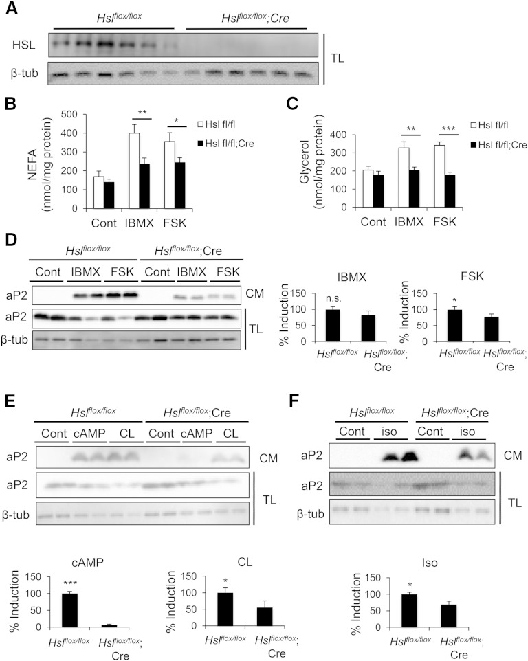
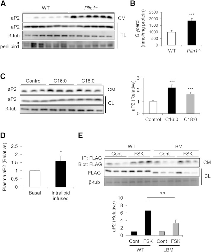
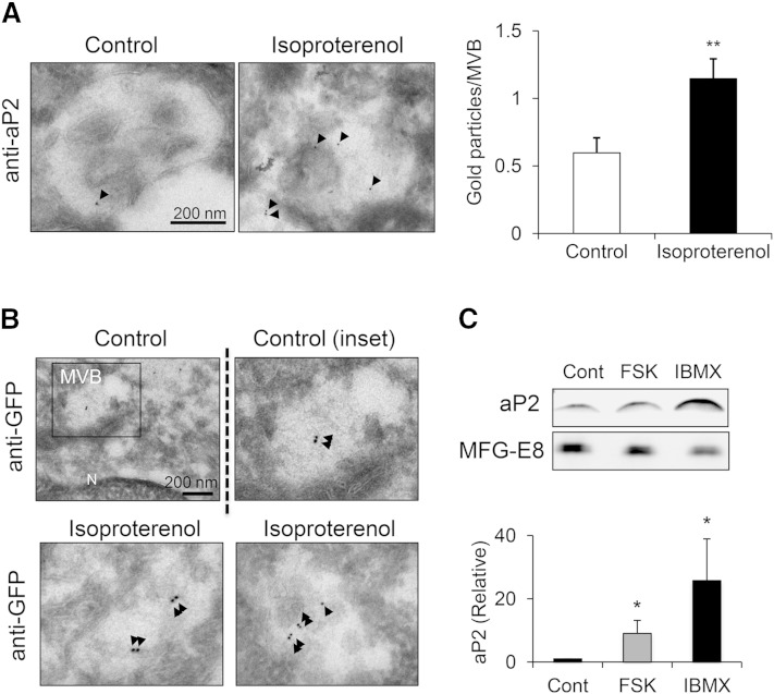
Adipocyte fatty acid binding protein 4, aP2, contributes to the pathogenesis of several common diseases including type 2 diabetes, atherosclerosis, fatty liver disease, asthma, and cancer. Although the biological functions of aP2 have classically been attributed to its intracellular action, recent studies demonstrated that aP2 acts as an adipokine to regulate systemic metabolism. However, the mechanism and regulation of aP2 secretion remain unknown. Here, we demonstrate a specific role for lipase activity in aP2 secretion from adipocytes in vitro and ex vivo. Our results show that chemical inhibition of lipase activity, genetic deficiency of adipose triglyceride lipase and, to a lesser extent, hormone-sensitive lipase blocked aP2 secretion from adipocytes. Increased lipolysis and lipid availability also contributed to aP2 release as determined in perilipin1-deficient adipose tissue explants ex vivo and upon treatment with lipids in vivo and in vitro. In addition, we identify a nonclassical route for aP2 secretion in exosome-like vesicles and show that aP2 is recruited to this pathway upon stimulation of lipolysis. Given the effect of circulating aP2 on glucose metabolism, these data support that targeting aP2 or the lipolysis-dependent secretory pathway may present novel mechanistic and translational opportunities in metabolic disease.

Keywords: adipokine; adipose triglyceride lipase; hormone; hormone-sensitive lipase; lipolysis; obesity.


Figures

Fig. 1.
Fig. 1.
Chemical manipulation of the lipolytic pathway inhibits aP2 secretion. A: Canonical lipolytic pathway and inhibitors used to block steps in this pathway. B–D: Differentiated 3T3-L1 adipocytes were pretreated with the PKA inhibitor H89 (B), ATGL inhibitor Atglistatin (C), HSL inhibitor 76-0079 (D), or HSL-MAGL dual inhibitor CAY10499 (E), followed by IBMX stimulation. Secreted aP2 was measured by Western blot analysis of CM. Glycerol release into CM was measured to assess lipolysis and normalized to total protein content (shown below or at right in each panel). Atgli, Atglistatin; CAY, CAY10499; CL, cell lysate; CM, conditioned media; Cont, control; I, IBMX; 76, 76-0079. Statistical analysis was done using Student’s t-test. * P < 0.05, *** P < 0.001. Bars indicate SEM. Western blots are representative of at least three independent experiments.
Fig. 2.
Fig. 2.
Genetic deficiency of ATGL blocks aP2 secretion ex vivo. A: Confirmation of Atgl deletion in adipose tissue explants from Atglflox/flox;Cre mice compared with Atglflox/flox mice. Cre, Adipoq-Cre. B–F: Atglflox/flox;Cre and control adipose explants were prepared and treated with various lipolytic stimulants. NEFA (B) and glycerol (C) levels in CM were measured and normalized to protein content to assess lipolysis. Secreted aP2 was measured by Western blot analysis of CM upon lipolysis stimulation with IBMX (1 mM) or FSK (20 μM) (D), dibutyryl-cAMP (db-cAMP, 1 mM) or CL 316,243 (CL, 10 μM) (E), or isoproterenol (iso, 10 μM) (F). CM, conditioned medium; TL, tissue lysate. Quantification of % induction for aP2 secretion in CM (presented on the right or bottom of each panel) was calculated based on two to five independent experiments, and representative Western blots are shown. Statistical analysis was done with Student’s t-test. * P < 0.05, ** P < 0.01, *** P < 0.001. Bars indicate SEM.
Fig. 3.
Fig. 3.
Genetic deficiency of HSL decreases aP2 secretion ex vivo. A: Confirmation of Hsl deletion in adipose tissue explants from Hslflox/flox;Cre mice compared with Hslflox/flox mice. Cre, Adipoq-Cre. B–F: Hslflox/flox;Cre and control adipose explants were prepared and treated with various lipolytic stimulants. NEFA (B) and glycerol (C) levels in CM were measured and normalized to protein content to assess lipolysis. Secreted aP2 was measured via Western blot analysis of CM upon lipolysis stimulation with IBMX (1 mM) or FSK (20 μM) (D), dibutyryl-cAMP (db-cAMP, 1 mM) or CL 316,243 (CL, 10 μM) (E), or isoproterenol (iso, 10 μM) (F). CM, conditioned medium; TL, tissue lysate. Quantification of % induction for aP2 secretion in CM (presented on the right or bottom of each panel) was calculated based on three independent experiments, and representative blots are shown. n.s., not significant. Statistical analysis was done with Student’s t-test. * P < 0.05, ** P < 0.01, *** P < 0.001. Bars indicate SEM.
Fig. 4.
Fig. 4.
Free fatty acid availability increases aP2 secretion. A: Perilipin1-deficient (Plin1−/−) and WT adipose explants were prepared and incubated for 16 h under basal conditions. Secreted aP2 was measured via Western blot analysis of CM. Perilipin1 deficiency was confirmed by Western blot analysis of TL. Arrow indicates the perilipin1 band. B: Higher basal lipolysis in Plin1−/− explants was confirmed by measurement of glycerol levels in CM and normalization with protein content. C: Differentiated 3T3-L1 adipocytes were treated with vehicle, 500 µM palmitic acid (C16:0), or stearic acid (C18:0) for 24 h. Secreted aP2 was measured by Western blot analysis of CM. Quantification of aP2 secretion in CM (right panel) was calculated based on at least four independent experiments, and a representative Western blot is shown. D: Plasma was collected from WT mice at the basal state, and after intralipid infusion (5 ml/kg/h) for 5 h, aP2 levels were measured using ELISA (n = 8). E: In-house aP2−/− differentiated adipocytes were electroporated with cytomegalovirus (CMV) promoter-driven FLAG-aP2 expression constructs that are either WT or lipid binding mutant (LBM). The extent of aP2 secretion was tested after lipolysis stimulation with FSK. FLAG-aP2 was immunoprecipitated from CM, and levels were determined by immunoblotting for FLAG. Quantification of aP2 secretion in CM (bottom panel) was calculated based on three independent experiments, and a representative Western blot is shown. CL, cell lysate; CM, conditioned medium; n.s., not significant; TL, tissue lysate. Statistical analysis was done with Student’s t-test. * P < 0.05, *** P < 0.001. Bars indicate SEM.
Fig. 5.
Fig. 5.
aP2 secretion is mediated by an exosome-like pathway. A: Differentiated 3T3-L1 adipocytes were infected with lentivirus to express GFP-fused aP2. Cells were treated with isoproterenol (10 μM) for 1 h and stained with lipidTOX and DAPI, and confocal images were taken. Arrows indicate peripheral localization of aP2 (green: FLAG-tagged-GFP-aP2; red: lipidTOX, lipid droplet; blue, DAPI, nuclear staining). B: Differentiated 3T3-L1 adipocytes were treated with IBMX or vehicle, and a microsomal fraction was prepared at indicated time points. aP2 levels were assessed by Western blotting. PDI is used as microsomal marker. C, control; I, IBMX. C: 3T3-L1 adipocytes infected with lentivirus to express FLAG-tagged-GFP-aP2 were immunogold labeled against GFP and examined by electron microscopy. Negative control is uninfected cells. A lower magnification image is shown in top panel. G, Golgi; M, mitochondria. GFP labeling is indicated with arrows. D and E: EVs isolated from 3T3-L1 adipocyte CM were layered on a continuous sucrose gradient (0.25–2 M), ultracentrifuged for 20 h at 100,000 g for density-based separation. Fractions (1 ml) were collected, and densities were measured with a refractometer (shown on top, g/ml). Fractions were diluted with PBS and ultracentrifuged at 100,000 g to pellet vesicles in the fractions followed by immunoblotting for aP2 or exosomal markers CD63, ALIX, and TSG101 (D) or MFG-E8 and ALIX (E). F: Isolated EVs from WT or FABP−/− (aP2−/−; mal1−/−) differentiated adipocyte CM were mounted on a grid for immunogold labeling against aP2. Arrows indicate some of the aP2 labeling.
Fig. 6.
Fig. 6.
aP2 is recruited to the nonclassical exosome-like pathway upon lipolysis. A: 3T3-L1 adipocytes were treated with isoproterenol (10 μM), fixed, and immunogold labeled for aP2. Right panel: Gold particles per MVB were quantified from at least 62 randomly selected MVBs in each treatment. aP2 labeling is indicated with arrows. B: 3T3-L1 adipocytes expressing FLAG-tagged-GFP-aP2 were treated with isoproterenol (10 μM) or vehicle, immunogold labeled against GFP, and examined by electron microscopy. Lower magnification of control is shown at top left panel. N, nucleus. GFP labeling is indicated with arrows. C: EVs were isolated from 3T3-L1 adipocyte CM treated with vehicle or lipolytic stimuli, and aP2 was measured by Western blot. MFG-E8 was used as vesicular marker. Western blots were quantified (lower panel) using seven independent experiments. * P < 0.05, ** P < 0.01. Bars indicate SEM.

References

    1. Kershaw E. E., Flier J. S. 2004. Adipose tissue as an endocrine organ. J. Clin. Endocrinol. Metab. 89: 2548–2556. - PubMed
    1. Scherer P. E. 2006. Adipose tissue: from lipid storage compartment to endocrine organ. Diabetes. 55: 1537–1545. - PubMed
    1. Furuhashi M., Hotamisligil G. S. 2008. Fatty acid-binding proteins: role in metabolic diseases and potential as drug targets. Nat. Rev. Drug Discov. 7: 489–503. - PMC - PubMed
    1. Hertzel A. V., Bernlohr D. A. 2000. The mammalian fatty acid-binding protein multigene family: molecular and genetic insights into function. Trends Endocrinol. Metab. 11: 175–180. - PubMed
    1. Hotamisligil G. S., Johnson R. S., Distel R. J., Ellis R., Papaioannou V. E., Spiegelman B. M. 1996. Uncoupling of obesity from insulin resistance through a targeted mutation in aP2, the adipocyte fatty acid binding protein. Science. 274: 1377–1379. - PubMed

Publication types