

TAp73 opposes tumor angiogenesis by promoting hypoxia-inducible factor 1α degradation
- PMID: 25535359
- PMCID: PMC4291637
- DOI: 10.1073/pnas.1410609111
TAp73 opposes tumor angiogenesis by promoting hypoxia-inducible factor 1α degradation
Abstract
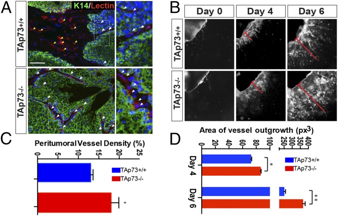
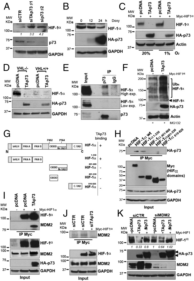
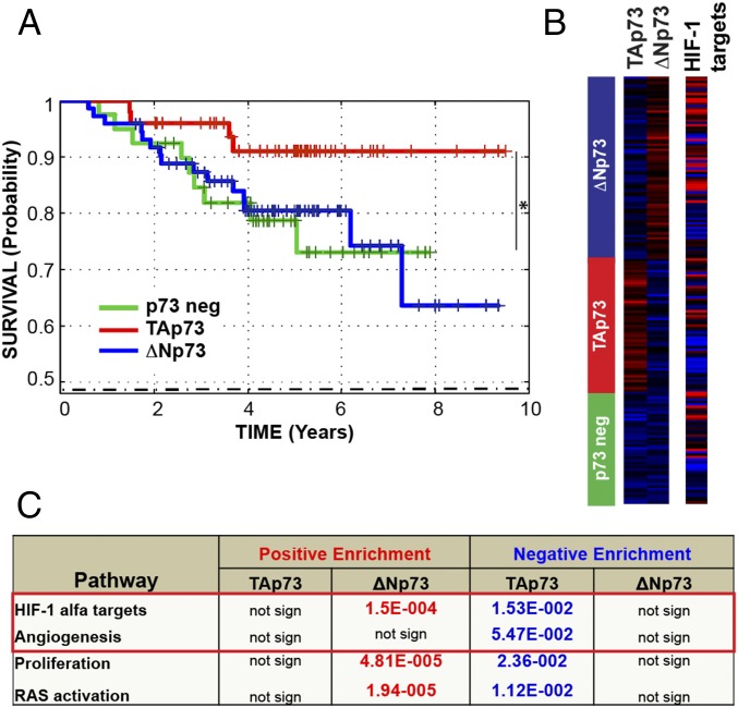
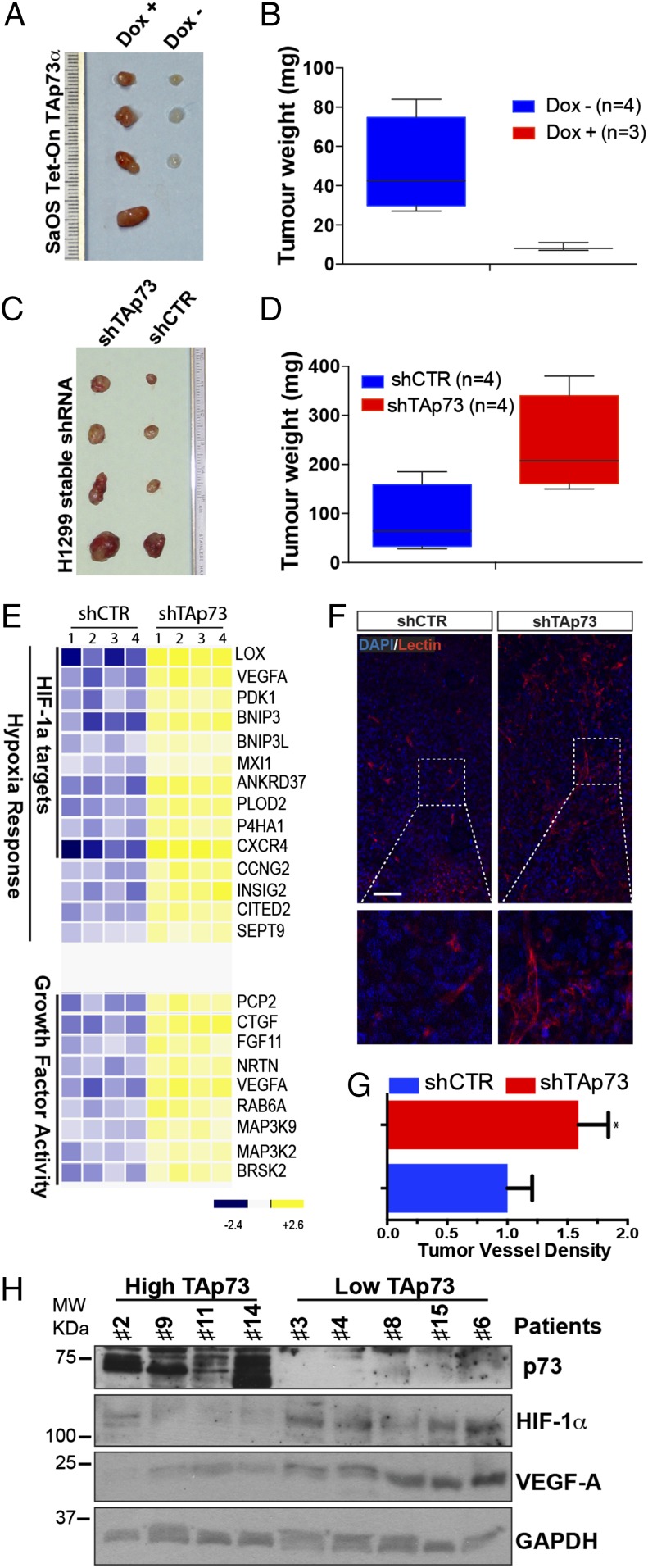
Tumor hypoxia and hypoxia-inducible factor 1 (HIF-1) activation are associated with cancer progression. Here, we demonstrate that the transcription factor TAp73 opposes HIF-1 activity through a nontranscriptional mechanism, thus affecting tumor angiogenesis. TAp73-deficient mice have an increased incidence of spontaneous and chemically induced tumors that also display enhanced vascularization. Mechanistically, TAp73 interacts with the regulatory subunit (α) of HIF-1 and recruits mouse double minute 2 homolog into the protein complex, thus promoting HIF-1α polyubiquitination and consequent proteasomal degradation in an oxygen-independent manner. In human lung cancer datasets, TAp73 strongly predicts good patient prognosis, and its expression is associated with low HIF-1 activation and angiogenesis. Our findings, supported by in vivo and clinical evidence, demonstrate a mechanism for oxygen-independent HIF-1 regulation, which has important implications for individualizing therapies in patients with cancer.
Keywords: VEGF; p53 family; p73; tumor progression; tumor vascularization.
Conflict of interest statement
The authors declare no conflict of interest.
Figures
Similar articles
-
Hypoxia-induced DNp73 stabilization regulates Vegf-A expression and tumor angiogenesis similar to TAp73.Cell Cycle. 2015;14(22):3533-9. doi: 10.1080/15384101.2015.1078038. Cell Cycle. 2015. PMID: 26267146 Free PMC article.
-
Hypoxia-inducible TAp73 supports tumorigenesis by regulating the angiogenic transcriptome.Nat Cell Biol. 2015 Apr;17(4):511-23. doi: 10.1038/ncb3130. Epub 2015 Mar 16. Nat Cell Biol. 2015. PMID: 25774835
-
TAp73 suppresses tumor angiogenesis through repression of proangiogenic cytokines and HIF-1α activity.Proc Natl Acad Sci U S A. 2015 Jan 6;112(1):220-5. doi: 10.1073/pnas.1421697112. Epub 2014 Dec 22. Proc Natl Acad Sci U S A. 2015. PMID: 25535357 Free PMC article.
-
p73: a Positive or Negative Regulator of Angiogenesis, or Both?Mol Cell Biol. 2015 Dec 28;36(6):848-54. doi: 10.1128/MCB.00929-15. Mol Cell Biol. 2015. PMID: 26711266 Free PMC article. Review.
-
Hypoxia-induced angiogenesis during carcinogenesis.J Biochem Mol Biol. 2003 Jan 31;36(1):120-7. doi: 10.5483/bmbrep.2003.36.1.120. J Biochem Mol Biol. 2003. PMID: 12542982 Review.
Cited by
-
The Undervalued Avenue to Reinstate Tumor Suppressor Functionality of the p53 Protein Family for Improved Cancer Therapy-Drug Repurposing.Cancers (Basel). 2020 Sep 22;12(9):2717. doi: 10.3390/cancers12092717. Cancers (Basel). 2020. PMID: 32971841 Free PMC article. Review.
-
Repair of Critical-Sized Mandible Defects in Aged Rat Using Hypoxia Preconditioned BMSCs with Up-regulation of Hif-1α.Int J Biol Sci. 2018 Mar 11;14(4):449-460. doi: 10.7150/ijbs.24158. eCollection 2018. Int J Biol Sci. 2018. PMID: 29725266 Free PMC article.
-
Involvement of E3 Ligases and Deubiquitinases in the Control of HIF-α Subunit Abundance.Cells. 2019 Jun 15;8(6):598. doi: 10.3390/cells8060598. Cells. 2019. PMID: 31208103 Free PMC article. Review.
-
p53/p73 Protein Network in Colorectal Cancer and Other Human Malignancies.Cancers (Basel). 2021 Jun 9;13(12):2885. doi: 10.3390/cancers13122885. Cancers (Basel). 2021. PMID: 34207603 Free PMC article. Review.
-
Isoform-specific disruption of the TP73 gene reveals a critical role for TAp73γ in tumorigenesis via leptin.Elife. 2023 Aug 31;12:e82115. doi: 10.7554/eLife.82115. Elife. 2023. PMID: 37650871 Free PMC article.
References
Publication types
MeSH terms
Substances
Grants and funding
LinkOut - more resources
Full Text Sources
Other Literature Sources
Molecular Biology Databases
Research Materials
Miscellaneous
