

In vivo albumin labeling and lymphatic imaging
- PMID: 25535368
- PMCID: PMC4291643
- DOI: 10.1073/pnas.1414821112
In vivo albumin labeling and lymphatic imaging
Abstract
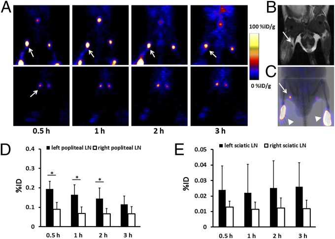
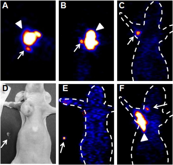
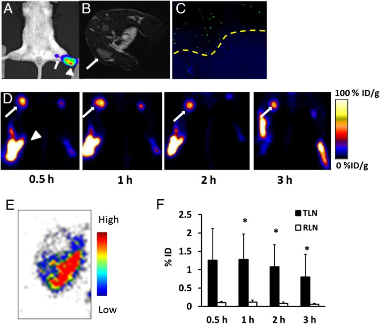
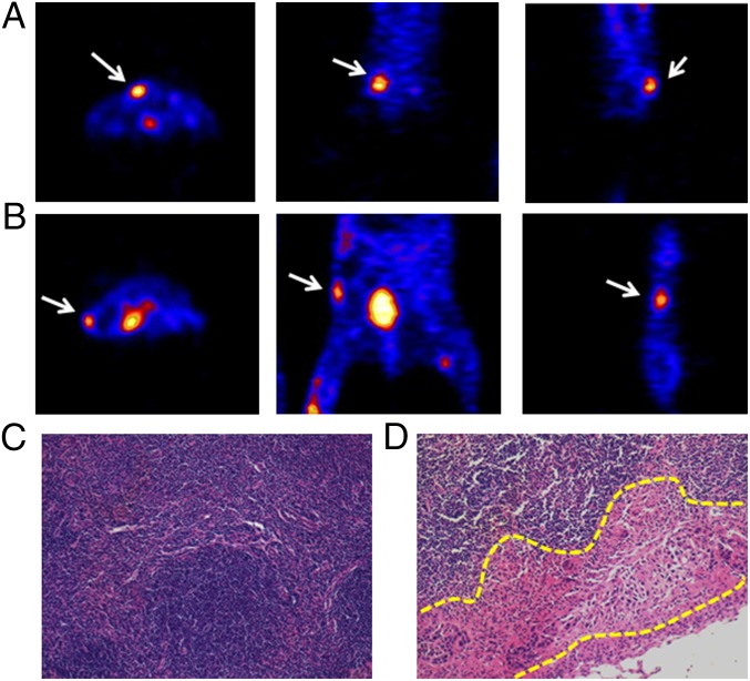
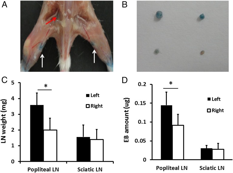
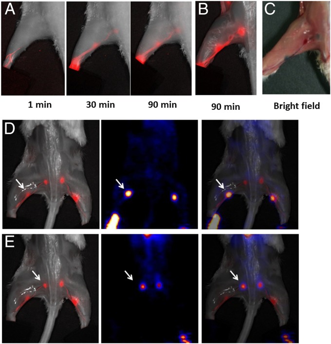
The ability to accurately and easily locate sentinel lymph nodes (LNs) with noninvasive imaging methods would assist in tumor staging and patient management. For this purpose, we developed a lymphatic imaging agent by mixing fluorine-18 aluminum fluoride-labeled NOTA (1,4,7-triazacyclononane-N,N',N''-triacetic acid)-conjugated truncated Evans blue ((18)F-AlF-NEB) and Evans blue (EB) dye. After local injection, both (18)F-AlF-NEB and EB form complexes with endogenous albumin in the interstitial fluid and allow for visualizing the lymphatic system. Positron emission tomography (PET) and/or optical imaging of LNs was performed in three different animal models including a hind limb inflammation model, an orthotropic breast cancer model, and a metastatic breast cancer model. In all three models, the LNs can be distinguished clearly by the apparent blue color and strong fluorescence signal from EB as well as a high-intensity PET signal from (18)F-AlF-NEB. The lymphatic vessels between the LNs can also be optically visualized. The easy preparation, excellent PET and optical imaging quality, and biosafety suggest that this combination of (18)F-AlF-NEB and EB has great potential for clinical application to map sentinel LNs and provide intraoperative guidance.
Keywords: Evans blue; PET; albumin; lymph node; optical imaging.
Conflict of interest statement
The authors declare no conflict of interest.
Figures
Similar articles
-
Clinical Translation of an Albumin-Binding PET Radiotracer 68Ga-NEB.J Nucl Med. 2015 Oct;56(10):1609-14. doi: 10.2967/jnumed.115.159640. Epub 2015 Aug 6. J Nucl Med. 2015. PMID: 26251416 Free PMC article.
-
In Vivo Labeling of Serum Albumin for PET.J Nucl Med. 2014 Jul;55(7):1150-6. doi: 10.2967/jnumed.114.139642. Epub 2014 May 19. J Nucl Med. 2014. PMID: 24842890 Free PMC article.
-
Imaging tumor-induced sentinel lymph node lymphangiogenesis with LyP-1 peptide.Amino Acids. 2012 Jun;42(6):2343-51. doi: 10.1007/s00726-011-0976-1. Epub 2011 Jul 19. Amino Acids. 2012. PMID: 21769497 Free PMC article.
-
The role of lymphatics in cancer as assessed by near-infrared fluorescence imaging.Ann Biomed Eng. 2012 Feb;40(2):408-21. doi: 10.1007/s10439-011-0476-1. Epub 2011 Dec 3. Ann Biomed Eng. 2012. PMID: 22139396 Free PMC article. Review.
-
Evans Blue Dye: A Revisit of Its Applications in Biomedicine.Contrast Media Mol Imaging. 2018 Apr 22;2018:7628037. doi: 10.1155/2018/7628037. eCollection 2018. Contrast Media Mol Imaging. 2018. PMID: 29849513 Free PMC article. Review.
Cited by
-
Metastatic status of sentinel lymph nodes in breast cancer determined with photoacoustic microscopy via dual-targeting nanoparticles.Light Sci Appl. 2020 Sep 16;9:164. doi: 10.1038/s41377-020-00399-0. eCollection 2020. Light Sci Appl. 2020. PMID: 33014359 Free PMC article.
-
A Comparison of Evans Blue and 4-(p-Iodophenyl)butyryl Albumin Binding Moieties on an Integrin αvβ6 Binding Peptide.Pharmaceutics. 2022 Mar 30;14(4):745. doi: 10.3390/pharmaceutics14040745. Pharmaceutics. 2022. PMID: 35456579 Free PMC article.
-
Direct loading of CTL epitopes onto MHC class I complexes on dendritic cell surface in vivo.Biomaterials. 2018 Nov;182:92-103. doi: 10.1016/j.biomaterials.2018.08.008. Epub 2018 Aug 6. Biomaterials. 2018. PMID: 30107273 Free PMC article.
-
Chemical Conjugation of Evans Blue Derivative: A Strategy to Develop Long-Acting Therapeutics through Albumin Binding.Theranostics. 2016 Jan 1;6(2):243-53. doi: 10.7150/thno.14322. eCollection 2016. Theranostics. 2016. PMID: 26877782 Free PMC article.
-
In and out: Leishmania metastasis by hijacking lymphatic system and migrating immune cells.Front Cell Infect Microbiol. 2022 Aug 12;12:941860. doi: 10.3389/fcimb.2022.941860. eCollection 2022. Front Cell Infect Microbiol. 2022. PMID: 36034709 Free PMC article.
References
-
- Swartz MA. The physiology of the lymphatic system. Adv Drug Deliv Rev. 2001;50(1-2):3–20. - PubMed
-
- Picker LJ, Butcher EC. Physiological and molecular mechanisms of lymphocyte homing. Annu Rev Immunol. 1992;10:561–591. - PubMed
-
- Tammela T, Alitalo K. Lymphangiogenesis: Molecular mechanisms and future promise. Cell. 2010;140(4):460–476. - PubMed
-
- Veronesi U, et al. Sentinel-node biopsy to avoid axillary dissection in breast cancer with clinically negative lymph-nodes. Lancet. 1997;349(9069):1864–1867. - PubMed
Publication types
MeSH terms
Substances
Grants and funding
LinkOut - more resources
Full Text Sources
Other Literature Sources
Medical
