. 2015 Jan 6;112(1):256-61.
doi: 10.1073/pnas.1417878112. Epub 2014 Dec 22.

Autophagic flux without a block differentiates varicella-zoster virus infection from herpes simplex virus infection

Affiliations

Autophagic flux without a block differentiates varicella-zoster virus infection from herpes simplex virus infection

Erin M Buckingham et al. Proc Natl Acad Sci U S A. .

Abstract

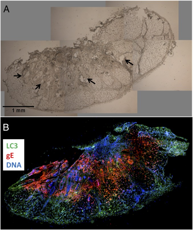
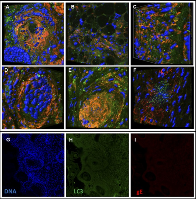
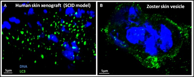
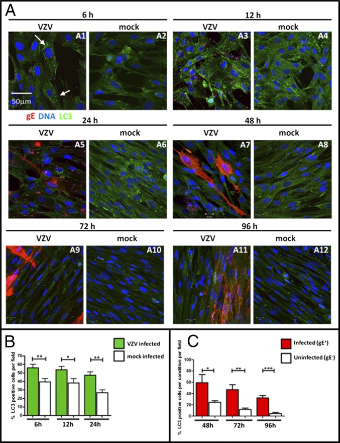
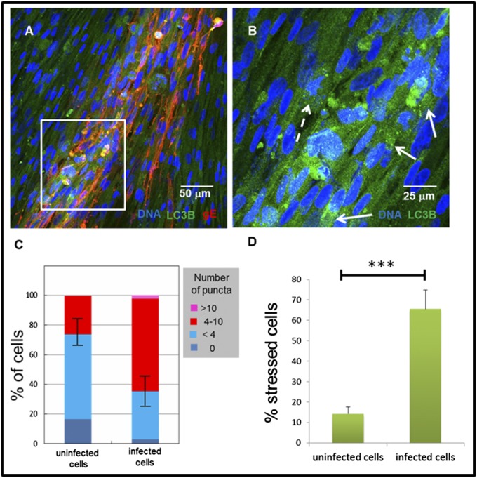
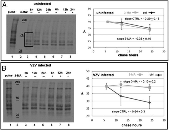
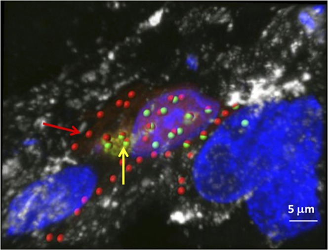
Autophagy is a process by which misfolded and damaged proteins are sequestered into autophagosomes, before degradation in and recycling from lysosomes. We have extensively studied the role of autophagy in varicella-zoster virus (VZV) infection, and have observed that vesicular cells are filled with >100 autophagosomes that are easily detectable after immunolabeling for the LC3 protein. To confirm our hypothesis that increased autophagosome formation was not secondary to a block, we examined all conditions of VZV infection as well as carrying out two assessments of autophagic flux. We first investigated autophagy in human skin xenografts in the severe combined immunodeficiency (SCID) mouse model of VZV pathogenesis, and observed that autophagosomes were abundant in infected human skin tissues. We next investigated autophagy following infection with sonically prepared cell-free virus in cultured cells. Under these conditions, autophagy was detected in a majority of infected cells, but was much less than that seen after an infected-cell inoculum. In other words, inoculation with lower-titered cell-free virus did not reflect the level of stress to the VZV-infected cell that was seen after inoculation of human skin in the SCID mouse model or monolayers with higher-titered infected cells. Finally, we investigated VZV-induced autophagic flux by two different methods (radiolabeling proteins and a dual-colored LC3 plasmid); both showed no evidence of a block in autophagy. Overall, therefore, autophagy within a VZV-infected cell was remarkably different from autophagy within an HSV-infected cell, whose genome contains two modifiers of autophagy, ICP34.5 and US11, not present in VZV.

Keywords: Epstein–Barr virus; ICP34.5; SCID-mouse; autophagosome; autophagy.


Conflict of interest statement

The authors declare no conflict of interest.

Figures

Fig. 1.
Fig. 1.
VZV induces extensive LC3 puncta indicative of autophagosomes in human skin cells in the xenografts in the SCID mouse model. VZV-infected human skin xenografts at 21 dpi were immunolabeled with antibodies to VZV gE (red) and to LC3 (green), as well as the Hoescht 33342 DNA stain (blue). Brightfield (A) and fluorescent (B) images of successive, whole sections of human skin xenografts showed extensive disruption of the tissue with prominent VZV gE expression as well as LC3 staining of autophagosomes. Areas of tissue disruption caused by VZV infection in A are noted by arrows.
Fig. 2.
Fig. 2.
VZV infection induces extensive autophagosome formation in human skin xenografts. (AF) Sections from 21 dpi human skin xenografts were immunolabeled with MAb 3B3 (anti-VZV gE, red) and anti-LC3 antibody [Santa Cruz (AE) or Epitomics (F), green] primary antibodies as well as H33342 (DNA stain, blue). Z-stack images were analyzed using the Imaris software to create 3D reconstructions that show LC3-positive autophagosomes as green spots and nuclei as blue spots. Abundant autophagosomes are noted in both samples. gE staining (red) is displayed flat to show diffuse and abundant staining in infected cells, often most highly concentrated in the ER/Golgi. Images shown are from either 400× total magnification (A and B) or 630× total magnification (CF). (GI) These three images each show a single fluorescent channel of the same 2D confocal image of mock infected human skin implants from a SCID-hu mouse at 630× total magnification. Mock infection showed no gE staining (I) and minimal LC3 staining (H).
Fig. 3.
Fig. 3.
VZV infection induces comparable autophagy in human skin and in a SCID mouse model. This comparison included 3D reconstructions of confocal microscopy images of a VZV-infected (21 dpi) human skin xenograft from a SCID mouse (A) and an impression of cells from a skin vesicle of a zoster patient (B). Both samples were immunolabeled with antibodies against LC3 (green) as well as a DNA stain (H33342, blue). Z-stack images at 630× total magnification were analyzed using the Imaris software to create 3D reconstructions that show LC3-positive autophagosomes as green spots and nuclei as blue spots. Abundant autophagosomes were noted in both samples. The gE staining (red) is displayed flat to show diffuse and abundant staining in infected cells, often most highly concentrated in the ER/Golgi.
Fig. 4.
Fig. 4.
Cell-free VZV infection of fibroblasts leads to autophagosome formation at early times post infection. MRC-5 cells were infected with a high input of cell free VZV-32 or were mock infected. Infected cells were fixed and permeabilized at 6, 12, 24, 48, 72, and 96 hpi, and stained with antibodies against VZV gE (red) and LC3 (green), as well as a DNA stain (blue). Samples were viewed by confocal microscopy. (A) Representative images of cells at each time point, both mock and VZV-infected. Arrows in panel A1 point to typical cells scored as LC3 positive due to the presence of four or more LC3 puncta. Images shown are 400× total magnification. (Scale bar, 50 μm.) (B) Quantitative analysis of LC3 positive cells per field. Confocal images were viewed in ImageJ and LC3-positive cells were identified as cells that contained four or more LC3 puncta. There were significantly more LC3-positive cells in the infected cultures than in the mock cultures. (**6 h, P < 0.008; *P < 0.024; **24 h, P < 0.001; n ≥ 9 images). (C) Quantitative analysis of LC3-positive cells in infected versus uninfected cells within the VZV-infected cultures. Confocal microscopy images of cells from VZV-infected cultures at 48, 72, and 96 hpi were analyzed using ImageJ. Cells were labeled as infected if they were gE-positive by immunolabeling and uninfected if they contained no gE. Significantly more infected cells were LC3-positive than uninfected cells. (*P < 0.033; **P < 0.001; ***P ≤ 0.0001; n = 10 images.)
Fig. 5.
Fig. 5.
Individual cells within a focus of infection after cell-free VZV infection exhibited LC3 puncta similar to cells infected with cell-associated VZV. MRC-5 cells were infected with a high-input of cell-free VZV-32 and fixed at 72 and 96 hpi. (A) Infected cells at 72 hpi (VZV gE; red) are apparent in the representative image at 400×. (B) Higher magnification (800×) images showing LC3 (green) in the area outlined by the white rectangle in panel A. Representative LC3 puncta indicative of autophagosomes are indicated by solid white arrows; similar puncta in a newly infected cell are indicated by a dashed white line. (C) Stacked bar chart showing the percentage of cells with a given number of LC3 puncta for both uninfected cells and infected cells from cultures infected with cell free VZV for 72–96 h. Blue colors indicate levels of puncta in unstressed cells, whereas red and pink colors indicate levels of puncta in stressed cells. (D) Different presentation of data from panel C, showing percent of stressed cells (cells that contain four or more LC3 puncta) in populations of uninfected (no gE staining) or infected (gE positive) cells. Cells infected with cell-free VZV exhibited more LC3 puncta indicative of autophagosomes than uninfected cells (***P < 0.007) and at levels similar to cells infected with cell-associated virus (1).
Fig. 6.
Fig. 6.
Autophagic flux during VZV infection of fibroblasts. VZV-infected monolayers (by cell-associated virus) were pulse labeled with 35S-labeled Met/Cys and then lysates saved at chase times 6, 12, and 24 h with and without 3-MA treatment. Reduced lysates at all pulse and chase time points were electrophoresed, transferred to PVDF membranes, and exposed to film. The darkness of a selected area of each lane was measured with ImageJ and plotted relative to the pulse value. The procedure was duplicated three times. (A) Uninfected MRC-5 cells. 3-MA treatment made little difference to the overall rate of degradation in uninfected cells. The area used to measure pixel density is shown as a box. (B) VZV-infected cells. 3-MA treatment significantly decreased protein degradation in VZV-infected cells, indicating increased autophagic flux in infected cells.
Fig. 7.
Fig. 7.
Analysis of autophagic flux with a tandem red/green fluorescent protein-tagged LC3 plasmid. MRC-5 cells transfected with the tandem fluorescent tagged LC3 plasmid were inoculated with VZV-infected cells for 72 h then immunolabeled. Samples were examined by confocal microscopy, and analyzed with Imaris software for 3D reconstruction. LC3 puncta are illustrated by spheres that are green and red (yellow arrow), showing LC3 expression in autophagosomes, or red only (red arrow), showing LC3 expression in autolysosomes, where GFP fluorescence has been extinguished. Nuclei (blue), VZV gE (white). (Scale bar, 5 μm.)

Similar articles

Cited by

References

    1. Buckingham EM, Carpenter JE, Jackson W, Grose C. Autophagy and the effects of its inhibition on varicella-zoster virus glycoprotein biosynthesis and infectivity. J Virol. 2014;88(2):890–902. - PMC - PubMed
    1. Jackson W, Yamada M, Moninger T, Grose C. Visualization and quantitation of abundant macroautophagy in virus-infected cells by confocal three-dimensional fluorescence imaging. J Virol Methods. 2013;193(1):244–250. - PMC - PubMed
    1. Kabeya Y, et al. LC3, a mammalian homologue of yeast Apg8p, is localized in autophagosome membranes after processing. EMBO J. 2000;19(21):5720–5728. - PMC - PubMed
    1. Tra T, et al. Autophagy in human embryonic stem cells. PLoS ONE. 2011;6(11):e27485. - PMC - PubMed
    1. Moffat JF, et al. Attenuation of the vaccine Oka strain of varicella-zoster virus and role of glycoprotein C in alphaherpesvirus virulence demonstrated in the SCID-hu mouse. J Virol. 1998;72(2):965–974. - PMC - PubMed

Publication types

MeSH terms

Substances