. 2015 Jan 6;112(1):148-53.
doi: 10.1073/pnas.1412089112. Epub 2014 Dec 22.

Activation of Rab8 guanine nucleotide exchange factor Rabin8 by ERK1/2 in response to EGF signaling

Affiliations

Activation of Rab8 guanine nucleotide exchange factor Rabin8 by ERK1/2 in response to EGF signaling

Juanfei Wang et al. Proc Natl Acad Sci U S A. .

Abstract

Exocytosis is tightly regulated in many cellular processes, from neurite expansion to tumor proliferation. Rab8, a member of the Rab family of small GTPases, plays an important role in membrane trafficking from the trans-Golgi network and recycling endosomes to the plasma membrane. Rabin8 is a guanine nucleotide exchange factor (GEF) and major activator of Rab8. Investigating how Rabin8 is activated in cells is thus pivotal to the understanding of the regulation of exocytosis. Here we show that phosphorylation serves as an important mechanism for Rabin8 activation. We identified Rabin8 as a direct phospho-substrate of ERK1/2 in response to EGF signaling. At the molecular level, ERK phosphorylation relieves the autoinhibition of Rabin8, thus promoting its GEF activity. We further demonstrate that blocking ERK1/2-mediated phosphorylation of Rabin8 inhibits transferrin recycling to the plasma membrane. Together, our results suggest that ERK1/2 activate Rabin8 to regulate vesicular trafficking to the plasma membrane in response to extracellular signaling.

Keywords: ERK; Rab GTPases; Rab8; guanine nucleotide exchange factor; phosphorylation.


Conflict of interest statement

The authors declare no conflict of interest.

Figures
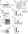
Fig. 1.
Fig. 1.
ERK1/2 phosphorylate Rabin8 in vitro and in cells. (A) Schematic representation of Rabin8 domains. Asterisks indicate the potential phosphorylation sites at Serine 16, 19, 247, and 250. (B) ERK2 phosphorylates Rabin8, but not GST, GST-Rab11a, or GST-Rab8a in vitro. Purified GST, GST-Rab11a, Rabin8, or GST-Rab8a were incubated with ERK2-CA in the presence of [32P]γ-ATP in an in vitro kinase assay. The samples were subjected to SDS/PAGE and autoradiography. Molecular weights (“MW”) in kDa are indicated to the left. (C) Rabin8 serine 16, 19, 247, and 250 are involved in ERK2 phosphorylation in vitro. Recombinant Rabin8 and Rabin8-4A mutant (S16/19/247/250A) were incubated with ERK2-CA and ERK2-KD. Samples were analyzed by SDS/PAGE and autoradiography. The upper panel is a Coomassie Blue-stained gel showing the purified wild-type and mutant Rabin8 used in the kinase assay. The lower panel shows the phosphorylation of Rabin8 by autoradiogram. (D) The levels of Rabin8 in vitro phosphorylation were quantified and normalized to the levels of Rabin8 proteins. The numbers in the y axis indicate the relative signal intensities on the autoradiogram. Error bar, SD. **P < 0.01; ***P < 0.001; n = 3. (E) EGF stimulates the phosphorylation of Rabin8 in cells. HEK293T cells expressing Flag-Rabin8 were treated with EGF for 5 min or treated with U0126 for 30 min before EGF treatment. Flag-Rabin8 proteins were immunoprecipitated from cell lysates and detected by Western blotting using anti-Flag antibody or anti-ERK1/2 phospho-substrate antibody. The levels of total ERK1/2 and phospho-ERK1/2 were also determined by Western blotting. (F) Quantification of the levels of Rabin8 phosphorylation in the above experiments. A.U., arbitrary units. Error bar, SD. *P < 0.05; n = 3. (G) The four serine residues on Rabin8 (S16, S19, S247, and S250) are required for phosphorylation by ERK1/2 in HEK293T cells. HEK293T cells were transfected with Flag vector control, Flag-tagged Rabin8, or Rabin8-4A. Immunoprecipitation was performed using the anti-Flag antibody. The immunoprecipitated proteins were analyzed with the anti-ERK1/2 phospho-substrate antibody and the anti-Flag antibody. Flag-Rabin8-4A had a significantly reduced level of phosphorylation compared with wild-type Rabin8. (H) Quantification of the levels of Rabin8 phosphorylation. Error bar, SD. **P < 0.01; n = 3.
Fig. 2.
Fig. 2.
Phosphorylation stimulates Rabin8 GEF activity. (A) Coomassie Blue-stained gel showing purified Rab8, Rabin8, Rabin8-4A, and Rabin8-4D used in the GEF assay. MW is indicated to the left. (B) The percentage of [3H]GDP bound to Rab8 was measured over time. Phosphorylation of Rabin8 by ERK2-CA significantly enhanced the GEF activity of Rabin8 toward Rab8. (C) Analysis of the release of [3H]GDP from Rab8 catalyzed by Rabin8, Rabin8-4A, or Rabin8-4D. Rabin8-4D was more potent than Rabin8 (squares) or Rabin8-4A (triangles) in promoting GDP release from Rab8. Results are representative of three independent experiments.
Fig. 3.
Fig. 3.
Rab8 is activated in cells in response to EGF. (A) HeLa cells were serum-starved overnight and then treated with EGF for 5 min or incubated with U0126 before EGF treatment. Cell lysates were incubated with purified GST-JFC1 fusion protein. The amounts of GTP-Rab8 bound to GST-JFC1 were analyzed by Western blotting with anti-Rab8 antibody. The cell lysates were also analyzed for total Rab8, ERK1/2, and phospho-ERK1/2. (B) Quantification of GTP-Rab8 levels in the above experiments. The amounts of GTP-Rab8 were normalized to the control level. The intensity of the bands was quantified by ImageJ and analyzed using the Student t test. Values are presented as mean ± SD. *P < 0.05; **P < 0.01; n = 3. (C) GTP-Rab8 was pulled down by GST-JFC1 from HeLa cells stably expressing GFP-tagged wild-type Rabin8, Rabin8-4A, or Rabin8-4D. (D) Quantification of GTP-Rab8. The levels of GTP-Rab8 are normalized to the level of GTP-Rab8 in cells expressing GFP-Rabin8 (n = 3).
Fig. 4.
Fig. 4.
Phosphorylation by ERK relieves the autoinhibition of Rabin8. (A) Schematic diagram showing the use of BRET in analyzing Rabin8 autoinhibition. NanoLuc (BRET donor) and HaloTag (BRET acceptor) were fused to the N and C terminus of Rabin8, respectively. If Rabin8 is in a closed conformation, BRET will occur owing to the close proximity between the donor and acceptor. However, the BRET signal will be significantly decreased when Rabin8 switches to an “open” conformation. (B and C) Intramolecular BRET signals detected for syntaxin-4 (“STX4”) and Rabin8 in the range of 590–650 nm. STX4 and Rabin8 were expressed using the pNLHT vector and purified from E. coli. STX4 was used as a positive control. Both Rabin8 and STX4 show positive BRET signals in the assay (red curves). As negative controls, STX4 and Rabin8 fail to be excited (blue curves) upon HaloTag removal by TEV protease. (D) BRET analysis of Rabin8 and Rabin8Δ(300-305). The luminescence intensities at 460 nm and 620 nm were measured for the donor and acceptor, respectively. The excitation was shown as a normalized ratio of RLU620nm/RLU460nm of substrate/ligand group minus the RLU620nm/RLU460nm of substrate-only group (Materials and Methods). The BRET ratios were normalized. Error bars, SD. *P < 0.05; n = 3. (E) Purified Rabin8-NLHT proteins were first incubated with ERK2-KD or ERK2-CA and then used in the BRET assay. BRET ratios were normalized and compared. **P < 0.01; n = 5. (F) Protein samples in E were subjected to SDS/PAGE, followed by Western blotting with the anti-Rabin8 antibody and the anti-ERK1/2 phospho-substrate antibody. (G) The luminescence intensities of Rabin8 and Rabin8-4D were measured. The BRET ratios were calculated and compared. **P < 0.01; n = 3.
Fig. 5.
Fig. 5.
Phosphorylation of Rabin8 promotes its binding to Rab8 but inhibits its interaction with Rab11. (A) Purified GST-Rabin8 was treated with ERK2-KD or ERK2-CA and then conjugated to glutathione Sepharose to test the binding to GFP-Rab8a[T22N] expressed in HEK293T cells. Ponceau S staining shows the GST-Rabin8 or GST on beads. MWs are shown to the left. The proteins bound to the beads were detected using anti-ERK1/2 phospho-substrate antibody or anti-GFP monoclonal antibody. (B) Quantification of GFP-Rab8a[T22N] bound to GST-Rabin8 with different treatments. Error bars, SD. *P < 0.05; n = 3. (C) Purified Rab11a or Rab11a[Q70L] was incubated with GST-Rabin8 that was pretreated with either ERK2-CA or ERK2-KD. The phosphorylation of Rabin8 and the bound Rab11a were analyzed by Western blotting. Ponceau S staining shows the amounts of GST-Rabin8 and GST used in the binding assay. MWs are shown to the right. (D) Quantification of Rab11a bound to GST-Rabin8 with different treatments. *P < 0.05; **P < 0.01; n = 3. (E) The binding of recombinant Rab11a[Q70L] to Rabin8, Rabin8-4A, and Rabin8-4D was examined. Coomassie Blue staining shows the GST-tagged Rabin8 variants, and GST was used as negative control. The bound Rab11a[Q70L] was analyzed by Western blotting using anti-Rab11a antibody.
Fig. 6.
Fig. 6.
Rabin8 phosphorylation regulates transferrin recycling. (A) Rabin8 regulates Tf recycling. hTERT-RPE1 cells were treated with either Luciferase siRNA or Rabin8 siRNA and then used in Tf recycling assay. Cells were fixed at indicated time points for microscopy. (Scale bar, 10 µm.) (B) Western blotting showing the knockdown of Rabin8 in cells. Rab8 and actin were used as controls. (C) The amounts of Tf retained in cells were quantified using ImageJ. Error bars, SD. One hundred cells were analyzed in each experiment (n = 3). *P < 0.05. (D) Tf was retained in recycling endosomes (colocalized with Rab11) in Rabin8 knockdown cells but exocytosed in control cells after chase for 60 min. Higher-magnification views of the boxed areas are shown under each image. (Scale bar, 10 µm.) (E) Fluorescence intensity of Rab11 and Tf signals along the line were analyzed using ImageJ. Tf partially colocalized with Rab11. (Scale bar: 10 μm.) (F) hTERT-RPE1 cells stably expressing siRNA-resistant, GFP-tagged Rabin8, Rabin8-4A, or Rabin8-4D were treated with siRNA against endogenous Rabin8 and subjected to transferrin recycling assay. Exocytosis of Tf was delayed in cells expressing Rabin8-4A. (Scale bar, 10 µm.) (G) The amounts of cell-associated Tf in F were quantified using ImageJ and plotted in the bar graph. Three independent experiments were performed, and 100 cells were analyzed for each experiment. *P < 0.05.

Similar articles

Cited by

References

    1. Stenmark H. Rab GTPases as coordinators of vesicle traffic. Nat Rev Mol Cell Biol. 2009;10(8):513–525. - PubMed
    1. Barr F, Lambright DG. Rab GEFs and GAPs. Curr Opin Cell Biol. 2010;22(4):461–470. - PMC - PubMed
    1. Mizuno-Yamasaki E, Rivera-Molina F, Novick P. GTPase networks in membrane traffic. Annu Rev Biochem. 2012;81:637–659. - PMC - PubMed
    1. Pfeffer SR. Rab GTPase regulation of membrane identity. Curr Opin Cell Biol. 2013;25(4):414–419. - PMC - PubMed
    1. Cherfils J, Zeghouf M. Regulation of small GTPases by GEFs, GAPs, and GDIs. Physiol Rev. 2013;93(1):269–309. - PubMed

Publication types

MeSH terms

Substances