. 2015 Jan 6;112(1):112-7.
doi: 10.1073/pnas.1417910112. Epub 2014 Dec 22.

Engineering an improved light-induced dimer (iLID) for controlling the localization and activity of signaling proteins

Affiliations

Engineering an improved light-induced dimer (iLID) for controlling the localization and activity of signaling proteins

Gurkan Guntas et al. Proc Natl Acad Sci U S A. .

Abstract

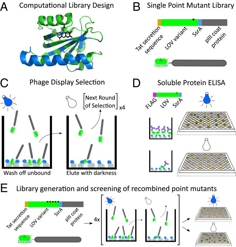
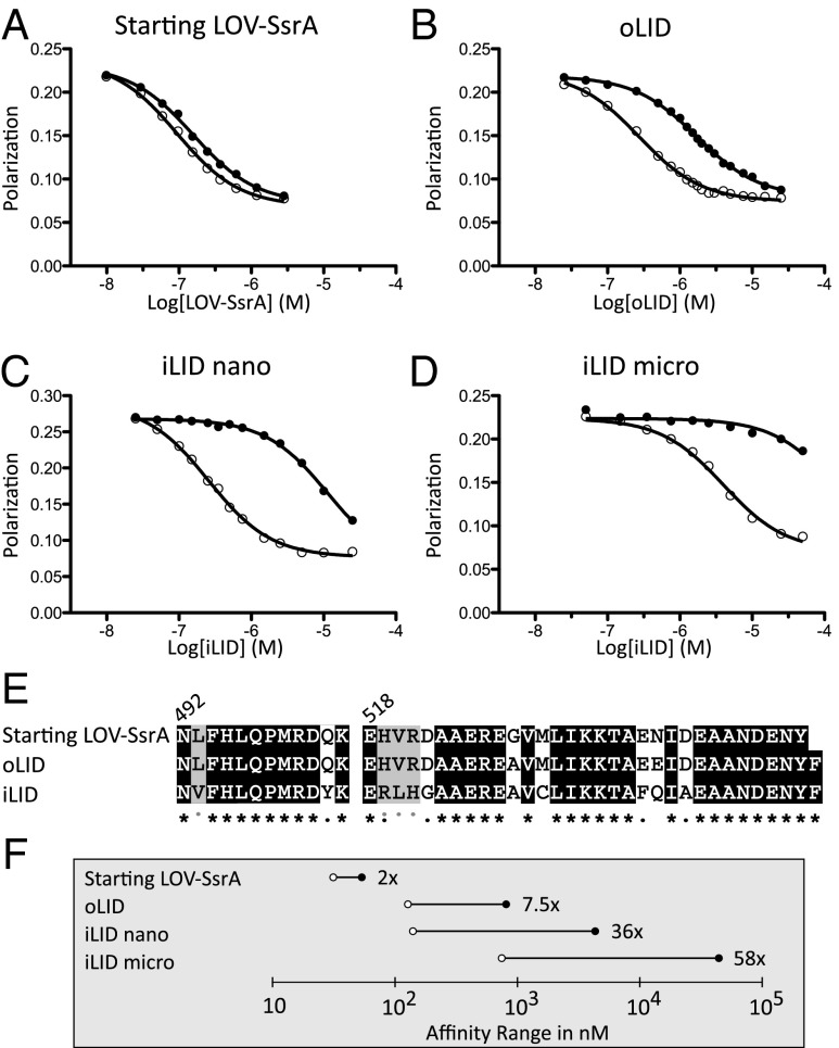
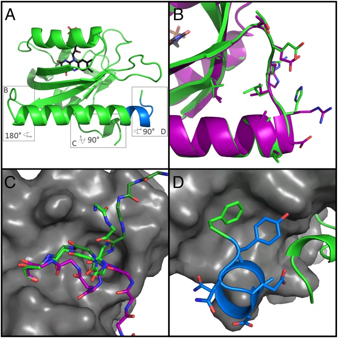
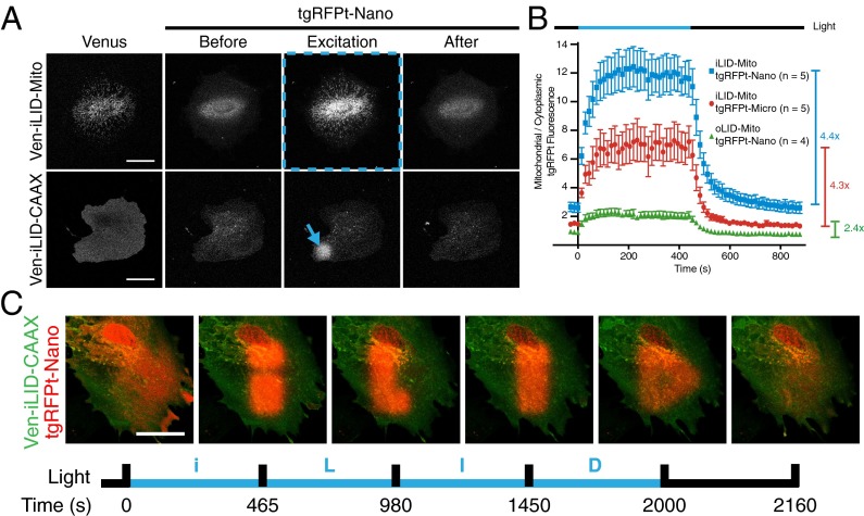
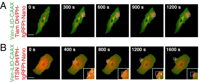
The discovery of light-inducible protein-protein interactions has allowed for the spatial and temporal control of a variety of biological processes. To be effective, a photodimerizer should have several characteristics: it should show a large change in binding affinity upon light stimulation, it should not cross-react with other molecules in the cell, and it should be easily used in a variety of organisms to recruit proteins of interest to each other. To create a switch that meets these criteria we have embedded the bacterial SsrA peptide in the C-terminal helix of a naturally occurring photoswitch, the light-oxygen-voltage 2 (LOV2) domain from Avena sativa. In the dark the SsrA peptide is sterically blocked from binding its natural binding partner, SspB. When activated with blue light, the C-terminal helix of the LOV2 domain undocks from the protein, allowing the SsrA peptide to bind SspB. Without optimization, the switch exhibited a twofold change in binding affinity for SspB with light stimulation. Here, we describe the use of computational protein design, phage display, and high-throughput binding assays to create an improved light inducible dimer (iLID) that changes its affinity for SspB by over 50-fold with light stimulation. A crystal structure of iLID shows a critical interaction between the surface of the LOV2 domain and a phenylalanine engineered to more tightly pin the SsrA peptide against the LOV2 domain in the dark. We demonstrate the functional utility of the switch through light-mediated subcellular localization in mammalian cell culture and reversible control of small GTPase signaling.

Keywords: PER-ARNT-SIM domain; Rosetta molecular modeling suite; computational library; optogenetic tool; phage display.


Conflict of interest statement

The authors declare no conflict of interest.

Figures

Fig. 1.
Fig. 1.
Schematic overview of selection and screening protocol for improved LOV variants. (A) Using Rosetta’s pmut_scan, we generated a library of point mutations at 49 positions within the AsLOV2 domain (blue residues). (B) Phage-display construct illustration. (C) The phage library was added to plates coated with MBP-SspB (gray-blue) in the presence of blue light and washed. Plates were moved to the dark and eluted phages were collected. (D) Top single-mutation sequences were recloned as flag tag fusions and individually expressed. Binding of soluble protein to MBP-SspB coated plates was measured after exposure to blue light and sequestration in the dark by ELISA. (E) Mutations with the highest dynamic range were recombined to generate a new library of LOV variants, which was screened using the procedure shown in C and D.
Fig. 2.
Fig. 2.
Characterization of iLID nano and iLID micro. Competitive fluorescence polarization binding assays measure affinities of heterodimerization under blue light (○) and in the dark (●). (A) Incorporation of SsrA into final turn of the AsLOV2 Jα helix. (B) Addition of helix-strengthening mutations (G528A, N538E) and C-terminal phenylalanine. (C) Top sequence from phage display and screening, iLID, has a 36-fold change in affinity for SspB because of light. (D) A mutation to SspB (R73Q) yields a heterodimer pair that switches over the micromolar range of affinities with a 58-fold change in affinity. (E) Sequence alignment of starting, oLID, and iLID. Black, unmutated; gray, similar amino acid mutation. Gray dots indicate iLID mutations that converged in all top four sequences. (F) Comparison of lit (○) and dark (●) affinities between the original heterodimer pairs and our two new pairs, iLID nano and iLID micro.
Fig. 3.
Fig. 3.
Structure of iLID yields insight to role of mutated residues. (A) Overall topology iLID (green) remains unchanged from AsLOV2. Boxes surround three areas of interest to be shown in B, C, and D. (B) Alignment with AsLOV2 (PDB ID code 2V0U, purple) reveals no major backbone rearrangement of the hinge region due to mutations. (C) Close up view of A'α helix from iLID (green), AsLOV2 (purple), and PA-Rac1 (PDB ID code 2WKP, dark green), show different orientation in caged LOV variants from uncaged. (D) SsrA epitope (blue) contains an extra helical turn before wrapping back and interacting with the PAS fold. Designed C-terminal phenylalanine (green) packs nicely into a hydrophobic pocket of the LOV domain.
Fig. 4.
Fig. 4.
iLID provides improved local recruitment in cell culture. (A) IA32 fibroblasts expressing membrane (CAAX) and mitochondrial anchored Venus-iLID and cytoplasmic TagRFP-T-Nano. Localized activation (denoted by blue markings) caused relocalization of TagRFP-T-Nano. (Scale bar, 50 μm.) (B) A ratio of mitochondrial to cytoplasmic TagRFP-T signal intensity during a time course of whole-cell activation as shown in row 1 of A for each pair of mitochondria anchored switches. (C) Patterned activation of Venus-iLID-CAAX shows tight spatial and temporal control of TagRFP-T-Nano localization within a cell. (Scale bar, 50 μm.)
Fig. 5.
Fig. 5.
Spatial control of GEF DH/PH domains by iLID produces localized control of Rac and Cdc42 activity. IA32 cells expressing Venus-iLID-CAAX and (A) Tiam DH/PH-TagRFP-T-Nano or (B) ITSN DH/PH-TagRFP-T-Nano. iLID was activated in the regions highlighted in blue. White arrowheads mark vesicles. Images are representative (n > 5). (Scale bars, 50 μm.)

References

    1. Rivera VM, et al. A humanized system for pharmacologic control of gene expression. Nat Med. 1996;2(9):1028–1032. - PubMed
    1. Yazawa M, Sadaghiani AM, Hsueh B, Dolmetsch RE. Induction of protein–protein interactions in live cells using light. Nat Biotechnol. 2009;27(10):941–945. - PubMed
    1. Levskaya A, Weiner OD, Lim WA, Voigt CA. Spatiotemporal control of cell signalling using a light-switchable protein interaction. Nature. 2009;461(7266):997–1001. - PMC - PubMed
    1. Kennedy MJ, et al. Rapid blue-light-mediated induction of protein interactions in living cells. Nat Methods. 2010;7(12):973–975. - PMC - PubMed
    1. Konermann S, et al. Optical control of mammalian endogenous transcription and epigenetic states. Nature. 2013;500(7463):472–476. - PMC - PubMed

Publication types

MeSH terms

Associated data